
Contents
 In my last column, I discussed some of the unique features that help birds to fly — from hearts that pump five times their volume per second, to their respiratory system, which allows them to aerate more quickly with a larger blood volume compared to that of mammals. Their shape reduces turbulence, and their organs are lighter in weight. Yet one organ that is relatively heavy is their intestines — their guts. So let’s look at the structure and function of this complex organ, and at adaptations birds use to make the most of it.
From Beak to Crop & Into The Esophagus
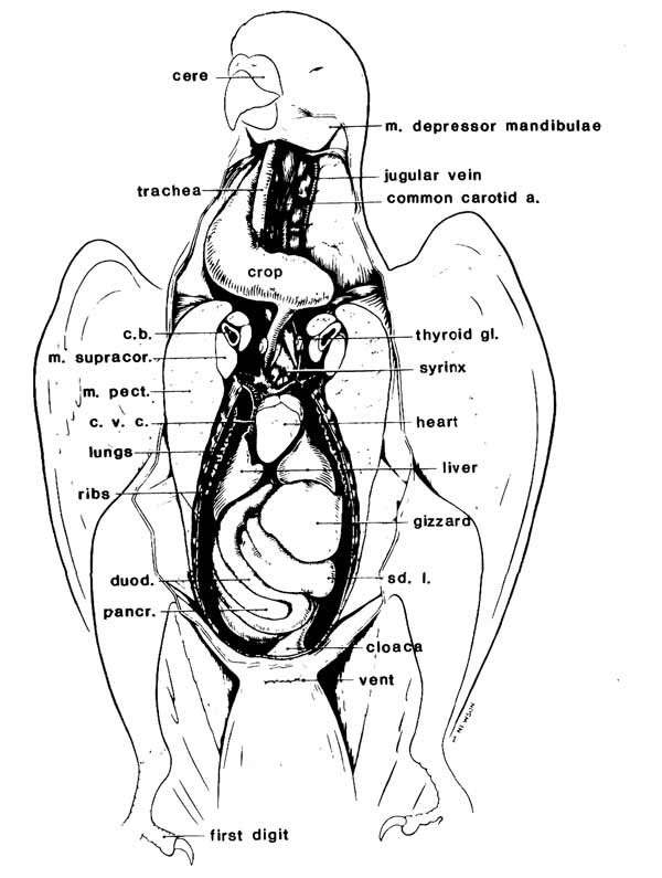
The gut, or GI tract, starts anatomically at the bird’s beak and ends at its vent. The proximal portion of the GI tract consists of the beak, oropharynx, cervical esophagus, crop and thoracic esophagus. The esophagus of birds is distensible and sits on the right side of the neck — just the opposite of what occurs in mammals. Some birds, but not all, have a distensible portion of the esophagus, which is the crop or ingluvies.
The crop is an esophageal diverticulum and varies in size depending on the species and the age. Young chicks have a much larger crop than adult birds of the same species. In addition, the size of the crop decreases in size with sick birds that are not eating normal volumes of food. This is important when determining the volume to be fed when gavage-feeding critically ill birds. In psittacine birds (parrots), the crop normally extends from the right side of the neck at the base of the thoracic inlet to the left before narrowing into the thoracic esophagus. From the thoracic esophagus, which lies on the left, the GI tract continues into the stomach.
The Stomach — A Rotatory Grinder
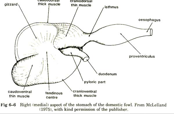
The stomach of birds consists of the proventriculus, isthmus and ventriculus or the gizzard. It lies on the cranial left side of the abdominal cavity. Since birds do not have a diaphragm, it is termed the abdominal coelom. The proventriculus is the proximal portion and is the glandular portion that secretes the digestive enzymes. There is a narrow isthmus or intermediate portion followed by a saclike to highly muscular gizzard.
The gizzard acts to grind food, making it more readily available for the enzymes to digest. In psittacine species, there is a rotatory motion of the food in the stomach with food moving from the proventriculus to the ventriculus for grinding and then back into the proventriculus. This normal pattern can be disturbed by a variety of disease processes, with the most noted being proventricular dilitation disease or PDD.
The Small Intestine — Reduced Length & A Clever Strategy
The anatomy of the small intestine includes the duodenum, jejunum and ilium. The majority of the pancreas lies between the 2 limbs of the duodenum. The splenic portion is located near the back of the bird and is not as closely associated with the GI tract. This portion contains more of the endocrine portion of the pancreas. The bile duct and pancreatic ducts open near the terminal portion of the ascending duodenum.
In most species of birds, the jejunum and ileum are composed of a number of U-shaped loops along the edge of the dorsal mesentery, which is the membrane that takes origin from the back and hangs into and supports the intestines. From our perspective for understanding flight, birds have a limited length to their small intestines and have a unique peristalsis-retroperistalsis pattern to digest and absorb nutrients.
What that means is that they reduce the length and hence the weight of the intestines — a brilliant move! Then they run the digesting foods back (retroperistalsis) and forth (peristalsis) in this shorter segment until digestion and absorption has been completed—so they don’t need to move a bunch of guts around like mammals!
From Ceca to Vent
The next physiologic principle involves the last part of the GI tract. This final segment of the GI tract includes the large intestine or rectum and the cloaca. The large intestine begins at the level of the paired ceca. The large intestine consists of a short and straight segment and is probably homologous to the mammalian rectum.
The ceca — if the species of bird has them — arise at the junction of the end of the small intestine and the beginning of the large intestine or rectum. Parrots, however, don’t have ceca or they are rudimentary, and the ceca of passerines are small. Poultry and ostrich have very large ceca. The large intestine of birds functions similarly to mammals; its major task is to reabsorb water. That is an important factor for flight as well. To see how that works we need to explore the cloaca.
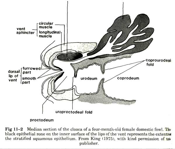
The cloaca of birds is divided into the coprodeum, urodeum and proctodeum. Its basic organization is similar between species, except for the possible phallus in waterfowl and ratites. The coprodeum is the cranial-most compartment and it stores fecal material. The coprodeum is continuous with the large intestine, but is separated from the urodeum by the coprourodeal fold.
The urine and urates are stored in the urodeum prior to evacuating the cloaca, as the ureters open into this receptacle. The oviducts or ductus deferens open into the urodeum. The more caudal fold is the uroproctodeal fold that separates the urodeum from the central and distal-most cavity; the proctodeum. The cloacal bursa opens into the proctodeum. The vent is the opening of the proctodeum to the outside.
The Role Of Retroperistalsis In Water Reabsorption
Interestingly, urine from the ureters flow into the urodeum. The urine of birds is not as concentrated as that of mammals so that means that there may be a larger volume and hence weight of the water. But birds have solved that problem because they can move that urine by retroperistalsis from the urodeum into the coprodeum to the large intestine.
Remember, the large intestine is designed for the reabsorption of water. So birds have again solved a problem using a physiologic function — retroperistalsis! This ability to run fluids and gut contents backwards help solve the weight problem for flight. The shorter GI tract works more efficiently while reducing weight. Retroperistalsis of the urine from the kidneys help to reduce carrying water around and allows water to be used more efficiently. Birds are their own miracle! How brilliant from a physiologic perspective!
Birds And The Miracle Of Flight
Humans have often marveled at the ability of birds to fly and sometimes suffered terribly in our initial efforts to join them through technology. Birds have solved the problem through aerodynamic design of their own, streamlined bodies, reduced weight, a powerful heart and an efficient respiratory system, among other adaptations.
Also important are their adaptations to their guts that allow rotatory digestion in the stomach, peristalsis and retroperistalsis in shortened small intestines, and water absorption that allows effective water management. While their GI tract is relatively short, it works efficiently to fuel the flight that thrills us. From starling to eagle, they’re all pretty impressive from a flight perspective.
Wow I never thought about everything that a bird needs to fly!!! Right down to water conservation!!