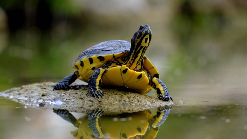
Photo: Engin_Akyurt/pixabay
Abstract
Respiratory disease in chelonians bears many similarities to respiratory disease in any animal. The disease can be infectious, inflammatory, neoplastic, or toxic. Infectious causes can be bacterial, fungal or viral. From a practical point-of-view for the clinician, what matters is the differences presented by chelonians. Slight adjustments to the normal case workup are required due to differences in anatomy, physiology, and specific agents of disease. Otherwise, the workup is similar to other species. By that logic, understanding the differences is the most important thing to offering good quality of care for the affected animals in a veterinary practice.
There are numerous anatomical differences from the mammalian species commonly taught in veterinary medical school curricula. In mammals, inhalation and exhalation are accomplished by expansion and contraction of the rib cage and the diaphragm. The most obvious difference with chelonians is the presence of the shell. Rigid fusion of the ribs and sternum varies greatly from the flexible rib cage structure of mammals. They also lack a diaphragm. The lungs don’t terminate in alveolar sacs, but are rather divided into multiple chambers with a reticulated structure of the lung parenchyma housing the alveoli. The trachea has complete cartilaginous rings through at least a portion of the structure, which branches cranially compared to most mammalian species. Finally, the glottis is more cranial in the oral cavity in most chelonians by comparison to mammalian counterparts.
What do these differences mean? Let’s go through a typical workup to get at the practical differences from the perspective of clinical practice. Without a flexible rib cage and a diaphragm, inhalation and exhalation are both active processes depending upon movement of the limbs and various abdominal muscles. Respiratory rate is normally counted by looking at the inward and outward movement of the forelimbs. The rigid shell is also a barrier to auscultation of the lungs with a stethoscope. However, even if one could hear the lungs, the differing structure would mean that the lung sounds would differ in these animals. External nares are rigid in most chelonians, so nostril flare cannot be considered a reliable indication of dyspnea. The oral cavity is harder to open, but once opened, the clinician is usually rewarded with good visualization of the internal nares and the glottis.
For diagnostic testing, the lack of diaphragm leads to superimposition of viscera with standard radiographic positioning. Horizontal beam is a minimum requirement for truly diagnostic images in all views, but computed tomography should be considered the standard means for effectively imaging the lower airways. Pulmonary wash or endoscopic views are possible with adjustments to the sampling methods.
For infectious agents, cultures and next-generation DNA sequencing can be used. For tortoises with upper respiratory disease, PCR for mycoplasma should be considered. Primary viral pathogens are most commonly herpesvirus and ranavirus.
Outline
|
 |
- Diagnostic testing
- Imaging
- Radiographs – horizontal beam
- CT
- Culture, cytology, and biopsy
- Endoscopy
- Imaging
- Specific pathogens and agents of disease
- Mycoplasma
- Viral
- Herpesvirus
- Ranavirus
- Treatment considerations
- Oral versus injectable
- Nebulization
About the presenter
Mike Corcoran is a certified aquatic veterinarian and a board-certified specialist in reptile and amphibian practice through the American Board of Veterinary Practitioners. Dr. Corcoran currently works as an exotic animal medicine consultant in Southern California. He also works part-time at the Arizona Exotic Animal Hospital, where he mentors reptile and amphibian residents…Dr. Corcoran has served on the Board of the Association of Reptile and Amphibian Veterinarians (ARAV) for more than 5 years. He also served as a Past President of ARAV and the Chair of the ARAV Legislation and Animal Welfare Committee. [MORE]
Webinar recording
Transcript
Download a transcript of Dr. Mike Corcoran’s recording, available as both a Word document and PDF.
![]()
Post-test
Take the brief post-test to earn 1 hour of continuing education credit. With a passing grade, you will receive a continuing education certificate in jurisdictions that recognize AAVSB RACE approval.
Expert Q&A
Virtually all questions were addressed during the live event, two remaining questions were answered by email and are shared below:
Have you had a situation where it was risky to even sedate to do hands-on diagnostics? What do you do in that situation – Oxygen cage? Nebulize with aminophylline?
Yes, placing [these patients] in oxygen and nebulizing would be appropriate if you have one in distress, but once they have time in oxygen, remember that sedation will reduce anxiety and therefore reduce the oxygen demand in most cases. Once you have reduced distress with oxygen, I will often give a dose of midazolam…and return later. A complete exam may not happen on the first day in bad cases.
In tortoises with oral/pharyngeal plaques – we used to say this was herpes- and would treat with acyclovir. Is this now a ranavirus? Have you used antivirals in any of [these] chelonians?
I haven’t used any antivirals in reptiles. I know some use it with papillomas in sea turtles, but I never saw those in practice, only the cold-stunned sea turtles. The papillomas occur in warmer waters. I vaguely remember a paper addressing its use in pet turtles with herpes, but now I can’t find it, so it may only be in proceedings.
Editor’s note: Although not the paper to which Dr. Corcoran refers, the manuscripts listed below discuss the use of antivirals in chelonians:
Okoh GR, Horwood PF, Whitmore D, Ariel E. Herpesviruses in reptiles. Front Vet Sci. 2021;8:642894. doi: 10.3389/fvets.2021.642894. PMID: 34026888; PMCID: PMC8131531.
Gandar F, Marlier D, Vanderplasschen A. In vitro and in vivo assessment of eprociclovir as antiviral treatment against testudinid herpesvirus 3 in Hermann’s tortoise (Testudo hermanni). Res Vet Sci. 2019;124:20-23. doi: 10.1016/j.rvsc.2019.02.001. Epub 2019 Feb 16. PMID: 30782569.
Marschang RE, Gravendyck M, Kaleta EF. Herpesviruses in tortoises: investigations into virus isolation and the treatment of viral stomatitis in Testudo hermanni and T. graeca. Zentralbl Veterinarmed B. 1997;44(7):385-94. doi: 10.1111/j.1439-0450.1997.tb00989.x. PMID: 9323927.
RACE approval
This program is approved by the American Association of Veterinary State Boards (AAVSB) Registry of Approved Continuing Education (RACE) for 1 hour of continuing education credit for veterinarians and veterinary technicians in jurisdictions that recognize AAVSB RACE approval.
References
References
Caron M, Kanda I, Mitchell M, Boyer T. Effectiveness of nasal flush treatments in Mycoplasma PCR- or DNA sequencing–positive tortoises. Journal of Herpetological Medicine and Surgery. 2025;35(1):40-44. doi: 10.5818/JHMS-D-24-00029.
Lyson TR, Schachner ER, Botha-Brink J, et al. Origin of the unique ventilatory apparatus of turtles. Nat Commun. 2014;5:5211. doi: 10.1038/ncomms6211. PMID: 25376734.
Mans C, Drees R, Sladky KK, Hatt JM, Kircher PR. Effects of body position and extension of the neck and extremities on lung volume measured via computed tomography in red-eared slider turtles (Trachemys scripta elegans). J Am Vet Med Assoc. 2013;243(8):1190-1196. doi: 10.2460/javma.243.8.1190. PMID: 24094268.
Pardo MA, Divers S. Jugular central venous catheter placement through a modified Seldinger technique for long-term venous access in chelonians. J Zoo Wildl Med. 2016;47(1):286-290. doi: 10.1638/2015-0143.1. PMID: 27010289.
Rieppel O. Turtle origins. Science. 1999;283(5404):945-946(1999). doi:10.1126/science.283.5404.945.
Corcoran M. Respiratory disease in chelonians. LafeberVet website. April 14, 2025. Available at https://lafeber.com/vet/respiratory-disease-in-chelonians/