Abstract
Radiography of exotic pets can be challenging. Small patient size makes restraint and positioning difficult. The small size and unique anatomy of small exotic patients can also cause confusion when interpreting images, and even the slightest amount of motion artifact can complicate interpretation. Familiarity with normal anatomy is the basis for radiographic interpretation in any species.
The choice between manual restraint or sedation for radiographs should be determined by several factors. For some exotic pets, manual restraint is effective for radiographic imaging, however, the overall condition of the patient, including its stress level, as well as the speed of the X-ray generating equipment, should all be considered. Perhaps most importantly, the expertise level of the staff restraining the patient must also be considered. Dyspneic animals should almost always be anesthetized for radiography.
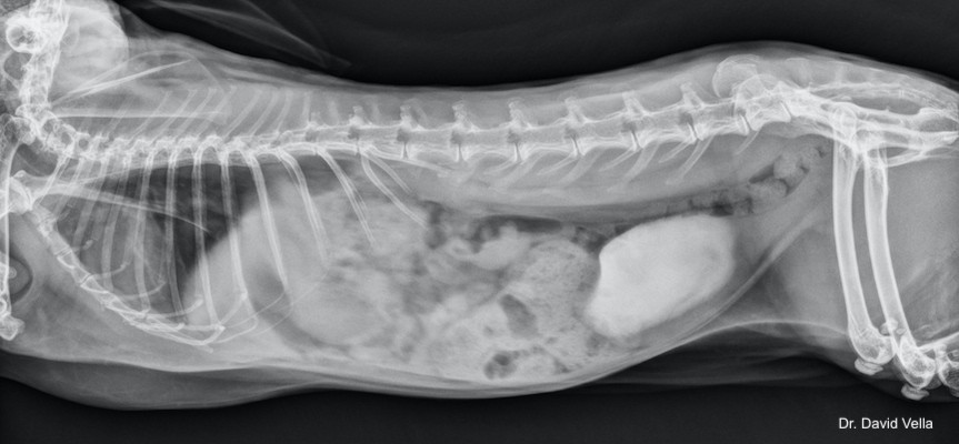
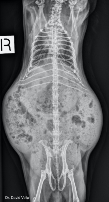
This presentation reviews the normal radiographic and sonographic anatomy of ferrets (Mustela putorius furo), rabbits (Oryctolagus cuniculus), and popular rodents in detail, while also touching upon some unique anatomic features of the sugar glider (Petaurus breviceps) and hedgehog (Atelerix albiventris). Common pathologic conditions and typical radiographic findings are also explored. Case examples are used to introduce these concepts.
Outline
 |
|
|
Download a PDF of a detailed presentation outline. 
About the presenter
Dr. Natalie Antinoff is the owner of Antinoff Veterinary Services, which provides veterinary relief and consulting for various practices, primarily specialty facilities. Dr. Antinoff also regularly provides scheduled per diem care for non-traditional pets at Texas Avian & Exotic Animal Hospital in Grapevine, Texas and Mountain West Veterinary Specialists in Layton, Utah. Dr. Antinoff has also been a consultant for the Veterinary Information Network since 1997. [Learn more].
Webinar recording
Post-test
With a passing grade of 70% or higher, you will receive a continuing education certificate for 2 hours of continuing education credit in jurisdictions that recognize American Association of Veterinary State Boards (AAVSB) Registry of Approved Continuing Education (RACE) approval.
RACE approval
This program is approved for 2 hours of continuing credit for veterinarians and veterinary technicians in jurisdictions that recognize AAVSB RACE approval.
Reference
Boehmer E, Crossley D. Objective interpretation of dental disease in rabbits, guinea pigs and chinchillas: Use of anatomical reference lines. Tierärztl Prax 2009;37(K):250-260. Available at http://www.medirabbit.com/GE/Zahnkrankheit/Publications/rabbit_dental_lines.pdf.
Antinoff N. Small mammal imaging and radiographic cases. LafeberVet website. Sep 30, 2021. Available at https://lafeber.com/vet/small-mammal-imaging-and-radiographic-cases/