Introduction
The bird egg, sometimes called a “miracle of packaging”, can be intimidating for the avian veterinarian. This amazing structure comes with a plethora of new vocabulary terms, complex anatomy, and intricate physiology (Fig 1). LafeberVet’s “Understanding the Avian Egg” describes the basics of egg anatomy from the eggshell to the embryo with a brief description of hatch.

Figure 1. Schematic illustrating egg anatomy by de:Benutzer: Horst Frank via Wikimedia Commons. (1) Eggshell (2) Outer shell membrane (3) Inner membrane (4) Chalaza (5) Outer thin albumen (6) Inner thick albumen (7) Vitelline membrane (8) Nucleus of pander (9) Germinal disc (blastoderm) (10) Yellow yolk (11) White yolk (12) Internal albumen (13) Chalaza (14) Air cell (15) Cuticula. Click image to enlarge
Cuticle
The outermost component of the eggshell is called the “cuticle”. Composed of protein, polysaccharide, and lipid, the thin, waxy cuticle protects the egg from water evaporation as well as microbial invasion. The cuticle is found on most, but not all, bird eggs (Johnson 1999).
Testa
The testa is the largest portion of the eggshell consisting primarily of calcite, the crystalline form of calcium carbonate. The testa can be further divided into external, spongy, and mammillary layers.
The external layer, also known as the ‘outermost crystal layer’ is present in many, but not all birds. This dense crystalline layer measures anywhere from 3-8 μm in thickness and serves as a transitional zone to the outer surface of the egg.
The spongy or palisade layer makes up approximately two-thirds of the shell. Approximately 200 μm thick, the palisade layer contains calcified crystals intermixed with an organic matrix, that consists of a series of protein layers and mucopolysaccharides.
The thin, inner cone or mammillary layer consists of organic cores and cone-shaped knobs embedded within the outer shell membrane below. Cores are made primarily of protein, but contain carbohydrates and mucopolysaccharides. Deposition of calcium carbonate crystals begins upon these organic cores (Johnson 1999).
Normal areas of incomplete crystallization create thousands of funnel-shaped openings or pores within the eggshell. The number of pores varies with the species and has been shown to correlate with metabolic demands. The number of pores increases as egg weight decreases. The diameter of the pores ranges from 0.3-0.9 μm. The pores serve as a major site of gas exchange (water and carbon dioxide) as well as water vapor transport across the eggshell. The exchange of gases and water occurs primarily by passive diffusion.
When eggs are pigmented, variable amounts of blue-green biliverdin and/or red-brown porphyrin crystals are deposited within the inorganic portion of the testa and sometimes the cuticle (Fig 2).

Figure 2. Robin (Turdus migratorius) eggs are a beautiful blue color. Photo credit: Soltrcy. Click to enlarge.
Shell membranes
If you’ve ever peeled a boiled egg, you’ve noted the presence of membranes beneath the eggshell. Semipermeable shell membranes serve as a barrier against microbial invasion and they also play a minor role in egg respiration (Nys 2007).
The outer shell membrane is in contact with the mammillary layer of the testa, while the inner shell membrane is in direct contact with albumen (see below). The inner and outer shell membranes are firmly attached except in a small region, usually at the blunt end of the egg.
Air cell
The air cell, located at the blunt end of the egg, is created by separation of the inner and outer shell membranes. The air cell is created as the egg cools after oviposition or egg laying. Creation of the air cell is a dangerous time in egg development, as this is the most likely time for microbes to be drawn into the egg.
Go to Hatch below for more information on the air cell.
Chorion
The chorion is a highly vascular outer embryonic membrane that is found in not
only birds, but also reptiles and mammals. The chorion is another
semipermeable membrane that participates in gaseous exchange.
Amnion
The amnion is a fluid filled sac that surrounds and cushions the embryo. The amnion serves as a source of protein, water, and minerals throughout embryological development. Albumen drains into amnion, allowing this structure to serve as a protein source.
There are four distinct layers of albumen (Johnson 1999):
- Outer thin (fluid) layer
- Outer thick layer
- Inner thin (liquid) layer
- Inner thick chalaziferous layer
Chalazae
The inner thick layer of albumen or the chalaziferous layer, consists of a dense,
gel-like albumen that holds the yolk within the center of the egg. Chalaze
consists of a double strand at the sharp end of the egg and a single
strand at the rounded end.
Allantois
The allantois is another vascular fetal membrane of reptiles, birds, and mammals that forms as a pouch from the hindgut.
Eggs need to be turned regularly, and the reason for this relates to the existence of the allantois and amnion. Regular turning stirs nutrients and waste material within the egg, while preventing fetal membranes from adhering to the embryo. Psittacine eggs pulled for artificial incubation are typically turned every 1 to 2 hours. Inadequate turning rates have an adverse effect on embryonic development and are associated with increased mortality rates.
Yolk
The yolk membrane, or vitelline membrane, consists of four membranes that separate the yolk sac from albumen. Yolk is another important nutrient, providing protein and lipids in late development and during the first few days of life.
Not all yolk is the same. Yellow yolk is higher in fat, while white yolk is higher in protein. The latebra is a region of white yolk that can be seen during candling. The latebra consists of small round center, a neck, and a cone-shaped disc that lies beneath the germinal disc.
Candling is a technique used to evaluate the egg (Box 1). Candling is used to look for evidence for:
- Fertility or infertility
- Cracks
- Shell thickness
- Membrane and vessel integrity
- Air cell size and shape
- Yolk size
- Chick size, position, and/or movement
Visit the University of California Division of Agriculture and Natural Resources Publication 8134: Egg Candling and Breakout Analysis for additional information and illustrative color photographs.
| Box 1. Distinguishing fertile and infertile eggs during candling | ||
|---|---|---|
| Appearance of… | Fertile egg | Infertile egg |
| Blood vessels | Radiate out like a spider | Blood ring (blood vessels disintegrate and blood diffuses into embryonic membranes |
| Germinal disc | Blastoderm: White spot on yolk consisting of an area opaca (distinct ring) surrounded by the area pellucida (clear region) | Blastodisc: Lacks organization, looks like a tuft of cotton |
Fetal position
The normal chicken embryo is positioned with the head closer to the blunt end of the egg and the air cell. The beak normally sits beneath the right wing tip (Fig 3). Because of their shorter, thicker necks, the psittacine beak merely lies close to the wing.
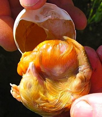
Figure 3. Chick embryo. Photo credit: Crispin Semmens. Click to enlarge.
For more information on malpositions, go to Chick Embryo Malpositions by the University of Florida’s IFAS Extension.
Hatch
The process of hatch begins with drawdown when the air cell expands and extends down the side of the egg. Drawdown occurs approximately 24 to 48 hours prior to internal pip.
Internal pip is caused by rising carbon dioxide (CO2) levels within the egg. When CO2 levels reach a critical level, the overdeveloped muscles on the back of the (otherwise) scrawny chick’s neck begin to twitch. Repeated twitching allows the egg tooth to penetrate the inner shell membrane, allowing the chick’s head to pops into the air cell. This is a crucial moment as the chick’s respiration now changes from chorioallantoic to pulmonary. Once internal pip has occurred, the egg no longer needs to be turned and the chick can actually be heard peeping within the egg.
Eventually the chick uses up the small amount of oxygen present within the air cell. As CO2 levels begin to rise again, muscular twitching causes the egg tooth to penetrate the eggshell. Twitching of abdominal muscles also causes the yolk sac to be pulled within the coelom. Also the time frame is species-specific, external pip or breakout generally occurs within 36-72 hour of internal pip. Assisted hatch or ovotomy should never be initiated prematurely as there are a host of possible complications including bacterial infection, iatrogenic damage to the chick, and unabsorbed yolk sac. If associated vasculature has not receded the chick can also suffer fatal hemorrhage.
References
References
Ernst RA, Bradley FA, Abbott UK, Craig RM. Egg candling and breakout analysis. University of California Division of Agriculture and Natural Resources Publication 8134. Available at http://animalscience.ucdavis.edu/avian/pfs32.htm. Accessed on March 23, 2013.
Johnson AL. Reproduction in the female. In: Whittow CG (ed). Sturkie’s Avian Physiology, 5th ed. Academic Press: San Diego: 1999. Pp. 586-596.
King AS, McLelland J (eds). Form and Function in Birds. London, UK: Academic Press; 1989.
Joyner KL. Theriogenology. In: Avian Medicine: Principles and Applications. In: Ritchie BW, Harrison GJ, Harrison LR (eds). Avian Medicine: Principles and Application. Lake Worth, FL: Wingers Publishing, Inc.; 1994. Pp. 753-755.
Nys Y, Gautron J. Structure and formation of the eggshell. Bioactive Egg Compounds Springer; City: 2007. Pp. 99-102.
UCMP. Eggshell morphology and structure. University of California Museum of Paleontology website. Available at http://www.ucmp.berkeley.edu/science/eggshell/eggshell2.phpaz. Accessed on March 23, 2013.
Pollock C. Understanding the avian egg: From outside to in. March 23, 2013. LafeberVet Web site. Available at https://lafeber.com/vet/understanding-the-avian-egg-from-outside-to-in/