Key Points
- Enterocolitis is the most important cause of morbidity and mortality in hamsters.
- “Wet-tail” is a non-specific clinical sign that can result from enterocolitis and resultant diarrhea in pet hamsters.
- The anaerobic obligate intracellular bacterium, Lawsonia intracellularis, is an important cause of wet-tail, however other bacteria and parasites may also serve as etiologic agents.
- Many of the potential pathogens of “wet-tail” are zoonotic.
- Clinical outbreaks of diarrhea in hamsters may be precipitated by stress, including high temperatures or humidity, overcrowding, malnutrition, dietary changes, shipping, or underlying diseases, such as endoparasitism.
- Prevention and treatment of “wet’-tail”, which centers around the use of antimicrobials, husbandry practices, selective breeding, and culling, has generally proved unrewarding.
Introduction
Diarrhea is the most common presenting problem in Syrian hamsters (Mesocricetus auratus).2,3,5,14 In a survey of two large American commercial breeding facilities, approximately 3% of shipped hamsters developed diarrhea.2 Diarrhea caused by enterocolitis can occur in hamsters of any age or breed and is commonly known as “wet-tail” (Fig 1).2,3,14 Clinical signs in weanlings usually include fetid diarrhea, anorexia, ruffled hair, dehydration, weight loss, and death.2,5,14 The mortality rate is often highest in 3-10 week old hamsters, however adult hamsters may also suffer debilitating disease and death.2,3,5,14 Potential sequelae in surviving hamsters can include intestinal obstruction, intussusception, or rectal prolapse.3,5
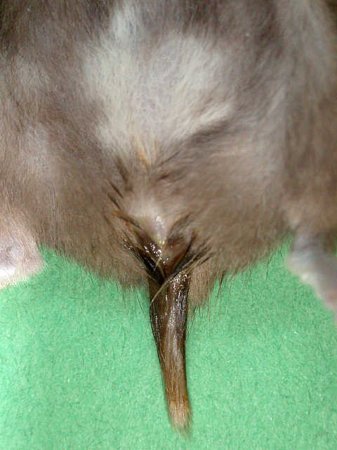
Figure 1. “Wet-tail” or diarrhea in a hamster (Mesocricetus auratus). Photograph by Dr. Lauren Richey. Click image to enlarge.
Etiology
The cause of enterocolitis can be multifactorial in hamsters, involving bacterial and parasitic etiologic agents. A number of the pathogens documented in hamsters are also transmissible to humans. Clinical outbreaks may be precipitated by stress, such as high temperatures or humidity, overcrowding, malnutrition, dietary changes, shipping, or underlying disease, such as endoparasitism.3
Bacteria
Historically, enterocolitis or “wet-tail” has been associated with proliferative ileitis caused by Lawsonia intracellularis, a highly fastidious, anaerobic obligate intracellular bacterium.5,9,14 Multiple studies have confirmed that hamsters experimentally infected with L. intracellularis can develop the clinical signs of proliferative ileitis as well as the gross and histopathological lesions of disease.3,20 This organism infects the small intestine and less frequently the large intestine (Fig 2).9 Malabsorption is considered the main mechanism involved in the physiopathology of diarrhea in L. intracellularis-infected animals.14,20
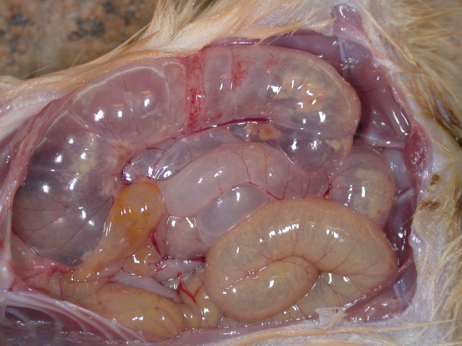
Figure 2. Gross appearance of enterocolitis in a hamster (Mesocricetus auratus). Photograph provided by Dr. Lauren Richey. Click image to enlarge.
Despite the importance of L. intracellularis, other bacteria that have been implicated in “wet-tail” include Clostridium spp., E. coli, Campylobacter jejuni, Helicobacter spp., Salmonella spp., and Pasteurella pneumotropica.1,3,4,7,8,11,12,15-17,19 Campylobacter and Clostridium are most commonly associated with clinically significant infection.4,7,11,19 Clostridium piliforme is a Gram-negative, spore-forming, obligate intracellular bacterium and the causative agent of Tyzzer’s disease.15 Clostridium difficile has also been associated with enterotoxemia, typhlitis and enteritis in hamsters (Fig 3).3

Figure 3. Histologic appearance of Tyzzer’s disease in a hamster (Mesocricetus auratus). Photograph provided by Dr. Lauren Richey. Click image to enlarge.
Parasites
Concurrent mild to severe infestation of the tapeworm, Hymenolepis nana, is very common in hamsters with bacterial enterocolitis (Fig 4). As the degree of disease and debilitation increases, so does the tapeworm burden and in some individuals, the tapeworm load is high enough to nearly obstruct the intestines.3 The protozoa Giardia muris, Entamoeba histolytica, Spironucleus muris, Cryptosporidium spp., and trichomonads are also variably found in individuals with enterocolitis.3,10,18
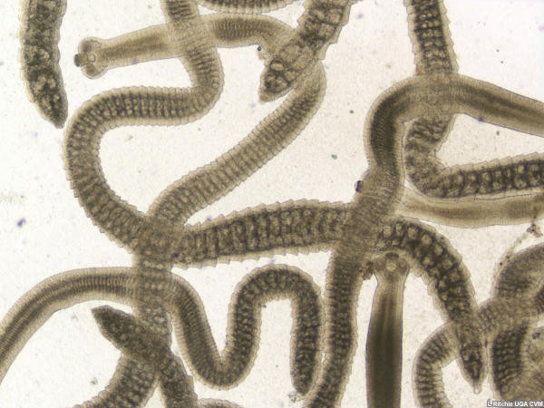
Figure 4. Gross appearance of the tapeworm, Hymenolepsis nana, from a hamster (Mesocricetus auratus). Photograph provided by Dr. Lauren Richey. Click image to enlarge.
Management
Prevention and treatment of “wet’-tail” has historically centered around antimicrobial therapy, husbandry practices, selective breeding, and culling. Unfortunately treatment is rarely rewarding because of the intracellular nature of L. intracellulare and C. piliforme.13
Clostridial bacterial spores are quite stable and can remain infectious for 1 to 2 years. Therefore when Clostridium is been identified as the causative agent, Control of enterocolitis in a commercial breeding facility would ideally involve depopulation, rigorous disinfection, and repopulation with C. piliforme-free breeding stock.
Care of the individual patient relies upon antibiotic therapy and aggressive supportive care, including fluid therapy and nutritional support.2,14 Begin oral antibiotics, such as sulfa-trimethoprim (30 mg/kg per os every 12 hours for 5-7 days), tetracycline (400 mg/L of drinking water for 10 days or 10 mg/kg PO q12 hours for 5–7 days), or enrofloxacin (5-10 mg/kg PO or IM q 12 hours for 5–7 days).2,14 If diarrhea persists, add bismuth subsalicylate as a form of symptomatic treatment.14
Theorizing that high worm burdens make the gut more vulnerable to bacterial infection, Barron et al also treated weanling hamsters with praziquantel (20 mg/kg PO, repeat in 10d) and fenbendazole (20 mg/kg PO q24h x 5d) prior to shipping.2 Unfortunately this treatment regimen made no significant difference when compared to control animals.2
Note: Napa Nectar™ Plus is a useful and easy way to administer fenbendazole to large hamster colonies.2
Diagnosis
Definitive diagnosis often depends on necropsy and histologic examination. Infection is characterized by marked thickening of the ileal wall with excessive epithelial hyperplasia, necrosis, and inflammation.3,5,9 Peritonitis is also possible.5 The presence of L. intracellularis can also be confirmed by immunohistochemistry of intestinal lesions or fecal polymerase chain reaction assays.6,14,21
Summary
“Wet-tail” is a non-specific clinical sign that can result from enterocolitis and resultant diarrhea in pet hamsters. Unfortunately enterocolitis is the most important cause of morbidity and mortality in hamsters. The causative agent is often Lawsonia intracellularis, however the development of enterocolitis may be multifactorial in hamsters of all ages and may include other bacteria, such as Clostridium spp. and Campylobacter jejuni, and parasites, particularly Hymenolepis nana. Clinical outbreaks of diarrhea in hamsters may be precipitated by stress, including high temperatures or humidity, overcrowding, malnutrition, dietary changes, shipping, or underlying diseases, such as endoparasitism. Prevention and treatment of “wet’-tail”, which centers around the use of antimicrobials, husbandry practices, selective breeding, and culling, have generally proved unrewarding.
Acknowledgement: I thank Dr. Lauren Richey who was instrumental in the study referenced below (Barron et al) and who also provided the photographs provided above.
References
References
- Alworth L, Simmons J, Franklin C, Fish R. Clostridial typhlitis associated with topical antibiotic therapy in a Syrian hamster. Lab Anim. 2009; 43(3):304-309.
- Barron HW, Richey L, Hernandez-Divers S, Ritchie B. Etiology, pathology, and control of enterocolitis in a group of hamsters. Proc Annu Conf Assoc Exotic Mammal Vet. 2007. Pp. 123-126.
- Barthold SW, Griffey SM, Percy DH (eds). Hamsters. In: Pathology of Laboratory Rodents and Rabbits, 4th Ames: Wiley Blackwell; 2016. Pp. 180-193.
- Chang J, Rohwer RG. Clostridium difficile infection in adult hamsters. Lab Anim Sci. 1991;41(6):548-52. PMID: 1667195.
- Cooper TK, Meyerholz DK, Beck AP, et al. Research-relevant conditions and pathology of laboratory mice, rats, gerbils, guinea pigs, hamsters, naked mole rats, and rabbits. ILAR J. 2021;62(1-2):77-132. doi: 10.1093/ilar/ilab022. PMID: 34979559; PMCID: PMC9291387.
- Cooper DM, Swanson DL, Gebhart CJ. Diagnosis of proliferative enteritis in frozen and formalin-fixed, paraffin-embedded tissues from a hamster, horse, deer and ostrich using a Lawsonia intracellularis-specific multiplex PCR assay. Vet Microbiol. 1997;54(1):47-62. doi: 10.1016/s0378-1135(96)01264-3. Erratum in: Vet Microbiol 1998;59(2-3):247. Erratum in: Vet Microbiol 1999;67(2):159. PMID: 9050170.
- Dillehay DL, Paul KS, Boosinger TR, Fox JG. Enterocecocolitis associated with Escherichia coli and Campylobacter-like organisms in a hamster (Mesocricetus auratus) colony. Lab Anim Sci. 1994;44(1):12-6. PMID: 8007654.
- Frisk CS, Wagner JE, Owens DR. Hamster (Mesocricetus auratus) enteritis caused by epithelial cell-invasive Escherichia coli. Infect Immun. 1981;31(3):1232-8. doi: 10.1128/iai.31.3.1232-1238.1981. PMID: 7014460; PMCID: PMC351447.
- Karuppannan AK, Opriessnig T. Lawsonia intracellularis: Revisiting the disease ecology and control of this fastidious pathogen in pigs. Front Vet Sci. 2018;5:181. doi: 10.3389/fvets.2018.00181. PMID: 30140680; PMCID: PMC6095029.
- Kunstýr I, Poppinga G, Friedhoff KT. Host specificity of cloned Spironucleus originating from the European hamster. Lab Anim. 1993;27(1):77-80. doi: 10.1258/002367793781082430. PMID: 8437440.
- Lentsch RH, McLaughlin RM, Wagner JE, Day TJ. Campylobacter fetus subspecies jejuni isolated from Syrian hamsters with proliferative ileitis. Lab Anim Sci. 1982;32(5):511-4. PMID: 7144126.
- Lesher RJ, Jeszenka EV, Swan ME. Enteritis caused by Pasteurella pneumotropica infection in hamsters. J Clin Microbiol. 1985;22(3):448. doi: 10.1128/jcm.22.3.448-.1985. PMID: 4044801; PMCID: PMC268431.
- McNeil PE, Al-Mashat RR, Bradley RA, Payne AP. Control of an outbreak of wet-tail in a closed colony of hamster (Mesocricetus auratus). Vet Rec. 1986; 119(11):272-3. doi: 10.1136/vr.119.11.272. PMID: 2877518.
- Miwa Y, Mayer J. Hamsters and gerbils. In: Quesenberry KE, Orcutt CJ, Mans C, Carpenter JW (eds). Ferrets, Rabbits, and Rodents: Clinical Medicine and Surgery. St. Louis: Elsevier, 2021.
- Motzel SL, Gibson SV. Tyzzer’s disease in hamsters and gerbils from a pet store supplier. J Am Vet Med Assoc. 1990; 197:1776-1778.
- Nambiar PR, Kirchain SM, Courmier K, et al. Progressive proliferative and dysplastic typhlocolitis in aging syrian hamsters naturally infected with Helicobacter : a spontaneous model of inflammatory bowel disease. Vet Pathol. 2006;43(1):2-14. doi: 10.1354/vp.43-1-2. PMID: 16407482.
- Patterson MM, Schrenzel MD, Feng Y, Fox JG. Gastritis and intestinal metaplasia in Syrian hamsters infected with Helicobacter aurati and two other microaerobes. Vet Pathol. 2000;37(6):589-96. doi: 10.1354/vp.37-6-589. PMID: 11105948.
- Sheppard BJ, Stockdale Walden HD, Kondo H. Syrian hamsters (Mesocricetus auratus) with simultaneous intestinal Giardia, Spironucleus sp., and trichomonad infections. J Vet Diagn Invest. 2013;25(6):785-90. doi: 10.1177/1040638713505286. Epub 2013 Sep 30. PMID: 24081933.
- Stills HF Jr, Hook RR Jr. Experimental production of proliferative ileitis in Syrian hamsters (Mesocricetus auratus) by using an ileal homogenate free of Campylobacter jejuni. Infect Immun. 1989;57(1):191-5. doi: 10.1128/iai.57.1.191-195.1989. PMID: 2462537; PMCID: PMC313067.
- Vannucci FA, Borges EL, de Oliveira JS, Guedes RM. Intestinal absorption and histomorphometry of Syrian hamsters (Mesocricetus auratus) experimentally infected with Lawsonia intracellularis. Vet Microbiol. 2010;145(3-4):286-91. doi: 10.1016/j.vetmic.2010.03.027. Epub 2010 Apr 1. PMID: 20418028.
- Yeh JY, Kim TJ, Park SY, et al. Isolation of Lawsonia intracellularis in Korea and reproduction of proliferative enteropathy in pigs and hamsters. J Vet Med Sci. 2006;68(5):499-501. doi: 10.1292/jvms.68.499. PMID: 16757895.
Further reading
Cooper DM, Swanson DL, Barns SM, Gebhart CJ. Comparison of the 16S ribosomal DNA sequences from the intracellular agents of proliferative enteritis in a hamster, deer, and ostrich with the sequence of a porcine isolate of Lawsonia intracellularis. Int J Syst Bacteriol. 1997;47(3):635-9. doi: 10.1099/00207713-47-3-635. PMID: 9226893.
Fox JG, Shen Z, Muthupalani S, et al. Chronic hepatitis, hepatic dysplasia, fibrosis, and biliary hyperplasia in hamsters naturally infected with a novel Helicobacter classified in the H. bilis cluster. J Clin Microbiol. 2009;47(11):3673-81. doi: 10.1128/JCM.00879-09. Epub 2009 Sep 16. PMID: 19759229; PMCID: PMC2772605.
Manci EA, Heath LS, Leinbach SS, Coggin JH Jr. Lymphoma-associated ulcerative bowel disease in the hamster (Mesocricetus auratus) induced by an unusual agent. Am J Pathol. 1984 Jul;116(1):1-8. PMID: 6377905; PMCID: PMC1900378.
Stills HF Jr, Hook RR Jr, Kinden DA. Isolation of a Campylobacter-like organism from healthy Syrian hamsters (Mesocricetus auratus). J Clin Microbiol. 1989;27(11):2497-501. doi: 10.1128/jcm.27.11.2497-2501.1989. PMID: 2681250; PMCID: PMC267065.
Barron H, Pollock CG. Enterocolitis in hamsters. Updated March 20, 2024. LafeberVet Web site. Available at https://lafeber.com/vet/enterocolitis-in-hamsters/