Key Points
- The ferret is a carnivore with a short, simple gastrointestinal tract and a relatively rapid gastrointestinal transit time. The ferret lacks a cecum and an ileocolic junction.
- Diarrhea is the one of the most common clinical sign in ferrets. Weight loss and anorexia are also important signs of gastrointestinal disease in the ferret.
- Anorexia is the primary clinical sign of gastrointestinal foreign bodies in young ferrets (<2y)
- Important causes of diarrhea in young ferrets include coccidiosis, rotavirus, and stress or diet change induced ulceration or hypermotility.
- Ferrets of all ages may be afflicted with Helicobacter ulcerative gastritis and ferret enteric coronavirus (FECV).
- Middle-aged to older ferrets (>3y) may suffer from inflammatory bowel disease and intestinal lymphoma.
Introduction
The gastrointestinal tract of the domestic ferret (Mustela putorius furo) has been studied extensively as a model for several human gastrointestinal diseases including gastric and duodenal ulcers, gastroesophageal reflux, gastric carcinoma and lymphoma, and Helicobacter mustelae infection. Ferrets are also used as animal models for emesis because of a simple stomach for propulsion, a low tolerance for many chemicals, and a strong vagal reflex.
Gastrointestinal anatomy and physiology
The ferret is a carnivore with gastrointestinal anatomy and physiology that is similar to that seen in cats or dogs, but with unique species-specific features.
The ferret has a short gastrointestinal transit time of 148–219 minutes when fed a meat-based diet.
The ferret has a simple stomach, similar in shape to that of the dog. There is prominent vasculature in the stomach as well as a prominent lymph node lying in the lesser curvature. The stomach is innervated by parasympathetic fibers from the vagus nerve and sympathetic fibers via the celiacomesenteric plexus. The ferret stomach has considerable storage capacity, holding up to 100 ml over 10 minutes in the adult. Approximately 80% of a meal is stored in the proximal stomach.
The lower esophageal sphincter and the mechanisms of gastro-esophageal reflux in the ferret are used as an animal model in research. Gastric infusions of glucose, lipid, and gas all provoke gastroesophageal reflux in the ferret. The ferret stomach also secretes gastric acid in response to lipid, glucose, histamine, pentagastrin, and calcium. Hypoglycemia induced by insulin administration causes a sustained stimulation of acid secretion, which is particularly relevant for ferrets with pancreatic beta cell tumor or insulinoma. Therapy for ferrets with insulinoma should therefore include medications that decrease acid secretion.
The ferret intestine consists of the duodenum, jejunum, and ileum. The major duodenal papilla, containing the common opening for the bile and pancreatic ducts, is located about 3 cm distal to the pylorus. The minor duodenal papilla may be absent in the ferret. Brunner’s glands are present within the submucosa of the proximal duodenum. These glands produce only neutral mucosubstances, as seen in humans. The jejunal and ileal segments cannot be distinguished and may be referred to as the “jejunoileum” that ends at the ascending colon.
The large intestine is composed of the colon and rectum. There is no cecum and no ileocolic junction in the ferret, however anastomoses of the jejunal artery with the ileocolic artery are present at this junction. The large intestine consists of the ascending, transverse, and descending colon, with the largest segment being the descending colon. Unlike other carnivores, the entire gastrointestinal tract can secrete cholecystokinin, the hormone responsible for bile secretion into the small intestine in response to fats and food present anywhere within the gastrointestinal tract. This active secretion of bile contributes to the green coloration seen in ferret diarrhea.
Causes of gastrointestinal diseases in the ferret
Ferrets are used as laboratory models of esophageal acid reflux disease, Helicobacter gastritis, pyloric and intestinal ulceration, colitis, gastrointestinal neoplasia, and inflammatory bowel disease. All of these conditions may result in varying degrees of acute, chronic, or intermittent diarrhea: with or without visible hemorrhage, and with or without secondary bacterial or viral involvement. Diarrhea is the most common clinical sign with any of the above diseases in ferrets.
Bacterial disease
Helicobacter mustelae is a gastric, spiral-shaped bacterium of the domestic ferret. Although infection is often asymptomatic, Helicobacter spp. may overgrow with concurrent disease or other stressors leading to gastritis, peptic or pyloric ulcers in ferrets of all ages (Fig 1). Clinical signs frequently include anorexia and diarrhea, which may progress to melena, vomiting, and other signs of nausea and abdominal pain such as heavy drooling, pawing at the mouth, and teeth grinding (Fig 2 and Fig 3).

Figure 1. Helicobacter mustelae may overgrow leading to pyloric ulcers (as shown above), gastritis, or duodenal ulcers. Photograph provided by Cathy Johnson-Delaney.
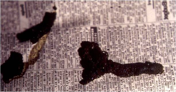
Figure 2. Dark, tarry stool or melena may be associated with clinical Helicobacter spp. infection in ferrets (Mustela putorius furo). Photograph provided by Cathy Johnson-Delaney. Click image to enlarge.

Figure 3. Pawing at the mouth is an important sign of nausea in the ferret (Mustela putorious furo). Photograph provided by Cathy Johnson-Delaney.
The literature describes Lawsonia intracellularis or Desulfovibrio, an intracellular Campylobacter-like organism, as the cause of proliferative bowel disease in young ferrets. In 30 years I have never had histopathology come back positive for Lawsonia spp. in pet ferrets of any age group.
Reported clinical signs may include large bowel diarrhea, rectal prolapse, weight loss, anorexia, lethargy, fever, and a palpably thick colon. Campylobacter jejuni has also been associated with diarrhea in ferrets, and there have also been rare reports of mycobacteriosis in ferrets greater than 2 years of age.
Viral disease
Important causes of viral diarrhea in the ferret include rotavirus, canine distemper virus, and coronavirus. Ferret enteric coronavirus (FEVC), formerly known as epizootic catarrhal enteritis (ECE), can be seen in any age ferret. Disease usually follows a stressful event or a gathering of ferrets. Disease may be mild and transient in kits, but tends to be severe and debilitating in adults. Clinical signs may include lethargy, anorexia, and diarrhea that may vary in appearance from soft and brown to green and mucoid, with large amounts of undigested food giving it a “bird-seed” component (Fig 4A and Fig 4B). Characteristic microscopic lesions within the intestinal tract include vacuolar degeneration and necrosis of villous enterocytes, lymphocytic inflammation, as well as villous atrophy, fusion, and blunting.
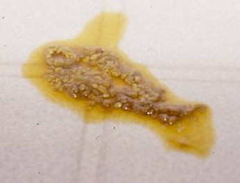
Figure 4a. Diarrhea with mucosal shreds is commonly seen in ferrets with enteric coronavirus. Photograph provided by Cathy Johnson-Delaney.

Figure 4b. Diarrhea with large amounts of mucus is also commonly seen with ferret enteric coronavirus. Photograph provided by Cathy Johnson-Delaney. Click image to enlarge.
Rotavirus is seen in neonatal and weanling ferrets. Affected kits may display distended abdomens and thin-walled small intestines that contained gas and fluid.
Canine distemper virus is primarily seen in young kits that did not complete their juvenile vaccine series. Initial clinical signs may indicate involvement of the gastrointestinal tract (vomiting and diarrhea), respiratory system (oculonasal discharge, cough, and lethargy), and the integumentary system (hyperkeratosis of the planum nasale and footpads and a papular rash that begins on the chin but may progress to a generalized form) (Fig 5). Animals that survive this early stage succumb to neurologic disease within several weeks.

Figure 5. Dermatitis in a ferret (Mustela putorius furo) infected with canine distemper virus. Photograph provided by Dr. Bernice Lopez. Click image to enlarge.
Parasitic disease
Protozoans are a common gastrointestinal parasite in young ferrets less than 1 year of age. Ferrets between 6 to 16 weeks of age most commonly shed Isospora spp. oocysts. Infected kits may be asymptomatic, however clinical signs may include stunted growth and bloody diarrhea (Fig 6).

Figure 6. Clinical signs of Isospora spp. infection may include stunted growth and bloody diarrhea. Photograph provided by Cathy Johnson-Delaney. Click image to enlarge.
Giardia spp. has also been detected in ferrets. Although the correlation between this organism’s presence and clinical disease is unclear, there may be some potential for zoonotic transmission.
Cryptosporidium spp. has been found in stressed ferrets, frequently coming from a pet store or shelter environment. At this time, supportive care and sanitation are the primary treatments although there are some new human antiprotozoal drugs that may be effective.
Inflammatory bowel disease
Inflammatory bowel disease (IBD) is usually seen in ferrets over 2 years of age. Affected ferrets initially suffer from intermittent, mild diarrhea that may also be associated with anorexia or a shift in food preferences. The consistency of the ferret’s stools may worsen over weeks to months. Histologic examination of the gastrointestinal tract often reveals lymphocytic-plasmacytic inflammation, or less commonly eosinophilic gastroenteritis may be seen. The underlying etiology is probably multifactorial however viruses, bacteria, dietary antigens, and toxins have all been implicated as potential causes of IBD. There may also be an underlying genetic component to IBD in the ferret. IBD is progressive involving more of the tract, inflammatory progressing to neoplastic changes of the mesenteric lymph nodes, and eventually intestinal lymphoma, either diffuse or localized.
Neoplasia
Lymphosarcoma is the most common malignancy in the domestic ferret. Intestinal lymphoma is usually seen in ferrets over 3 years of age, with a history of intermittent diarrhea or inflammatory bowel disease. The second most common neoplasm of the gastrointestinal system is tumors arising from the smooth muscular layers of the GI tract such as leiomyosarcoma. Helicobacter mustelae in ferrets has also been associated with both gastric mucosa-associated lymphoid tissue (MALT) lymphoma and gastric adenocarcinoma.
Trauma
Foreign body ingestion is usually seen in ferrets less than 2 years of age, but can occur with older ferrets particularly with concurrent gastrointestinal disease or pain resulting in abnormal ingestion of hair, materials, fabric, etc (Fig 7A and Fig 7B). Ferrets are notorious for chewing on rubber such as the soles of shoes or spongy material such as shoe liners or furniture stuffing. The most common clinical sign is anorexia, which frequently waxes and wanes. Affected ferrets rarely vomit. They will exhibit slow loss of weight and body condition. Most also show signs of abdominal pain including hunched posture, reluctance to play, and bruxism.

Figure 7a. Surgical removal of a foreign body in a ferret (Mustela putorious furo). Photograph provided by Cathy Johnson-Delaney. Click image to enlarge.

Figure 7b. Close-up of the trichobezoar removed. Photograph provided by Cathy Johnson-Delaney. Click image to enlarge.
Diagnostics
Obtain a detailed history to determine the course of action:
- Determine stool volume, color, consistency, and frequency, as well as the duration of the diarrhea.
- Record tenesmus, or accompanying borborygmus, vocalization, or flatulence.
- Teeth grinding may indicate abdominal pain. Anorexia may be sequelae to the pain.
- Learn the source of the ferret.
- How long has the ferret has been in the household? Are other ferrets and pets present?
- What type of litter is used and what is the sanitation program?
- Record the diet including treats.
- What toys are available?
- Do any human family members have gastrointestinal signs such as diarrhea?
- Has the patient had any access to commonly ingested items? Ferrets are notorious for licking soaps, chewing on stuffing dug out of furniture, and chewing shoes, shoe liners, and even perfume or shampoo bottles.
- Does the occurrence of diarrhea correlate with activity? For instance, does diarrhea occur around the clock or is it only seen after intense playtime? Does diarrhea only occur after a stressful event such as the vacuum cleaner being run near the ferret’s cage?
Perform a thorough physical examination, including auscultation of the abdomen, examination of the anus, and a complete dental exam. The minimum database may also include cytology, clinical pathology, and imaging:
- Parasite testing should include flotation and direct smear of fresh fecal material. Perform cytologic staining to assess the stool for bacteria and blood cells.
- The complete blood count (CBC) and biochemistry panel are frequently unremarkable. Anemia may indicate gastrointestinal hemorrhage. Serum lipase levels may elevate in cases of inflammatory bowel disease.
- Survey and contrast radiographs can be useful, however interpret results with caution. Many ferret gastrointestinal foreign bodies may consist of spongy, porous material that absorb contrast material allowing barium to pass, so trust your clinical impression based on signalment, history, and abdominal palpation.
- Use abdominal ultrasound when indicated to assess gastric motility, and to screen the ferret for other pathologic conditions. Ultrasound examination may illustrate gastric trichobezoar and in many cases find the foreign body. It needs to be done prior to administration of barium. Generally the ferret will need an analgesic as even gentle pressure on the abdomen may be painful.
Additional diagnostics may be performed as indicated including…
- Rectal culture and cytology
- Fecal occult blood testing: Although diagnosis of Helicobacter gastritis is often presumptive, fecal occult blood testing may sometimes prove helpful. Place the ferret on a diet that does not contain meat, such as Emeraid Carnivore, for at least 24–36 hours to avoid false positive results.
- Biopsy: Definitive diagnosis of IBD requires histologic examination of biopsy samples. Biopsy may also be used confirm the presence of Helicobacter gastritis. Biopsies may be collected endoscopically using the smallest biopsy cup or via laparotomy taking a full thickness wedge of the intestinal wall in multiple locations.
- PCR assay of the coronavirus causing FECV.
Therapy
Bacterial disease
Management of Helicobacter infection relies on combination therapy including antibiotics such as amoxicillin and metronidazole, gastroprotectants such as bismuth subsalicylate or sucralfate (Carafate Aventis Pharmaceuticals, Inc.), and histamine antagonists and/or proton pump inhibitors such as famotidine (Pepcid, Johnson & Johnson Merck Consumer Pharmaceuticals) 0.25-0.5 mg/kg PO, IM, IV q24h or short-term omeprazole (Prilosec, AstraZeneca Pharmaceuticals LP) 0.7 mg/kg PO q24h to reduce gastric acid secretion and provide pain relief. Additional supportive care including analgesics and fluid therapy, as well as hand-feeding of a liquid “soup” diet may be needed to keep the ferret eating.
The antimicrobial of choice in cases of proliferative colitis is oral chloramphenicol (50 mg/kg of body weight, q 12 h for 10 to 21 days).
Viral disease
Canine distemper virus is highly contagious and almost 100% fatal in ferrets. Treatment should not be attempted.
Ferrets suffering from coronavirus or rotavirus require aggressive supportive care in the form of fluid therapy, nutritional support, and prophylactic antibiotics. Gastroprotectants and histamine antagonists are also often indicated.
Parasitic disease
In addition to careful cleaning and disinfection of the environment, administer coccidiostats such as amprolium (Corid, Merck) (19 mg/kg PO q24h) or sulfadimethoxine (Albon, SmithKline) (20-25 mg/kg PO q24h).
Inflammatory bowel disease
Use the corticosteroid, prednisone (1-2.5 mg/kg PO q24h) to inhibit the inflammatory response in ferrets with IBD. Azathioprine (Imuran, GlaxoSmithKline) (0.9 mg/kg PO q24–72h) has also been used in ferrets that do not respond well to prednisone or in which prednisone is ineffective. Bone marrow suppression has been reported in several ferrets on azathioprine, so periodically monitor the CBC. Ivermectin has also been given to ferrets with eosinophilic gastroenteritis. Inflammatory bowel disease may be diffuse or segmental which makes monitoring and sampling for biopsies difficult. In most instances, the disease progresses to intestinal lymphoma. There are several additional oncologic agents currently under investigation for diffuse inflammatory bowel/lymphoma in the ferret.
In addition to immunomodulating medications, dietary changes are often of great benefit to IBD ferrets. Offer a highly digestible, easily absorbed diet such as Emeraid Intensive Care Carnivore. Prescription diets designed for food allergies such as hypoallergenic or novel protein feline diets have yet to be explored empirically in ferrets although there use has been described clinically. The grain or potato used in some of these commercial food formulations may make them too high in carbohydrates for the ferret.
Trauma
Treatment of gastrointestinal foreign bodies requires surgical or endoscopic, removal (Fig 8A and Fig 8B).

Figure 8a. Region of intestinal inflammation caused by ingestion of a foreign body. Photograph provided by Cathy Johnson-Delaney. Click image to enlarge.
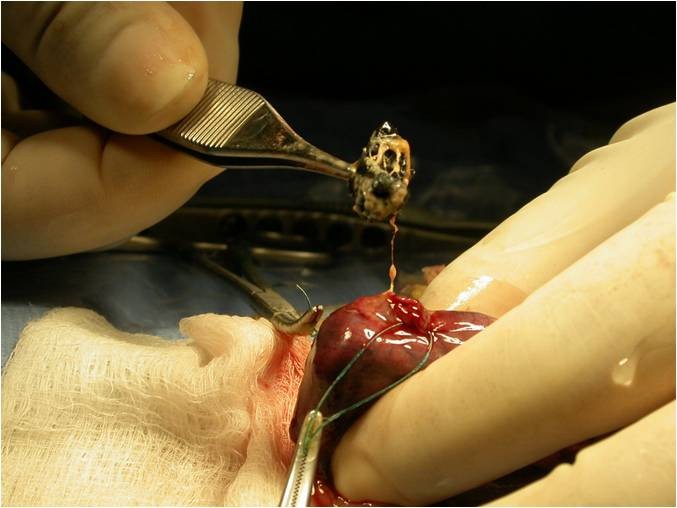
Figure 8b. Surgical removal of an intestinal foreign body. Photograph provided by Cathy Johnson-Delaney. Click image to enlarge.
Summary
The ferret is a carnivore with a short, simple gastrointestinal tract and a relatively rapid gastrointestinal transit time. The ferret lacks a cecum and an ileocolic junction. Important causes of diarrhea in young ferrets include coccidiosis, rotavirus, stress-induced diarrhea and proliferative bowel disease caused by Lawsonia intracellularis. Ferrets of all ages may be afflicted with Helicobacter gastritis and ferret enteric coronavirus, while middle-aged to older ferrets may suffer from inflammatory bowel disease and intestinal lymphoma. Diarrhea is the most common clinical sign in ferrets with gastrointestinal disease, with the exception of gastrointestinal foreign bodies where anorexia and weight loss are the primary presenting complaints. Although signalment, history, and physical exam findings may be sufficient to reach a tentative diagnosis, additional diagnostics may include cytology such as fecal parasite testing and imaging. Treatment will vary with the specific condition identified but frequently includes supportive care, antimicrobial therapy, gastroprotectants, and histamine blockers.
References
References
Abe N, Read C, Thompson RC, Iseki M. Zoonotic genotype of Giardia intestinalis detected in a ferret. Parasitol Res 9(1): 170-182, 2005.
Evans HE, Nguyen QA. Anatomy of the ferret. In: Fox JG (ed). Biology and Diseases of the Ferret, 2nd ed. Baltimore, MD: Williams & Wilkins; 1998:19–69.
Garner MM, Ramsell K, Morera N, et al. Clinicopathologic features of a systemic coronavirus-associated disease resembling feline infectious peritonitis in the domestic ferret (Mustela putorius). Vet Pathol 45(2):236-246, 2008.
Johnson-Delaney CA. Anatomy and physiology of the gastrointestinal system of the ferret and selected exotic carnivores. Proc Annu Conf Assoc Exotic Mammal Vet 2006. Pp. 29-38
Johnson-Delaney CA. The ferret gastrointestinal tract and Helicobacter mustelae infection. Vet Clin North Am Exotic Anim Pract 8(2):197-212, 2005.
Johnson-Delaney CA. A clinicians’s perspective on ferret diarrhea. Exotic DVM 6(3):27-28, 2004.
Lennox AM. Gastrointestinal diseases of the ferret. Vet Clin North Am Exotic Anim Pract 8(2): 213-226, 2005.
Saunders GK, Thomsen BV. Lymphoma and Mycobacterium avium infection in a ferret (Mustela putorious furo). J Vet Diagn Invest 18(5):513-515, 2006.
Williams BH, Klupel M, West KH et al. Coronavirus-associated epizootic catarrhal enteritis in ferrets. J Am Vet Med Assoc 217(4):526-530, 2000.
Williams BH. Pathology of the domestic ferret. www.ferrethealth.msu.edu/Diseases/Notes.pdf. Accessed Feb 17, 2010.
Wise AG, Kiupel M, Maes RK. Molecular characterization of a novel coronavirus associated with epizootic catarrhal enteritis (ECE) in ferrets. Virology 349(1):164-174, 2006.
Wise AG, Smedley RC, Kiupel M, Maes RK. Detection of group C rotavirus in juvenile ferrets (Mustela putorius furo) with diarrhea by reverse transcription polymerase chain reaction: sequencing and analysis of the complete coding region of the VP6 gene. Vet Pathol 46(5):985-491, 2009.
Johnson-Delaney C. Gastrointestinal disease in the ferret. June 1, 2010. LafeberVet Web site. Available at https://lafeber.com/vet/gastrointestinal-disease-in-the-ferret/