Key Points
- Endotracheal intubation is readily accomplished in most reptiles using a short, uncuffed endotracheal tube.
- Consider ventilating reptiles with room air using a bag valve mask as rising oxygen levels can suppress respiration in the reptile patient.
- Improper diet and inadequate husbandry are often major contributors to illness, and a thorough history is a critical part of clinical evaluation of the reptile patient.
- Many ill or injured reptiles that present on an emergency basis are hypothermic. House collapsed or debilitated reptiles in an incubator set at 82-85°F (28-29.4°C).
- Maintenance fluid requirements are estimated as 10-30 ml/kg/day. Always administer warmed fluids. Intraosseous catheters can be placed into the tibia or femur of the lizard, while the jugular vein can be accessed in chelonians.
- Most reptile pathogens are opportunistic, Gram-negative bacteria. Pending culture results, select antimicrobials with a primarily Gram-negative spectrum.
- Analgesic agents recommended for use in the reptile include non-steroidal anti-inflammatories, like meloxicam, and pure mu opioid receptor agonists, like morphine and hydromorphone.
- As a general rule, administer medications to the reptile by a parenteral route.
- Reptiles do not eat as frequently as birds or mammals due to their significantly lower metabolic rate. It is crucial to ensure that the patient is warm and hydrated before feeding begins.
Introduction
The unique challenges of reptile medicine must be balanced against the basic principles of critical care that are universal in all species. Use this emergency and critical care summary page to review the basic approach to the reptile patient and select additional links to supplement your knowledge base, as needed. This page includes two supplemental videos, which are not covered in the brief quiz.

This summary page is a part of the Emergency and Critical Care Teaching Module.
Airway/resuscitation
Reptiles lack an epiglottis and the glottis is ready visualized, making intubation readily accomplished in many species. The tracheal rings are complete in chelonians. Use of an inflated, cuffed endotracheal tube can lead to pressure necrosis because there is no elastic ligament to accommodate tracheal expansion. Although tracheal rings are incomplete in lizards and snakes, an uncuffed endotracheal tube is routinely used in most reptiles, particularly small patients (Video 1). Chelonians possess a cranial tracheal bifurcation, therefore the tube passed should also be relatively short. Secure the endotracheal tube with umbilical tape or a small strip of porous tape
For information on reptile intubation, view timestamp 58:06-59:38 of the R.A.C.E.-approved LafeberVet webinar Spotlight on Anesthesia and Analgesia in Reptiles by Dr. Javier Nevarez.
Reptiles can survive long periods using anaerobic metabolism, therefore it is possible to revive patients with cardiopulmonary arrest. Rising oxygen levels suppress respiration in the reptile, therefore administer low oxygen levels or ventilate with room air using a bag valve mask (i.e. Ambu® bag). The recommended rate for intermittent positive pressure ventilation ranges between 1-6 breaths per minute. Avoid overventilating the reptile patient because this can raise the partial pressure of oxygen (PO2) and further depress respiration. Reptiles should also be maintained at their preferred optimum temperature zone to trigger spontaneous respiration.
Visit Cardiopulmonary Resuscitation in Exotic Animals for additional information.
Signs of illness
Non-specific signs of illness in the reptile can include listlessness, inactivity, weight loss, and anorexia although the significance of a poor appetite can vary with age, season, and reproductive status in the reptile (Fig 1). Sometimes there is a physiologic cause for anorexia in the reptile. For instance, snakes normally go off feed before shedding. If the reptile is emaciated or dehydrated, wrinkled, inelastic skin and sunken eyes can be observed. Debilitated chelonians and lizards may also lack carpal or truncal lift, lying flat rather than lifting up on all four feet. Open-mouth breathing or increased respiratory effort can be observed with advanced lower respiratory tract disease, and an erythematous blush to the ventral belly scutes in squamates or the lower shell (plastron) in chelonians is often associated with septicemia.
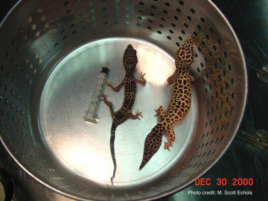
Figure 1. The tail base is a site of fat deposition in the normal leopard gecko (Eublepharis macularius). The gecko tail base can lose its fat depot in the emaciated patient (left). Source: Dr. M. Scott Echols. Click to enlarge
CHELONIANS
The debilitated turtle or tortoise may be unable to retract its head into the shell with the normal degree of strength. Respiratory disease is also an important problem in turtles and tortoises. The normal chelonian subtly moves its head and limbs with each breath, but this pumping movement is more pronounced when there is underlying respiratory disease. Breath sounds can also become audible and the chelonian may stretch the neck and gape its mouth while laboring to breathe. Aquatic turtles with pneumonia can exhibit uneven floating.
LIZARDS
Lizards with systemic disease, malnutrition, or exposure to cold temperatures can exhibit color change, appearing paler, darker, or even duller. Lizards can also appear dull when they are about to shed. Rapid color change is most highly developed in anoles and chameleons. The weak chameleon may be unable to climb or grasp. Musculoskeletal or ophthalmic disease can also prevent a chameleon from perching.
SNAKES
The weak, lethargic, or painful snake can lie listlessly in an uncoiled or “stretched out” position. There may be loss of tongue flicking or lack of interest in the environment. Severe weakness or neurologic deficits may manifest as a loss of the righting reflex. Evidence of retained shed or dysecdysis, including retained spectacles can be seen with ill thrift or deficient husbandry. Snakes can also appear dull when they are about to shed.
History
Improper diet and inadequate housing are often major contributors to illness in reptiles. For this reason, a thorough history is a crucial part of clinical evaluation of the reptile patient.
Listen to The Exotic Animal History podcast (or read the transcript) for additional information.
Obtaining a detailed history is only helpful, when the “correct” answers to your questions are known. The wide variety of reptiles seen in clinical practice can be daunting. Develop a collection of references, both online resources as well as textbooks and journals.
View LafeberVet’s collection of reptile Basic Information Sheets for additional information.
Restraint and handling
CHELONIANS
Most chelonians are easily held by the shell however some aquatic turtles will try to bite and their claws can inflict painful scratches.
LIZARDS
Never grasp any lizard patient by the tail. Some species, like iguanas and geckos, possess tail autotomy, a survival mechanism that allows the animal to escape from predators by dropping the distal tail.
SNAKES
For safety’s sake, there should be one handler for every 3 to 4 feet (0.9-1.2 m) of snake (minimum 5 feet or 1.5 m). Never allow a boa or python to form a complete loop around your neck. Large constrictors are incredibly strong. Simply by tightening muscles to maintain balance, a giant snake can cause injury by halting blood flow to the brain or cutting off air flow, resulting in loss of consciousness. Avoid coiling giant snakes around the torso as well.
Visit LafeberVet’s reptile handling and restraint series for additional information on chelonians, lizards, and snakes.
Housing
Many ill or injured reptiles that present on an emergency basis are hypothermic. Reptiles are ectotherms, which means they derive body heat from their environment. Each reptile species has its own unique preferred optimal temperature zone (POTZ) at which physiology functions optimally and the reptile’s ability to heal or convalesce is most effective. The POTZ is provided as a temperature gradient for the mobile patient, however heat must be provided more cautiously for the collapsed or debilitated reptile through an incubator or brooder. A temperature setting of 82-85°F (28-29.4°C) is suitable for many species.
Visit Exotic ICU: Nursing Care for Reptiles and Basic Husbandry: Hospitalizing Non-Traditional Species for additional information on caging the reptile patient.
Only reptiles of adequate and normal bone quality should be offered vertical space for climbing.
Vascular access
Debilitated reptiles are often dehydrated. Judiciously provide fluid therapy, replacing deficits over 48-96 hours. Maintenance fluid requirements are estimated at 1-3 % of body weight or 10-30 ml/kg/day. Always administer warmed fluids.
Depending on the size of the patient and the species of interest, intravenous and intraosseous catheters are viable options. Intraosseous catheters can be placed into the tibia or femur of the lizard, while the jugular vein is easily accessed in chelonians. Catheterization is challenging in snakes because there are no vessels that are easily accessible.
Visit Catheters in Reptiles for illustrative video and text.
Soaking is an effective way to supplement fluids as reptiles absorb fluids through the cloaca. Never soak a reptile that is so debilitated it is unable to lift its head above water. Subcutaneous fluids can be used to provide maintenance fluids to stable patients and to correct mild dehydration, however the subcutaneous space is relatively limited in reptiles. Only small volumes of subcutaneous fluids can be administered to reptile patients.
Go to the review article Fluid Administration in Reptiles for additional information.
Medical therapy
Most reptile pathogens are opportunistic, Gram-negative bacteria. Pending culture results, select antimicrobials that primarily have a Gram-negative spectrum. The parenteral route is typically used in reptile patients, particularly intramuscular injections. Injections are administered into the cranial half of the body to avoid the renal portal system. The reptile gastrointestinal tract is normally quiescent between meals. Oral medications should only be administered to reptile patients maintained at an appropriate temperature that eat frequently and demonstrate evidence of a functional gastrointestinal tract.
View the LafeberVet video Administration of Medication in Reptiles for additional information.
Analgesia
The painful reptile may exhibit the previously described signs of illness, as well as lameness, a hunched posture, aerophagia, and behavioral changes, such as aggression in a passive animal or apathy in a normally aggressive individual (Video 2).
For information on reptile analgesia, view timestamp 10:02-22:02 of the R.A.C.E.-approved LafeberVet webinar Spotlight on Anesthesia and Analgesia in Reptiles by Dr. Javier Nevarez.
Pain receptors in reptiles are poorly understood, however non-steroidal anti-inflammatory agents like meloxicam (0.5 mg/kg q24h) and opioids are commonly used (Antinoff 2016, Nevarez 2016). Research suggests pure mu opioid receptor agonists, like morphine (1.5-6.5 mg/kg SC), and hydromorphone (0.5 mg/kg SC), are the best choice in reptiles evaluated, although prominent respiratory depression can be observed. Species evaluated include the red-eared slider (Trachemys scripta elegans), green iguana (Iguana iguana), and bearded dragon (Pogona vitticeps) (Mans et al 2012, Kinney et al 2011, Sladky et al 2008). Prominent respiratory depression can be observed.
Nutritional support
Reptile metabolism is approximately one-tenth to one-third lower than similar-sized mammals. This significant lower metabolic rate means that reptiles do not eat as frequently as mammals. Therefore, never rush to feed a reptile patient. As in all species, ensure the patient is warm and hydrated before feeding begins. Nutritional support can be delivered via gavage tube short-term and many lizards can be syringe fed. Esophagostomy tube placement is a popular choice for long-term feeding of anorectic chelonians.
Visit LafeberVet’s Nutritional Support in Reptiles for additional information on feeding the reptile patient.
Test your knowledge
Take the brief quiz Test Your Knowledge: Reptile Critical Care
Common presenting problems
- Bite wounds
- Dystocia
View the R.A.C.E.-approved webinar Five Common Reptile Emergencies for a discussion of five common clinical presentations: trauma, reproductive disease, gastrointestinal foreign body, neurological deficits, and respiratory difficulty.
- Fractures
Fractures are often associated with nutritional secondary hyperparathyroidism and may often require temporary external coaptation.
- Hyperthermia
Hyperthermia can occur if a reptile is left in an aquarium in the sun without shade for as little as 5 minutes. Manage the patient with subcutaneous or intracoelomic fluids and cool, but not cold, water. If signs of coma or cerebral edema are observed, then steroids are indicated (Antinoff 2016).
- Hypothermia
Hypothermia can develop with power failures or when a reptile escapes. The hypothermic reptile is extremely lethargic and evidence of gastrointestinal stasis or bloating may be observed. Slowly warm the patient with warmed fluids and supplemental heat over 4-6 hours. Secondary infections or pneumonia can develop within days or weeks following the hypothermic episode (Antinoff 2016).
- Pneumonia
One of the driving parameters for reptile respiration is the partial pressure of oxygen; significantly elevated oxygen levels can inhibit breathing in the reptile. Therefore supplemental oxygen can actually compromise the breathing of an already ill reptile. If supplemental oxygen is provided, administer no more than 30-40% humidified oxygen.
- Prolapse
- Shell trauma
- Tail trauma or amputation
If a captive animal loses its distal tail and amputation is clean, all that is required is wound management. If bone is exposed, remove this bone segment manually with sedation.
- Thermal burns
Conclusion
The basic principles of emergency medicine and critical care are also the same for all species, however critically ill reptiles pose special challenges due to their unique anatomy, physiology, and behavior. Many ill or injured reptiles are hypothermic, and supplemental heat is mandatory. Provide a quiet, calm environment and an appropriate humidity level. House the collapsed or debilitated reptile in an incubator or brooder. A temperature setting of 28-29.4°C (82-85°F) is suitable for many reptiles.
To learn more…
The medical care of reptile patients is a complex and fascinating topic. To learn more, browse the content featured in LafeberVet’s emergency and critical care teaching module landing page as well as the reptile medicine page on LafeberVet,and continue your education with the Association of Reptilian and Amphibian Veterinarians.
References and further reading
References
Antinoff N. How to stabilize any reptile emergency. Annu Conf International Veterinary Emergency and Critical Care Society 2016: 183-186.
Ballard B, Cheek R (eds). In: Exotic Animal Medicine for the Veterinary Technician, 3rd ed. Ames, IA: Wiley Blackwell; 2017: 237-239, 327-329, 371.
Baker BB, Sladky KK, Johnson SM. Evaluation of the analgesic effects of oral and subcutaneous tramadol administration in red-eared slider turtles. J Am Vet Med Assoc 238(2):220-227, 2011.
Greenacre C, Takle G, Schumacher J, et al. Comparative antinociception of morphine, butorphanol, and buprenorphine versus saline in the green iguana (Iguana iguana) using electrostimulation. J Herpetol Med Surg 21(2-3):54-62, 2006.
Kinney ME, Johnson SM, Sladky KK. Behavioral evaluation of red-eared slider turtles (Trachemys scripta elegans) administered either morphine or butorphanol following unilateral gonadectomy. J Herpetol Med Surg 21(2-3):54-62, 2011.
Long SY. Approach to reptile emergency medicine. Vet Clin North Am Exotic Anim Pract 19(2):567-590, 2016.
Mans C, Lahner LL, Baker BB, et al. Antinociceptive efficacy of buprenorphine and hydromorphone in red-eared slider turtles (Trachemys scripta elegans). J Zoo Wildl Med 43(3):662-665, 2012.
Mitchell MA. Therapeutics. Mader DR (ed). Reptile Medicine and Surgery, 2nd ed. St. Louis; Saunders Elsevier; 2006: 631-639.
Music MK. Reptile critical care and common emergencies. In: Fordham M, Roberts BK (eds). Emergency and Critical Care. Veterinary Clinics of North America: Exotic Animal Practice 19(2), 2016: 341-368.
Nevarez J. Spotlight on anesthesia and analgesia in reptiles. LafeberVet website. Aug 4, 2016. Available at https://lafeber.com/vet/spotlight-anesthesia-analgesia-reptiles/.
Sladky KK, Kinney ME, Johnson SM. Analgesic efficacy of butorphanol and morphine in bearded dragons and corn snakes. J Am Vet Med Assoc 233(2):267-273, 2008