Key Points
- Lymphoma is one of the most common diseases in the domestic ferret.
- Although a juvenile and adult form have been described historically, today classification is based on localization and phenotype of infiltrates (e.g., lymphocytic or lymphoblastic, T- or B-cells).
- The most commonly involved organs are lymph nodes, spleen, liver, and gastrointestinal tract.
- Clinical disease is nonspecific and depends on the localization of lesions.
- Diagnostics should include hematology, serum biochemistry, diagnostic imaging, such as radiography and ultrasonography, and cytology of affected tissues.
- Definitive diagnosis relies upon histopathology of affected tissues.
- Chemotherapy is indicated in many cases. At this point, no conclusive information exists to indicate that any one treatment is superior for most cases, and controlled studies are decidedly lacking.
Introduction
Hemolymphatic tumors are the third most common neoplasm in ferrets (Mustela putorius furo), with lymphoma representing from 8.6% to 19.3% of ferret neoplasms in retrospective studies.1-4 Unlike humans and some domestic animals, causative genetic mutations and infectious or environmental triggers have not been identified as risk factors for lymphoma in ferrets. A viral origin has been speculated in a few research studies, and an association between Helicobacter infection and gastric lymphoma has been suggested.2,5,25
Ferrets of any age or sex can be affected, with lymphoma described in patients as young as 2 months.2 A recent retrospective found 5 years as the median age of diagnosis in ferrets treated for lymphoma, and was the same for small, intermediate, and large cell lymphomas.6 In this same study, the incidence of lymphoma seemed to decrease throughout the 18-year study period, with more than half the cases occurring during the first 5 years (1998-2003).6
Clinical forms of lymphoma
Historically, two forms of disease were described: a lymphoblastic form seen in young ferrets, less than 1 year of age, and a slow, progressive lymphocytic form, seen in ferrets older than 3 years. However, this age-related trend was not demonstrated in later case series.2, 5
Clinical forms of lymphoma are currently based on localization of the neoplastic infiltrates.5 Although any organ may be involved, lymphoma is most commonly found in lymph nodes (23% to 44%), spleen (17% to 20%), liver (23%) and/or gastrointestinal (GI) tract (14% to 45%) (Figs 1-4). Multicentric forms represented 11% to 40% of cases.1, 3, 7
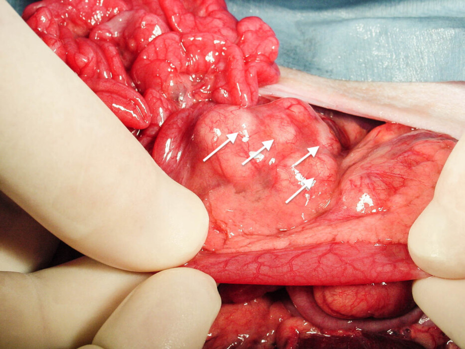
Figure 1a. Gross appearance of lymphoma involving mesenteric lymph nodes (arrows) in a ferret (Mustela putorius furo). Photograph provided by Dr. Peter Fisher. Click image to enlarge.
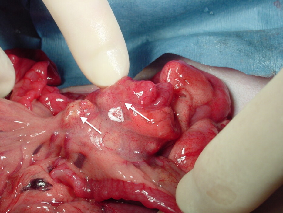
Figure 1b. Closer view of mesenteric lymph nodes (arrows) shown in Figure 1a. Photograph provided by Dr. Peter Fisher. Click image to enlarge.
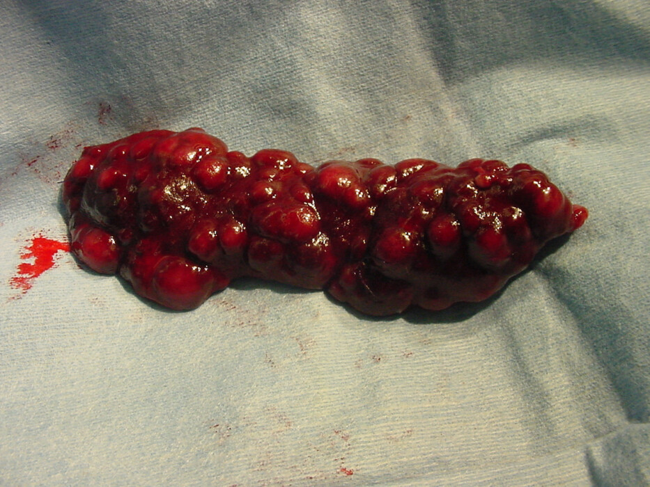
Figure 2a. Gross appearance of splenic lymphoma in a ferret (Mustela putorius furo). Photograph provided by Dr. Ruth Boll. Click image to enlarge.
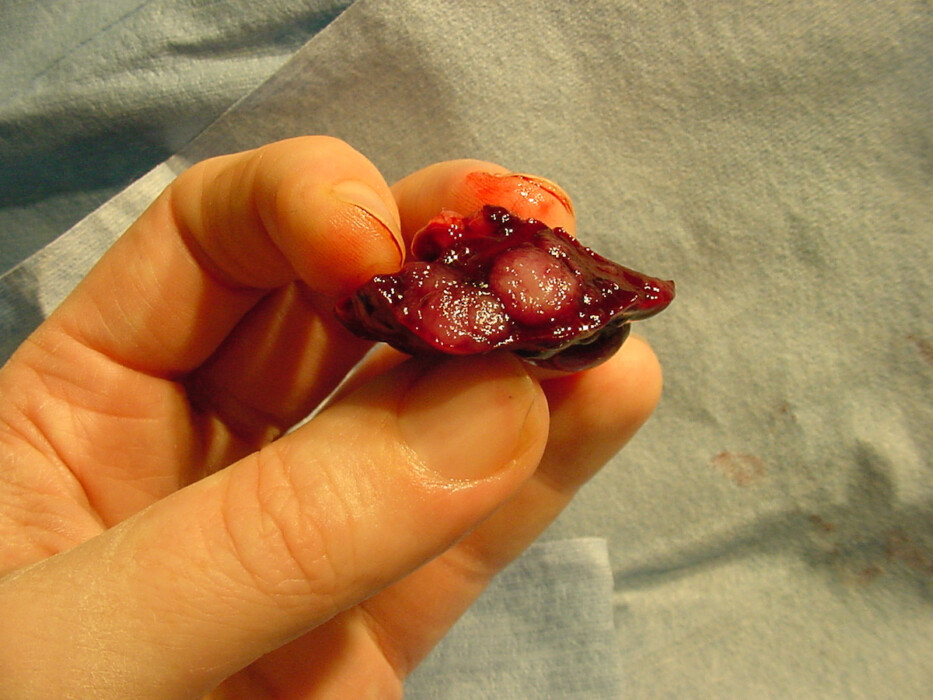
Figure 2b. Cut surface of the spleen shown in Figure 2a. Photograph provided by Dr. Ruth Boll. Click image to enlarge.
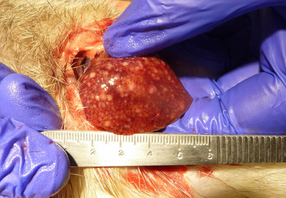
Figure 3. Gross appearance of metastatic lymphoma involving the liver in a ferret (Mustela putorius furo). Photograph provided by Dr. Peter Fisher. Click image to enlarge.
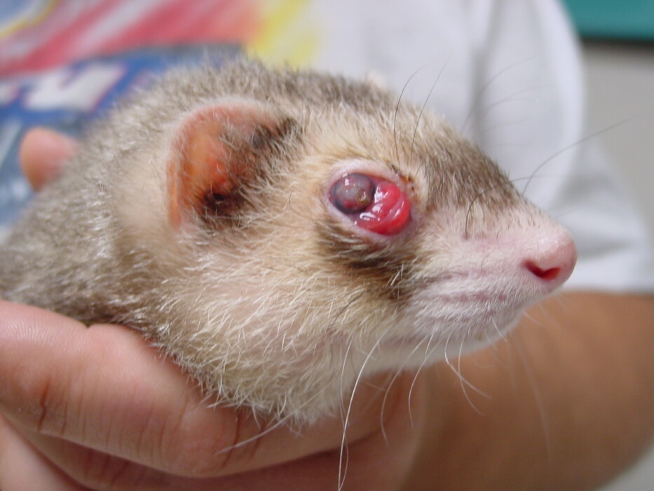
Figure 4. Ocular lymphoma in a ferret (Mustela putorius furo) causing prolapse of the lacrimal gland. Photograph provided by Dr. Peter Fisher. Click image to enlarge.
Ferrets with lymphoma have no specific clinical presentation, as clinical signs depend on the organ(s) involved. Patients may present with lethargy, anorexia, weakness, cutaneous masses or lesions, GI disorders, respiratory or cardiac signs, or neurologic deficits.2, 8, 9 Common abnormalities found during physical examination include abdominal mass(es), usually due to splenomegaly and/or lymphadenomegaly, and peripheral lymphadenomegaly (Fig 5).2 Lymphoma can also be an incidental finding in apparently healthy ferrets.
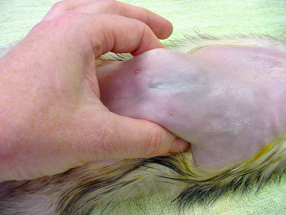
Figure 5. Profound splenomegaly in a ferret (Mustela putorius furo). Photograph provided by Dr. Ruth Boll. Click image to enlarge.
Although quite rare, cutaneous lymphoma can also be encountered (1% of cutaneous lesions and 5% to 7% of lymphoma cases depending on the study).1,3,5,7,10 Cutaneous epitheliotropic lymphoma commonly causes swollen, hyperemic, alopecic lesions on the feet or limbs, however lesions may also appear over the lumbar spine or even in a more diffuse, generalized pattern (Fig 6).27 These lesions tend to grow in size and multiple lesions will develop if left untreated.2, 10

Figure 6. Cutaneous lymphoma involving the foot in a ferret (Mustela putorius furo). Photograph provided by Dr. Véronique Mentré’. Click image to enlarge.
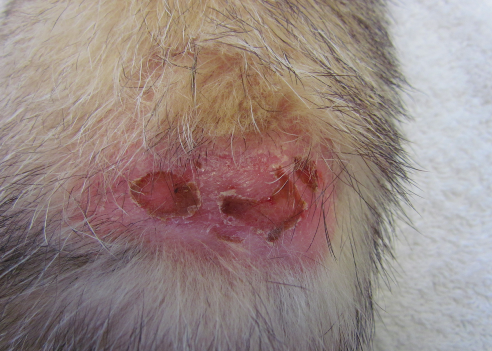
Figure 7. Cutaneous lymphoma involving the dorsal lumbar region in a ferret (Mustela putorius furo). Photograph provided by Dr. Véronique Mentré’. Click image to enlarge.
Diagnosis
Lymphoma is considered “the great impersonator”: it can look like almost any disease – and should always be kept on the differential list until a definitive diagnosis is reached (Table 1).
| Table 1. Important differential diagnoses for lymphoma in the ferret. | |
|---|---|
| Form of lymphoma | Differential diagnoses |
| Gastrointestinal | Other chronic gastrointestinal diseases including: Enzootic catarrhal enteritis (ferret enteric coronavirus) Gastrointestinal foreign body Helicobacter mustelae infection Inflammatory bowel disease Intussusception Other gastrointestinal tumors |
| Mediastinal | Chylothorax Congestive heart failure Dilated cardiomyopathy Hemothorax Other mediastinal tumors (thymoma, lipoma) |
| Cutaneous | Chronic inflammatory dermatitis Mast cell tumor or other cutaneous tumors |
A minimum database is necessary to ensure an accurate diagnosis and to properly stage lymphoma. This database should include blood work and imaging.
- Complete blood count
Anemia (packed cell volume <45%) is common and consistently nonregenerative.2 Leukemia is rare (7.4% of cases in one study). Reactive lymphocytosis is mentioned in up to 32% of cases 5, however lymphocytosis can be caused by chronic infections as well. Neutropenia is occasionally found; thrombocytopenia is rare.2
Blood smears can yield significant information. The presence of keratocytes, acanthocytes, and schistocytes showed a significant association with lymphoma, however, these abnormal red blood cell morphologies are not specific to lymphoma and are also seen with other disorders, such as GI disease (schistocytes) and adrenocortical disease (acanthocytes).11
- Complete biochemistry profile
Abnormalities depend on organ involvement. Hypoalbuminemia can be found in ferrets with GI forms of lymphoma; hyperproteinemia with hyperglobulinemia has been reported in rare occurrences,.2 Hypercalcemia has also been reported but is uncommon in the ferret and is seen in only about 7% of cases5, 12. When hypercalcemia is found, testing for intact parathyroid hormone (iPTH) and parathyroid hormone-related protein (PTHrP) can be performed to confirm humoral hypercalcemia of malignancy. Although the reference range for iPTH have been recently reported for ferrets, only canine/feline reference ranges are available for PTHrP.12, 13
- Urinalysis results are usually normal in ferrets with lymphoma but may be abnormal in cases with renal involvement and renal failure.
- Survey radiographs
Always obtain at least two views (ventrodorsal and lateral). Obtain right and left lateral views if thymic involvement is suspected. Potential findings on abdominal radiographs include hepatomegaly, ascites, mesenteric or sublumbar lymphadenopathy, nephromegaly, and splenomegaly.2 Thoracic radiographs may reveal sternal or tracheobronchial lymphadenopathy, a widened mediastinum, and/or pleural effusion, which was the most common thoracic abnormality in one study.2, 5, 14 Evaluate bones closely as skeletal lesions have been described in several case reports.2, 5, 15, 16,26
Abdominal ultrasound is often needed to identify affected viscera and lymph nodes. Mesenteric lymphadenomegaly, peritoneal effusion, and splenomegaly are often reported as the most common ultrasonographic findings.2, 5, 14 However these findings are not pathognomonic for lymphoma and can be observed with inflammatory bowel disease and other inflammatory conditions.2 Perform thoracic ultrasonography if indicated. Use fine needle aspirates of enlarged organs for diagnosis and staging. Sampling of free fluid may provide a presumptive diagnosis if organ aspirate is not possible.
Definitive diagnosis of lymphoma relies upon cytology or histology.
- In the hands of an experienced clinical pathologist, cytology of fine needle aspirates (FNA) can be diagnostic, although more commonly results may be suggestive of lymphoma. In one study, 66% of cases had a confirmed diagnosis with FNA.6 Depending on the FNA site, cytological findings might be insufficient, particularly when aspirating abdominal lymph nodes or liver as severe lymphoid infiltrate may also occur with inflammatory bowel disease. Moreover, evidence is lacking regarding the comparative diagnostic value and concordance of cytology versus biopsy.5 References are available for cytologic population of mesenteric lymph nodes aspirates in ferrets.17
Bone marrow aspirate cytology is indicated in cytopenic patients or if abnormal lymphocytes are seen on peripheral blood smears. The proximal femur or the proximal humerus may be used.2
- Histopathology of affected organ tissue is required for a definitive diagnosis, phenotyping, and staging.2, 18 Even with histology, lesions may be confused with severe chronic inflammatory bowel disease in the intestine and mesenteric lymph nodes, and immunohistochemistry may be advocated to ensure adequate diagnosis.2, 19 Thus, biopsies of peripheral lymph nodes might be better suited for diagnostic purposes.2
Prognosis
Overall mean survival times for untreated ferrets can range from 5.7 to 7 months.20 Some ferrets with small cell lymphoma have been reported to survive up to 2 years without any treatment.2 Staging may help to determine the prognosis more precisely. In a proposed staging system for ferrets, stage I neoplasia carries the best prognosis (Table 2).18
Table 2. Proposed staging system for lymphoma in ferrets18
| Stage I | The tumor involves only a single site |
| Stage II | Multiple sites are involved on the same side of the diaphragm |
| Stage III | The spleen is involved as well as lymph nodes on both sides of the diagram |
| Stage IV | Multiple sites on both sides of the diaphragm are involved |
One study found a median survival time of approximately 110 days with localized lymphoma versus 36 days with disseminated disease.7 Total remission has even been reported in several cases with localized disease.5
Pathologists may also grade lymphoma based on cytologic features. Low-grade neoplasms are typically characterized by small lymphocytes with few mitotic figures. Intermediate grade neoplasms show diffuse large cell lymphocytes; and high-grade lymphomas contain diffuse, immunoblastic cells with large numbers of mitotic figures. In some studies, small lymphocytic lymphoma has been associated with longer survival times than larger cell lymphoma.20,24
Although it may also be useful to characterize lymphoma as either B- or T-cell origin, data from the scientific literature is equivocal regarding the prognosis associated with phenotype. Classification based on immunohistochemistry found T-cell lymphomas accounted for 80% to 90% of cases, while B-cell lymphomas were uncommon.5 Depending on the study, B-cell lymphomas had either a better or worse survival time when compared with T-cell lymphomas.2,5,20
Cutaneous epitheliotropic lymphoma seems to have a fair prognosis, with prolonged survival times of up to 3 to 4 years reported, especially with rapid excision. Systemic involvement does not necessarily occur in ferrets with epitheliotropic lymphoma, as seen in dogs and humans.2, 10
Treatment options
The goal of treatment for the veterinary patient facing cancer is to improve the animal’s quality of life. At this point, no conclusive information exists to indicate that any one treatment is superior for most cases, and controlled studies are decidedly lacking in ferrets. In many cases, lymphoma is systemic in ferrets and chemotherapy is the recommended treatment. In the rare occurrence where a single organ is affected, surgery or radiation therapy may be successful and enable remission. In cases of cutaneous lymphoma, complete surgical excision is advised and can result in prolonged disease-free intervals. Patients with gastric lymphoma should also be managed for Helicobacter infection as an association has been suggested.2
COP or modified COP protocols
Cyclophosphamide, vincristine sulfate (Oncovin), and prednisone (COP); the COP protocol with doxorubicin hydrochloride or hydroxydaunorubicin (CHOP), and modified COP or CHOP are the main published protocols for treating lymphoma in ferrets.2 In a retrospective study, 15 ferrets treated with modified COP, with or without additional chemotherapeutics, had a median survival time of 429 days, higher than the overall median survival time of 124 days for all treatment protocols.6 Another author reported a mean survival time of 437 days in ferrets treated with “an aggressive chemotherapy protocol”.21 Based on data from feline and canine medicine, these treatment protocols are typically recommended for intermediate to high-grade lymphomas.2 The main limitation is that intravenous (IV) access is required. Ferret body surface area calculation has been described and should be used when calculating the chemotherapy dosing regimen, however the feline formula might also be adequate as they do not differ significantly.22
“Non-IV” protocols
Another commonly used regimen is the 27-week “non-IV” protocol from Tufts Cummings School of Veterinary Medicine. This protocol uses prednisone, L-asparaginase, cyclophosphamide, cytarabine, methotrexate, chlorambucil, and procarbazine. The main drawback of this protocol is that the oral drugs require compounding and cannot be administered by the owner on an outpatient basis.2 With this protocol, median survival time was lower (86 days) but most ferrets were lost to follow-up so this value might be underestimated.6
A protocol using chlorambucil and prednisone has been described for ferrets with lymphocytic lymphoma. Dosages were extrapolated from protocols used in cats.2
Corticosteroids alone
In numerous cases, ferrets with lymphoma are treated with corticosteroids alone. This treatment protocol should be considered palliative, is relatively inexpensive, and may reduce clinical signs and improve the quality of life. Median survival time with prednisolone alone is reportedly 97.5 days (range: 3-744 days).6 Long-term remission (more than 1 year) has even been described with prednisolone alone in a case of epitheliotropic gastrointestinal lymphoma23, however cutaneous epitheliotropic lymphoma respond variably to corticosteroids.2,10,27 Dosages have ranged between 0.5 to 2 mg/kg once to twice a day.2
Patient monitoring
Common adverse effects of chemotherapy include GI distress, hair loss, and myelosuppression. Complete blood counts and biochemistry panels should be regularly monitored throughout chemotherapy. Symptomatic treatment may be administered for GI side effects.2
Conclusion
Lymphoma is common in ferrets and has multiple clinical presentations. Diagnostic imaging, cytology and histology are the most useful diagnostic tools for this condition. Prognosis is poor, as the median survival time is about 4 to 6 months in studies, however some ferrets live more than 2 years after diagnosis. Chemotherapy is generally advocated; however controlled studies are lacking regarding chemotherapeutic protocols and survival time.
Acknowledgments: This article is an updated version of a manuscript shared by Sandra Mitchell, DVM and critically reviewed by Michael S. Kent, DVM, DACVIM (Oncology) DACVR
References
References
- Avallone G, Forlani A, Tecilla M, et al. Neoplastic diseases in the domestic ferret (Mustela putorius furo) in Italy: classification and tissue distribution of 856 cases (2000-2010). BMC Vet Res 2016;12(1):275. doi: 10.1186/s12917-016-0901-7. PMID: 27919252; PMCID: PMC5139086.
- Williams BH, Wyre NR. Neoplasia in ferrets. In: Quesenberry KE, Orcutt CJ, Mans C,Carpenter JW (eds.) Ferrets, Rabbits, and Rodents, 4th ed. Philadelphia: WB Saunders; 2020:92-108.
- Shiga T, Nakata M, Miwa Y, et al. A retrospective study (2006-2020) of cytology and biopsy findings in pet rabbits (Oryctolagus cuniculus), ferrets (Mustela putorius furo) and four-toed hedgehogs (Atelerix albiventris) seen at an exotic animal clinic in Tokyo, Japan. Journal of Exotic Pet Medicine 2021;38:11-17. doi: 10.1053/j.jepm.2021.03.008.
- Miwa Y, Kurosawa A, Ogawa H, et al. Neoplasitic diseases in ferrets in Japan: a questionnaire study for 2000 to 2005. J Vet Med Sci. 2009;71(4):397-402. doi: 10.1292/jvms.71.397.
- Huynh M, Chassang L, Zoller G. Evidence-based advances in ferret medicine. Vet Clin North Am Exot Anim Pract 2017;20(3):773-803. doi: 10.1016/j.cvex.2017.04.009.
- Webb JK, Graham JE, Burgess KE, et al. Presentation and survival time of domestic ferrets (Mustela putorius furo) with lymphoma treated with single- and multiagent protocols: 44 cases (1998–2016). Journal of Exotic Pet Medicine 2019;31:64-67. doi: 10.1053/j.jepm.2019.07.011.
- Onuma M, Kondo H, Ono S, et al. Cytomorphological and immunohistochemical features of lymphoma in ferrets. J Vet Med Sci. 2008;70(9):893-898. doi: 10.1292/jvms.70.893.
- Menicagli F, Lanza A, Sbrocca F, et al. A case of advanced second-degree atrioventricular block in a ferret secondary to lymphoma. Open Vet J. 2016;6(1):68-70. doi: 10.4314/ovj.v6i1.10. PMC4833871.
- Ingrao JC, Eshar D, Vince A, et al. Focal thoracolumbar spinal cord lymphosarcoma in a ferret (Mustela putorius furo). Can Vet J. 2014;55(7):667-671. PMC4060909.
- Kanfer S, Reavill DR. Cutaneous neoplasia in ferrets, rabbits, and guinea pigs. Vet Clin North Am Exot Anim Pract. 2013;16(3):579-598. doi: 10.1016/j.cvex.2013.05.006.
- Bau-Gaudreault L, Grimes C. Evaluation of erythrocyte morphology and prevalence of poikilocytes in peripheral blood of sick domestic ferrets (Mustela putorius furo). Journal of Exotic Pet Medicine 2019;31:86-90. doi: 10.1053/j.jepm.2019.07.014.
- Bean AD, Fisher PG, Reavill DR, et al. Hypercalcemia associated with lymphomas in the ferret (Mustela putorius furo): Four cases. Journal of Exotic Pet Medicine 2019;29:147-153. doi: 10.1053/j.jepm.2018.09.014.
- Cannizzo SA, Rick M, Harrison TM, et al. Parathyroid hormone, ionized calcium, and 25-hydroxyvitamin D concentrations in the domestic ferret (Mustela putorius furo). Journal of Exotic Pet Medicine 2017;26(4):294-299. doi: 10.1053/j.jepm.2017.07.004.
- Suran JN, Wyre NR. Imaging findings in 14 domestic ferrets (Mustela putorius furo) with lymphoma. Vet Radiol Ultrasound. 2013 Sep-Oct;54(5):522-31. doi: 10.1111/vru.12068.
PMID: 23738830; PMCID: PMC7169257.
- Long H, di Girolamo N, Selleri P, et al. Polyostotic lymphoma in a ferret (Mustela putorius furo). J Comp Pathol 2016;154(4):341-344. doi: 10.1016/j.jcpa.2016.03.003.
- Eshar D, Wyre NR, Griessmayr P, et al. Diagnosis and treatment of myelo-osteolytic plasmablastic lymphoma of the femur in a domestic ferret. J Am Vet Med Assoc 2010;237(4):407-414. doi: 10.2460/javma.237.4.407.
- Paul-Murphy J, O’Brien RT, Spaeth A, et al. Ultrasonography and fine needle aspirate cytology of the mesenteric lymph node in normal domestic ferrets (Mustela putorius furo). Vet Radiol Ultrasound. 1999 May-Jun;40(3):308-10. doi: 10.1111/j.1740-8261.1999.tb00366.x. PMID: 10519312.
- Mayer J, Burgess K. An update on ferret lymphoma: A proposal for a standardized classification of ferret lymphoma. Journal of Exotic Pet Medicine 2012;21(4):343-346. doi: 10.1053/j.jepm.2012.09.010.
- Watson MK, Cazzini P, Mayer J, et al. Histology and immunohistochemistry of severe inflammatory bowel disease versus lymphoma in the ferret (Mustela putorius furo). J Vet Diagn Invest. 2016;28(3):198-206. doi: 10.1177/1040638716641156. PMID: 27026106.
- Ammersbach M, Delay J, Caswell JL, et al. Laboratory findings, histopathology, and immunophenotype of lymphoma in domestic ferrets. Vet Pathol 2008;45(5):663-673. doi: 10.1354/vp.45-5-663.
- Antinoff N, Hahn K. Ferret oncology: diseases, diagnostics, and therapeutics. Vet Clin North Am Exot Anim Pract. 2004;7(3):579-625. doi: 10.1016/j.cvex.2016.07.004. PMID: 27890288.
- Jones KL, Granger LA, Kearney MT, et al. Evaluation of a ferret-specific formula for determining body surface area to improve chemotherapeutic dosing. Am J Vet Res. 2015;76(2):142-148. doi: 10.2460/ajvr.76.2.142.
- Sinclair KM, Eckstrand C, Moore PF, et al. Epitheliotropic gastrointestinal T-cell lymphoma with concurrent insulinoma and adrenocortical carcinoma in a domestic ferret (Mustela putorius furo). Journal of Exotic Pet Medicine 2016;25(1):34-43. doi: 10.1053/j.jepm.2015.12.011.
- Erdman SE, Brown SA, Kawasaki TA, et al. Clinical and pathologic findings in ferrets with lymphoma: 60 cases (1982-1994). J Am Vet Med Assoc. 1996;208(8):1285-9. PMID: 8635973.
- Erdman SE, Correa P, Coleman LA, et al. Helicobacter mustelae-associated gastric MALT lymphoma in ferrets. Am J Pathol 151(1):273-280, 1997. PMID: 9212752. PMCID: PMC1857920.
- Eshar D, Wyre NR, Griessmayr P, Durham A, Hoots E. Diagnosis and treatment of myelo-osteolytic plasmablastic lymphoma of the femur in a domestic ferret. J Am Vet Med Assoc. 2010;237(4):407-14. doi: 10.2460/javma.237.4.407. PMID: 20707751.
- Bernhard C, Flenghi L, Nicolier A, Mentré V. Cutaneous epitheliotropic T-cell lymphoma in ferrets (Mustela putorius furo): 3 cases (2014–2021). J of Exotic Pet Med. 2023;44:30-34. doi: 10.1053/j.jepm.2022.10.006.
Chassang L. Lymphoma in the ferret: An overview of diagnosis and treatment. April 3, 2023. LafeberVet Web site. Available at https://lafeber.com/vet/lymphoma-in-the-ferret-an-overview-of-diagnosis-and-treatment/