Key Points
- Heavy metal poisonings in birds most commonly occur from ingestion of substances containing lead or zinc.
- Lead that is ingested can be absorbed through the gastrointestinal tract and then taken up by soft tissues and eventually by bone.
- Lead affects all major organs and can cause damage to the gastrointestinal tract, red blood cells, kidneys and liver.
- Clinical signs of heavy metal toxicosis may include non-specific signs of illness, gastrointestinal signs, urinary tract problems and neurological deficits.
- Antemortem diagnosis of heavy metal toxicity relies on blood lead or zinc levels. In some BUT NOT ALL cases, ingested lead will also be visible on survey radiographs.
- Remove heavy metal from the bird’s tissues with a chelation agent such as calcium EDTA.
- Pieces of heavy metal within the digestive tract may also be removed endoscopically, surgically or via gastric lavage.
Introduction
Heavy metal poisoning in birds most commonly occurs from the ingestion of substances containing lead, or less commonly zinc.
Lead
Acute heavy metal toxicity is occasionally seen in companion parrots that ingest or chew on objects containing metal because of their curious nature and innate desire to forage. Chronic lead poisoning most frequently affects free-ranging wildlife, such as ducks, geese, swans, and loons (Elliott 2025) (Fig 1). Lead toxicity also occasionally occurs in upland game birds, such as mourning doves (Zenaida macroura), wild turkey (Meleagris gallopavo), pheasants and quail. Lead poisoning has also been reported in raptors from the ingestion of lead-contaminated prey. Bald eagles (Haliaeetus leucocephalus) have repeatedly been shown to be more sensitive to lead toxicity than other wild avian species, including red-tailed hawks (Buteo jamaicensis), swans (Cygnus spp), and turkey vultures (Cathartes aura) (Elliott 2025, McTee 2023, Fallon 2017).
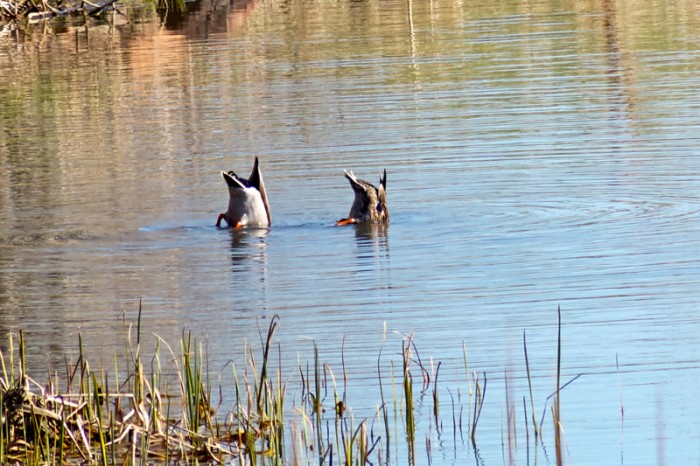
Figure 1. Waterfowl like these mallard ducks can be exposed to lead through the ingestion of spent lead shot, bullet fragments, and fishing sinkers. Photo credit: nature80020/Flickr Creative Commons. Click image to enlarge.
Lead toxicity in wild birds is most commonly seen during migration in the late fall and early spring (McTee 2023). In heavily contaminated areas, toxicity may be observed at any time of the year.
Sources of lead
Lead may be found in many household items (see Box 1):
|
|
An important source is lead from ammunition used by hunters. Raptors ingest lead in the form of gunshot and bullet fragments present within the viscera of prey and scavenge (including ingested shot) or embedded in tissues (Elliott 2025, Monoclús 2020)
Avian scavenger species, including birds of prey, like bald eagles (Haliaeetus leucocephalus) and vultures, are particularly prone to incidental ingestion of lead in spent ammunition or fragmented lead shot embedded within the tissues of carcasses or wounded animals (Elliott 2025, McTee 2023, Bassi 2021, Monoclus 2020).
Several nations have implemented national bans on lead shotgun ammunition, including Norway, the Netherlands, and Switzerland. In the United States, lead shot has been banned for hunting waterfowl, however, spent lead shot is still present in waterways (Aljohani 2023). Waterfowl ingest spent ammunition or lead tackle from fishing gear…when it is mistaken for grit in water sediment (Elliott 2025). Ingestion of one to three lead shotgun pellets has been reported to be lethal in waterfowl (Beasley 1999).
Lead is also widely distributed and persistent in the environment. Exposure to lead in the environment can also result from numerous sources, such as lead-based paint, fuel, batteries, fishing sinkers, mining activities, production of fertilizers, industrial activities, and waste disposal (Elliott 2025, Aljohani 2023, Monoclús 2020). Lead-contaminated soil is a persistent and widespread source of elevated blood lead (Gillings 2024).
Pathogenesis
Lead is relatively insoluble. Small amounts of this heavy metal are absorbed from the gastrointestinal tract after ingestion. Lead is absorbed in the small intestine, where it then enters systemic circulation (Elliott 2025). Lead is then transported around the body, reaching all organs and tissues, including the liver and kidneys, bones, and growing feathers (Monoclus 2020). Approximately 6% of systemic lead is stored in soft tissues, including blood, muscle, parenchymal tissues, and the nervous system, and the remaining 94% is stored in bone (Elliott 2025). The presence of grit within the ventriculus or gizzard increases the absorption of lead, which is first retained in soft tissues and eventually bone, which can retain elevated lead levels for years. The half-life of lead is weeks to months in soft tissues and approximately 2 weeks in blood (Monoclús 2020).
Lead causes endothelial damage while also inhibiting enzymes needed for cellular metabolism. Pathologic changes may include:
- Epithelial necrosis in the gastrointestinal system
- Increased erythrocyte fragility
- Bone marrow suppression leading to inhibition of erythrocyte production and function
- Damage to capillaries within the brain resulting in cerebral edema
Clinical signs of lead toxicosis
Clinical signs in birds vary with the dose and rate of lead exposure, and systemic effects can differ widely between individuals and species (Elliott 2025)
In cases of acute exposure, birds typically show non-specific signs of illness, such as bright green feces, labored breathing, as well as problems associated with the gastrointestinal tract, urinary tract and/or nervous system (Elliott 2025) (Box 2). It is also possible for acute toxicity to result in sudden death; the affected bird is usually in good body condition (Elliott 2025, Monoclús 2020). Neurologic signs, such as hind limb paresis, depressed mentation, and visual impairment, are most commonly observed in birds with higher blood lead levels (Elliott 2025).
| Non-specific signs of illness | Weakness or depression, pallor and anorexia |
| Gastrointestinal signs | Anorexia, crop stasis, vomiting or regurgitation, biliverdinuria and diarrhea (i.e. loose, dark or black stool) |
| Urinary signs | Hematuria or hemoglobinuria, particularly in Amazon parrots (Amazona spp.) |
| Neurologic signs | Twitching, circling, convulsions and/or blindness |
Chronic lead toxicity is often associated with gradual weight loss and poor body condition, weakness, anemia, and depression (Elliott 2025).
Diagnosis of lead toxicosis
A history of exposure to lead should significantly increase the index of suspicion. If lead toxicosis is suspected, perform a complete blood cell count or at minimum a packed cell volume. Mild to moderate anemia may be documented; however basophilic stippling is extremely rare in birds (Gillings 2024) (Box 3).
| History of exposure | Mild to moderate anemia |
| Hematology | Mild to moderate anemia (basophilic stippling rare) |
| Survey radiographs | Metallic density |
| Whole blood levels | Lead > 0.2 ppm |
Survey radiographs are also recommended since some, but NOT ALL cases of lead toxicosis will reveal a discrete, metallic density within the gastrointestinal tract (Fig 2). In a survey of mallards (Anas platyrhynchos) dosed with lead shot, only 1.2% had a pellet in their ventriculus at necropsy (Rodriguez 2010).

Figure 2. Metal density in the ventriculus. Photo credit: Dr. Greg Rich. Click on image to enlarge.
Collect heparinized whole blood to measure blood lead levels. Antemortem levels can be measured using various methods, with spectrometry considered the gold standard (Elliott 2025). The most direct route for laboratory heavy metal testing includes resources such as:
Portable point-of-care analyzers (LeadCare, LeadCare II; Magellan Diagnostics) can also be used on whole blood and have been applied in field studies and wildlife rehabilitation (Elliott 2025, Manning 2019, Herring 2018, Fallon 2017).
Previously established avian blood lead toxicosis categories include (Elliott 2025, Golden 2016):
- Background: < 20 μg/dL
- Subclinical: 20 to 60 μg/dL)
- Clinical: > 60 μg/dL)
- Severely elevated: > 100 μg/dL)
A bird with a blood lead level > 60 µg/dL is more likely to present with neurologic disease and have a poor case outcome (Elliott 2025). With chronic toxicity, blood with blood lead levels often range between 60 and 100 μg/dL (Wismer 2016). With acute toxicity, blood lead levels are often severely elevated (Elliott 2025, Wismer 2016).
Inhibition of the enzyme, δ-aminolevulinic acid dehydratase (ALAD) also occurs with lead toxicity and ALAD levels may be measured. Aminolevulinic acid dehydratase levels have been described primarily in wildlife studies (van den Heever 2024).
Lead primarily accumulates in the renal cortex, liver, reproductive organs, and lungs with chronic exposure, though it can also be found in the brain and splenic red pulp (Aljohani 2023, Bassi 2021, Torimoto 2021). In a study of 93 bald eagles with severe lead poisoning, the most common gross necropsy lesions involved the heart and included multifocal myocardial pallor and apex rounding (Manning 2019). Brain lesions, including petechiae or hemorrhagic necrosis, were also observed in some birds. Other frequently reported findings in wild birds include gallbladder enlargement, proventricular impaction, and a cracked or peeling ventricular lining, with or without lead shot. Liver and kidney tissues are often collected to assess tissue lead levels.
Treatment of lead toxicosis
There are three goals for treatment of heavy metal toxicity. First, stabilize the patient by providing supportive care, such as supplemental heat, fluids to prevent dehydration, and medication to stop tremors or seizures.
Then remove heavy metal from bodily tissues with a binding or chelating agent:
- Calcium EDTA (Calcium disodium versenate, 3M Pharmaceuticals): 35-50 mg/kg every 8-12 hours by intramuscular or subcutaneous route for 5 days (Elliott 2025, Wismer 2016)
- Dimercaptosuccinic acid (Succimer, Chemet): 25 mg/kg every 24 hours by mouth for 10 days. In a study evaluating cockatiels (Nymphicus hollandicus), both DMSA and Ca EDTA were effective chelating agents in. Because DMSA is administered orally, it may be easier than other chelating agents for bird owners to administer at home. However, the narrow margin of safety of DMSA indicates that this agent should be used with caution (Denver 2000).
- D-Penicillamine (Cuprimine, Merck): 30 mg/kg by mouth every 12 hours for 7 days minimum
Chelation therapy may be unnecessary if the lead source is promptly removed. If initiated, renal function should be monitored throughout treatment (Richardson 2006). Lead stored in bone and soft tissues can complicate therapy, causing fluctuating blood lead levels. Redistribution of tissue-stored lead has been observed with both CaEDTA and DMSA.”
If a source of heavy metal is seen on radiographs, removal of lead from the gastrointestinal tract via endoscopy, surgery, or gastric lavage may also be indicated. Lubricants such as mineral oil or corn oil, cathartics (i.e. magnesium sulfate), or bulk agents such as peanut butter, psyllium or oral cellulose products may also be used to remove heavy metal from the digestive tract.
Removal of particles with an iron base using a feeding catheter loaded with neodymium-ferro-borium alloy magnets has also been described.
Prevention of lead toxiciosis
Since companion parrots are curious by nature, it can be challenging to prevent chewing and ingestion of undesirable objects. Pet birds should always be supervised during their time outside of the cage, and owners should also remove all known sources of heavy metal or limit exposure to areas with heavy metals (Box 1).
Control of problem areas for wildlife consists of plowing to lessen the availability of spent shot to birds. The use of non-toxic steel or bismuth shot for waterfowl hunting is also now required in the United States. This switch from lead to non-toxic shot has reduced the number of birds dying from lead poisoning in America. The United States and Canada are also considering a ban on lead fishing sinkers.
Zinc
Zinc is a trace metal or mineral essential for health. Zinc is involved in cell replication and in development of cartilage and bone (McDonald 2006). The primary target organs in zinc toxicity are the kidneys and pancreas.
Sources of zinc
Zinc toxicity usually arises from the ingestion of zinc-coated wire or metallic foreign bodies such as pennies minted after 1983. One penny contains approximately 2440 mg of elemental zinc (Richardson 2006).
|
|
Galvanized wire is another common source of zinc intoxication in parrots, especially cheap, imported wire. Aviary birds are often housed in galvanized steel wire cages. Galvanized coatings can contain up to 99.9% zinc, however galvanized wire can also contain lead (Platt 2006). Galvanized dishes should never be used since zinc can leach into the water.
Powder coating is a protective coating for the cage. While formulas differ by manufacturers, most contain no zinc, however some imported powder-coated cages use zinc to expedite setting of powder coating (Richardson 2006).
Zinc can also leach out of zinc-coated iron shot into the environment. Waterfowl can then ingest vegetation and sediments contaminated by zinc (Platt 2006).
A number of websites report that the adhesive in paper towel and toilet paper rolls contains significant amounts of zinc, however this appears to be more urban legend than fact.
Once, when we had a look at the actual zinc content in the glue in those paper rolls, and then “borrowed” the nutritional requirements for zinc in chickens; presuming that the requirements in a parrot would be similar, we were able to show that if a parrot ate toilet paper exclusively with that zinc containing glue, there would still be a need for zinc supplementation to meet the nutritional requirements for the bird. (Speer 2010, Veterinary Information Network)
Clinical signs of zinc toxicosis
As with lead toxicity, signs of zinc toxicity can be vague and non-specific, but clinical signs are often related to disease of the gastrointestinal tract, pancreas, kidneys and/or central nervous system. Although this is a bit controversial and reports are anecdotal, zinc toxicity has also been associated with an “extreme” loss of plumage and feather damaging behavior (Box 5).
| Box 5. Clinical signs of zinc toxicity | |
|---|---|
|
|
Diagnosis of zinc toxicosis
The minimum database in zinc toxicosis may be relatively unremarkable. Mild to moderate regenerative anemia due to erythrocyte loss has been described with zinc toxicity (Box 6). Since zinc toxicosis typically results from chronic exposure to fine metal powder, a metallic density is rarely observed on survey radiographs.
| Box 6. Diagnostic testing for zinc toxicity | |
|---|---|
| Hematology | Mild to moderate anemia |
| Whole blood levels | Zinc > 2 ppm |
The normal blood zinc ranges are only weakly understood for a variety of species. Additionally, in a trial following induced zinc toxicity in cockatiels, blood zinc levels were extremely inconsistent as a diagnostic predictor (Howard 1992). Part of the confusion stems from the fact that zinc is an essential nutrient. Normal homeostasis regulates zinc levels based on variations in gastrointestinal content, bioavailability and individual nutritional needs. Significant diurnal variations in zinc values have also been documented in 15 adult psittacine birds (Rosenthal 2005).
Nevertheless in the presence of clinical signs, blood zinc levels exceeding 200 μg/dl (or 2 ppm) are suggestive of toxicity. Collect heparinized whole blood to measure zinc levels. Take care in how the blood sample is drawn and stored to avoid contamination.
- Although microtainers are typically used for companion birds, remember that the
rubber stoppers in red-topped tubes contain zinc. The presence of rubber can cause an artifactual elevation in zinc levels. - Royal blue-topped tubes are an alternative collection tube to test for zinc and
other metals.
The pancreas is the tissue of choice for postmortem zinc analysis (Box 7). Liver and kidney samples may also be collected to measure tissue zinc levels.
| Box 7. Pancreatic tissue zinc levels in cockatiels (Dumonceaux 1994) | |
|---|---|
| Normal | 26.11 μg/gram (dry weight basis) |
| Toxic | 312.4-2418 μg/gram |
Important pathologic lesions seen with zinc toxiciosis include ileus and focal mononuclear degeneration of the liver, kidneys and pancreas (LaBonde 1995). Necrotizing pancreatitis and erosive ventriculitis are other common manifestations. Histologically, the koilin layer is disrupted and there is ulceration of the underlying mucosa and dysplasia of the ventricular glands.
Treatment of zinc toxicosis
The goals for treatment are the same as with lead toxicity. The most important difference is that zinc is not stored in bone, and therefore blood and tissue levels equilibrate faster. This means that the response to chelation therapy is also faster (LaBonde 1995).
Prevention of zinc toxicosis
Scrubbing all new galvanized cage wiring with a mildly acidic solution such as vinegar, then drying carefully, may reduce zinc levels.
Copper
Copper toxicosis is rare in birds. Sources of copper include wire, pennies minted before 1982, copper sulfate, anti-fouling paints and possibly copper ammunition (Franson 2011). An important source of acute copper toxicosis in free-ranging waterfowl and other aquatic birds are acid metalliferous water bodies.
Clinical signs of copper toxicosis may include depression, weakness, anemia, convulsions and coma. Black discoloration of the parenchyma is an important gross finding. Common histopathological lesions include proventricular and ventricular necrosis, ventricular hemorrhage and/or congestion, erosion and ulceration of the koilin layer and duodenal hemorrhage (Isanhart 2011).
Like zinc, significant diurnal variations in blood copper levels have been documented (Rosenthal 2005).
Iron
Iron toxicosis in companion parrots can result from exposure to cast-iron feeding bowls with chipped enamel. Non-specific signs of illness predominate such as lethargy, emaciation and anorexia. Deferoxamine is the treatment of choice in mammals, but calcium EDTA also works well (LaBonde 1995).
For more information on iron overload, see Iron Storage Disease in Birds.
References and further reading
References
Altman RB, Clubb SL, Dorrenstein GM, Quesenberry K. Avian Medicine and Surgery. Philadelphia: W.B. Saunders; 1997.
Bassi E, Facoetti R, Ferloni M, et al. Lead contamination in tissues of large avian scavengers in south-central Europe. Sci Total Environ. 2021;778:146130. doi: 10.1016/j.scitotenv.2021.146130. Epub 2021 Mar 2. PMID: 33714099.
Beyer WN, Franson JC, Locke LN, et al. Retrospective study of the diagnostic criteria in a lead-poisoning survey of waterfowl. Arch Environ Contam Toxicol. 1998;35(3):506-512. doi: 10.1007/s002449900409.
Brown CS, Luebbert J, Mulcahy D, et al. Blood lead levels of wild Steller’s eiders (Polysticta stelleri) and black scoters (Melanitta nigra) in Alaska using a portable blood lead analyzer. J Zoo Wildl Med. 2007; 37(3):361-365, 2006.
Denver MC, Tell LA, Galey, FD, et al. Comparison of two heavy metal chelators for treatment of lead toxicosis in cockatiels. Am J Vet Res. 2000;61(8):935-940.
Dumonceaux G, Harrison GJ. Toxins. In: Ritchie BW, Harrison GJ, Harrison LR (eds). Avian Medicine: Principles and Application. Lake Worth, FL: Wingers. 1994. Pp. 1030–1052.
Elliott SA, Hawkins S, Lemley E, McCormick L, Mans C. Evaluation and treatment of lead toxicosis in rehabilitated avian species: 95 cases (2014-2023). J Am Vet Med Assoc. 2025;263(4):1-8. doi: 10.2460/javma.24.09.0592. PMID: 39793202.
Fallon JA, Redig P, Miller TA, Lanzone M, Katzner T. Guidelines for evaluation and treatment of lead poisoning of wild raptors. Wildl Soc Bull. 2017;41(2):205-211. doi:10.1002/wsb.762.
Gillings MM, Ton R, Harris T, Taylor MP, Griffith SC. Blood lead increases and haemoglobin decreases in urban birds along a soil contamination gradient in a mining city. Environ Res. 2024;257:119236. doi: 10.1016/j.envres.2024.119236. Epub 2024 May 27. PMID: 38810819.
Gupta R. Veterinary Toxicology: Basic and Clinical Principles. Waltham, MA: Elsevier Science; 2025.
Herring G, Eagles-Smith CA, Bedrosian B, et al. Critically assessing the utility of portable lead analyzers for wildlife conservation. Wildl Soc Bull. 2018;42(2):284-294. doi:10.1002/wsb.892.
Hoogesteijn AL, Raphael BL, Calle P, et al. Oral treatment of avian lead intoxication with meso-2,3-dimercaptosuccinic acid. J Zoo Wildl Med. 2003; 34(1):82-87. doi: 10.1638/1042-7260(2003)34[0082:OTOALI]2.0.CO;2.
Howard BR. Health risks of housing small psittacines in galvanized wire mesh cages. J Am Vet Med Assoc. 1992;200(11):1667-174.
Hunt WG, Parish CN, Orr K, Aguilar RF. Lead poisoning and the reintroduction of the California condor in northern Arizona. J Avian Med Surg. 2009;23(2):145-150.
Isanhart JP, Wu H, Pandher K, et al. Behavioral, clinical, and pathological characterization of acid metalliferous water toxicity in mallards. Arch Environ Contam Toxicol. 2011;61(4):653-667.
Kerr R, Holladay S, Jarrett T, et al. Lead pellet retention time and associated toxicity in northern bobwhite quail (Colinus virginianus). Environ Toxicol Chem. 2010;29(12):2869-2874.
LaBonde J. Toxicity in pet avian patients. Seminars in Avian and Exotic Pet Medicine. 1995;4(1):23-31.
Macintire DK, Drobatz KJ, Haskins SC, Saxon WD. Manual of Small Animal Emergency and Critical Care Medicine. Ames: Blackwell Publishing; 2006. Pp. 270-271.
Manning LK, Wünschmann A, Armién AG, et al. Lead intoxication in free-ranging bald eagles (Haliaeetus leucocephalus). Vet Pathol. 2019;56(2):289-299. doi: 10.1177/0300985818813099. PMID: 30556491.
Martinez-Haro M, Green AJ, Mateo R. Effects of lead exposure on oxidative stress biomarkers and plasma biochemistry in waterbirds in the field. Environ Res. 2011;111(4):530-538.
McTee M, Kean B, Pons A, et al. The seasonal threat of lead exposure in bald eagles. Sci Total Environ. 2023;889:164256. doi: 10.1016/j.scitotenv.2023.164256. Epub 2023 May 19. PMID: 37209742.
Monclús L, Shore RF, Krone O. Lead contamination in raptors in Europe: A systematic review and meta-analysis. Sci Total Environ. 2020;748:141437. doi: 10.1016/j.scitotenv.2020.141437. Epub 2020 Aug 8. PMID: 32818895.
Platt SR. Evaluating and treating the nervous system. In: Harrison GJ, Lightfoot TL (eds). Clinical Avian Medicine. Palm Beach, FL: Spix Publishing; 2006. P. 510.
Richardson JA. Implications of toxic substances in clinical disorders. In: Harrison GJ, Lightfoot TL (eds). Clinical Avian Medicine. Palm Beach, FL: Spix Publishing; 2006. Pp. 713-715.
Ritchie BW, Harrison GJ, Harrison LR. Avian Medicine: Principles and Applications. Lake Worth: Wingers Publishing, Inc; 1994.
Rodriguez JJ, Oliveira PA, Fidalgo LE, et al. Lead toxicity in captive and wild Mallards (Anas platyrhynchos) in Spain. J Wildl Dis. 2010;46(3):854-863.
Rosenthal KL, Johnston MS, Shofer FS, Poppenga RH. Psittacine plasma concentrations of elements: daily fluctuations and clinical implications. J Vet Diagn Invest. 2005;17(3):239-244.
Speer B. Are toilet paper rolls a potential source of zinc poisoning? Veterinary Information Network. June 26, 2010. Available at http://www.vin.com/Members/boards/discussionviewer.aspx?FirstMsg=1&LastMsg=20&DocumentId=4550946. Accessed on November 15, 2011.
Stauber E, Finch N, Talcott PA, Gay JM. Lead poisoning of bald (Haliaeetus leucocephalus) and golden (Aquila chrysaetos) eagles in the U.S. inland Pacific northwest region—an 18-year retrospective study: 1991-2008. J Avian Med Surg. 2010;24(4):279-287.
Torimoto R, Ishii C, Sato H, et al. Analysis of lead distribution in avian organs by LA-ICP-MS: Study of experimentally lead-exposed ducks and kites. Environ Pollut. 2021;283:117086. doi: 10.1016/j.envpol.2021.117086. Epub 2021 Apr 5. PMID: 33848898.
van den Heever L, Naidoo V, Coetzer T, et al. Sub-lethal impacts of lead poisoning on blood biochemistry, immune function and delta-aminolevulinic acid dehydratase (δ-ALAD) activity in Cape (Gyps coprotheres) and white-backed (G. africanus) vulture chicks. Environ Res. 2024;245:117926. doi: 10.1016/j.envres.2023.117926. Epub 2023 Dec 15. PMID: 38104912.
Wismer T. Advancements in diagnosis and management of toxicologic problems. In: Speer B (ed). Current Therapy in Avian Medicine and Surgery. St. Louis: WB Saunders; 2016. Pp. 589-599.
Wynee J, Stringfield C. Treatment of lead toxicity and crop stasis in a California condor (Gymnogyps californianus). J Zoo Wildl Med. 2007;38(4):588-590.
Pollock C, Chow C. Heavy metal poisoning in birds. April 29, 2025. LafeberVet Web site. Available at https://lafeber.com/vet/heavy-metal-poisoning-in-birds/