Introduction
Although the rare veterinarian routinely deals with large numbers of waterfowl on a regular basis, many avian veterinarians encounter waterfowl only sporadically as wildlife rehabilitation cases, backyard poultry, and/or zoo specimens (Fig 1). When consulting textbooks for help, often a dizzying array of waterfowl diseases are encountered. Some conditions such as “angel wing” and predator trauma are important in captive populations, while infectious diseases like fowl cholera can cause massive die-offs in free-ranging birds. Unless captive populations are exposed to wild birds, the incidence of infectious disease is relatively low, although birds less than 7 weeks of age are at greater risk when compared to adult birds.

Figure 1. Many avian veterinarians encounter waterfowl only sporadically as wildlife rehabilitation cases, backyard poultry, and/or zoo specimens. Photo credit: Getty Images.
Below you will find a collection of differential diagnoses for common clinical problems observed in the anseriform. These abbreviated lists should in no way replace professional judgment when evaluating your patient. This “cheat sheet” is merely designed as an aide or reminder system and should never be used for diagnostic decision-making. Particularly important or common conditions are bolded or linked to disease descriptions (Box 1 through Box 12).
Diseases featured in the ‘Cheat Sheet’
Sudden death
Major rule-outs for acute to peracute death in waterfowl can include:
- Trauma
- Pesticide poisoning
- Chronic renal disease, gout
Other potential causes of sudden death include:
- Aspergillosis (young birds can die peracutely)
- Colibacillosis (ducks)
- Fowl cholera
- Pasteurella anatipestifer
In new duck disease or duck septicemia caused by P. anatipestifer, ducklings usually die within 6-12 hours after the onset of clinical signs. Acute death is seen in older birds.
- Renal coccidiosis (Eimeria truncata)
- Leucocytozoan simondi
- Duck virus enteritis (duck plague)
- Duck virus hepatitis type 1 (death rates up to 100% in ducklings < 1 week)
- Blue-green algal toxins
- Amyloidosis
| Box 1. Fowl Cholera or Avian Cholera |
|---|
| Fowl cholera, or disease caused by Pasteurella multocida, has been reported in many bird species and occurs primarily in free-ranging populations. Dramatic outbreaks, sometimes killing thousands of birds, are reported annually in North America. Although losses can occur any time of the year, outbreaks typically occur during the winter or spring in northern California, Oregon, Nebraska, as well as the Texas panhandle. Bacteria are shed in feces and bodily secretions such as oculonasal discharge and oral discharge. Infection is transmitted by ingestion of contaminated food and water, inhalation, or less commonly inoculation of feet by contaminated debris. In the acute form of disease, some birds may be found dead. Clinical signs can also include:
Death rapidly follows the onset of clinical signs with birds frequently dying within 6 to 12 hours. Chronic fowl cholera can follow an acute outbreak or may arise from a less virulent strain. Clinical signs vary and are related to focal disease like septic arthritis, sinusitis, or respiratory infection. Chronically infected, free-flying birds are the likely source of infection for captive poultry and waterfowl. The classic necropsy finding for the acute form of disease is a bird in good flesh with abundant fat reserves.
Chronic fowl cholera lesions usually involve focal infections with caseous exudate. Important differential diagnoses include duck virus enteritis, erysipelas, E. coli and other bacteremias. Diagnosis relies upon consistent clinical signs, gross necropsy findings, and culture of P. multocida from intestinal fluid or bone marrow. Wright’s stain can also be used to demonstrate bipolar rods in heart blood or liver impression smears. Antimicrobials commonly used against P. multocida include beta-lactam antibiotics, oxytetracycline (50 mg IM followed by 500 g/ton feed x 30d), or enrofloxacin (5 mg/kg PO, IM, SC q12h). Management of disease also relies upon prompt removal and incineration of carcasses as well as cleaning the environment. Pasteurella multocida can live in the environment for up to 3 months, however the organism is easily destroyed by common disinfectants, sunlight, drying, and heat. Therefore good sanitation practices are an important part of disease prevention and control. Additional helpful measures include:
A bacterin is available for valuable flocks with persistent problems. |
| Box 2. Duck Plague or Duck Virus Enteritis (DVE) |
|---|
| Duck plague is an important, acutely contagious viral disease affecting ducks, geese, and swans. The etiologic agent is an alpha-herpesvirus. Duck plague was first documented in the United States in Pekin ducks in New York state. Since then sporadic outbreaks have been documented in commercial operations or park waterfowl. Most outbreaks occur during Apr, May, and June. Anseriforms of all ages can be affected. Highly susceptible species include blue-winged teals (Anas discors), wood ducks (Aix sponsa), and redheads (Aythya americana). The incidence of duck virus enteritis (DVE) is also higher in Muscovy ducks (Cairina moschata), Canada geese (Branta canadensis), and Northern pintails (Anas acuta). Outbreaks can occur in both wild and captive populations, but is disease is relatively rare in free-ranging birds. The herpesvirus that causes DVE is shed in feces and oral secretions. Transmission occurs primarily via ingestion of fecal-contaminated water sources. The virus can survive up to 60 days in water at 4°C (39.2°F) or 30 days at room temperature. Infected birds can be asymptomatic. Clinical disease is usually acute in onset. Both clinical patients and asymptomatic carriers may possess ulcers or cheesy plaques beneath the tongue. Clinical signs can also include:
At necropsy birds are usually in good flesh. Although the severity of lesions can vary, gross lesions often include:
A histopathologist will detect the presence of intranuclear inclusions. Necrosis is identified histologically in the liver, spleen, esophagus, and intestines. There will also be histologic evidence of gastrointestinal hemorrhage and necrosis. Presumptive diagnosis of DVE relies on the presence of consistent clinical signs and gross lesions. Definitive antemortem requires electron microscopy of fecal/oral samples or viral isolation. The liver and spleen are the best tissues for culture. Treatment of the individual patient relies upon aggressive supportive care. Management of an entire flock usually relies upon culling affected birds and disinfecting the premises. Decontaminate the environment by chlorinating water and raising soil pH. Control/prevention measures include:
|
| Box 3. Amyloidosis |
|---|
| Amyloidosis is common in captive waterfowl, but extremely rare in wild birds. In one survey of trumpeter swan (Cygnus buccinator) necropsies, amyloidosis was the primary disease process found. Amyloidosis includes a group of diseases characterized by the abnormal accumulation of amyloid proteins organized into beta-pleated sheets in various tissues. Most avian amyloidosis cases consist of reactive systemic amyloidosis with increased production of serum amyloid A proteins, a normal acute-phase reactant protein. The incidence of amyloidosis increases with chronic inflammatory conditions like pododermatitis, arthritis, and gout as well as aging and social stressors. At necropsy, amyloid deposition is most prevalent in the liver, spleen, and kidney followed by the pancreas and adrenal gland. Affected tissues often appear pale, yellow-brown, firm, and enlarged. |
Non-specific signs of illness
Non-specific signs of illness frequently include weakness, lethargy, reluctance to fly, an unsteady gait, loss of appetite, and fluffed and ruffled feathers. Failure to preen can lead to loss of plumage water repellency. There are a host of potential causes of non-specific signs of illness (Table 1). Important causes in waterfowl include yolk peritonitis, amyloidosis, fowl cholera, renal coccidiosis, traumatic injury, and heavy metal poisoning.
| Table 1. Potential Causes of Non-Specific signs of Illness in Waterfowl | ||
|---|---|---|
| Disease Category | Differential Diagnoses | |
| Metabolic |
|
|
| Nutritional |
|
|
| Inflammatory | ||
| Infectious | Bacterial |
|
| Viral |
|
|
| Protozoal |
|
|
| Toxin |
|
|
| Trauma |
|
|
Lead is a persistent contaminant in the environment, and waterfowl are susceptible to lead poisoning from ingestion of lead pellets and fishing weights. In addition to non-specific signs of illness, clinical signs can include bright green droppings or biliverdinuria, mucoid oral secretions, pallor, and variable neurological deficits, like leg paresis, ataxia, wing droop, and abnormal head position. Waterfowl, particularly Canada geese (Branta canadensis), may also exhibit subcutaneous edema of the head and eyelids.
Conditions affecting the gastrointestinal tract
Foreign body ingestion is a very common problem in waterfowl. Objects such as fish hooks, wire, fishing line, and gunshot are often incidental findings at necropsy but their presence can also be associated with morbidity and mortality.
Potential causes of oropharyngeal plaques include:
- Duck virus enteritis or duck plague
- Derzy’s disease (fibrous material beneath the tongue)
Rule-outs for diarrhea in waterfowl include:
- Fowl cholera
- Avian tuberculosis
- Colibacillosis (ducks)
- Enteritis, colitis caused by Salmonella spp. (most common in young birds)
- Intestinal coccidiosis (occasionally seen in young, wild or captive birds under crowded conditions)
- Acanthocephalans
| Box 4. Acanthocephalans |
|---|
| More than 50 species of thorny-headed worms or Acanthocephalans have been reported in waterfowl. Thorny-headed worms use a hooked proboscis to attach to the intestinal tract setting off an intense inflammatory response. With heavy infestations, the resultant granulomatous, hemorrhagic enteritis leads to malnutrition, morbidity, and even death. |
The appearance of a waterfowl’s stool can sometimes provide a clue to the underlying cause of disease.
- Green or bile-stained watery droppings:
- Lead poisoning
- New duck disease (P. anatipestifer)
- Avian chlamydiosis (domestic ducklings and goslings; rare in the United States but serious outbreaks have been seen in Europe)
- White diarrhea can be due to Derzy’s disease
- Profuse, occasionally bloody diarrhea, may be caused by duck plague
It is possible for Newcastle disease virus to cause watery diarrhea, but most ducks and geese are resistant to clinical disease unless exposed to a virulent strain. It has been theorized that some waterfowl develop chronic infection and may serve as a reservoir of disease.
Emaciation
Potential causes of severe weight loss in waterfowl include:
- Heavy metal poisoning
- Pesticide poisoning
- Oil toxicosis
- Gastrointestinal foreign body ingestion
- Gastrointestinal parasites, such as the nematodes Echinuria uncinata (common proventricular worm), Capillaria contorta, which causes thickening and necrosis of the esophagus, and Amidostomum sp. (ventricular worms)
- Renal coccidiosis (young birds)
- Aspergillosis (young birds)
- Avian tuberculosis
- Colibacillosis (ducks)
- Avian chlamydiosis (domestic ducklings and goslings; rare in the United States but serious outbreaks have been seen in Europe)
Diseases of the juvenile bird
Important diseases of juvenile waterfowl include angel wing and renal coccidiosis caused by Eimeria truncata (Fig 2). Important viral diseases include duck virus hepatitis (affecting ducklings less than 1-2 weeks of age) as well as Derzy’s disease (goslings less than 4-6 weeks). Additional conditions may include yolk sacculitis or omphalitis caused by Gram-negative bacterial overgrowth (e.g. E. coli or Salmonella spp.), Actinobacillus infection, and duck septicemia.
| Box 5. Renal Coccidiosis |
|---|
| Renal coccidiosis caused by Eimeria truncata is a serious disease in domestic ducks and geese. Infection is subclinical in adult birds, but can cause heavy mortality in ducklings and goslings. Flocks housed in crowded surroundings with poor sanitation are at increased risk. Pale, swollen kidneys with multifocal white lesions may be found at necropsy. Oocysts can be identified on renal impression smears and on histopathologic exam. Sulfonamides are the treatment of choice. Control and prevention relies upon strict sanitation and disinfection. It is also helpful to house juvenile birds separate from adults. |
| Box 6. Angel Wing |
|---|
| Angel wing is a conformational abnormality of the wing known by many terms including “airplane wing” and “oar wing”. Angel wing is classically seen in large, juvenile waterfowl as the weight of the growing flight feathers causes the weak muscles of the carpus to rotate the wing outward at the level of the carpus (Fig 2). Known underlying causes include:
The incidence of angel wing decreases when young, growing birds are allowed to swim and dive. The normal waterfowl is in the water as it goes through a molt, which prevents wing dragging from being an important issue. Early treatment of angel wing relies either upon encouraging the bird to swim or bandaging that elevates the wingtip enough to make the bird more comfortable and allow normal feather growth. Tape the wing to itself, and not to the body, for 3 to 5 days. |
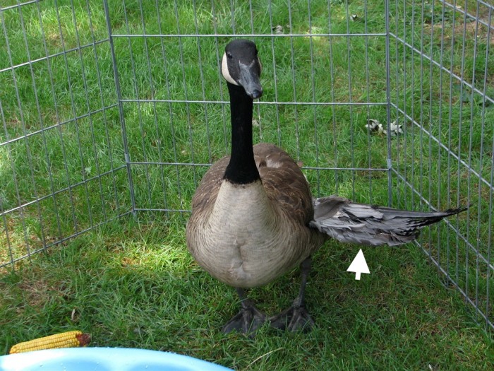
Figure 2. Outward rotation of the wing at the level of the carpus or “angel wing”. Photograph by audreyjm529. Click image to enlarge.
Aspergillosis is the most significant fungal disease of young birds. Birds usually become infected by inhalation of A. fumigatus spores in moldy or decaying organic matter.
Lameness
Important causes of lameness include septic arthritis and pododermatitis. Pododermatitis or “bumblefoot” frequently occurs in large-bodied birds such as swans housed on hard, rough surfaces. Obesity and lack of access to swimming facilities can predispose birds to bumblefoot. Most lesions occur on metatarsal pads. Septic arthritis may be caused by Salmonella spp., especially in young birds, and Mycoplasma synoviae.
| Box 7. Mycoplasma synoviae |
|---|
| Mycoplasma synoviae most commonly causes swelling of the hock joint or footpad. A presumptive antemortem diagnosis is based on consistent clinical signs and gross lesions. Definitive diagnosis requires culture or PCR testing. Gross necropsy findings include viscous to caseous exudate within joints and tendon sheaths. Lesions can extend into musculature and air sacs in advanced disease. Antimicrobials used against Mycoplasma spp. may include tylosin, tetracyclines, lincomycin, and spectinomycin. Treatment does NOT eliminate Mycoplasma spp. from a flock. Surgical intervention may be necessary but the prognosis is guarded. It is also important to move the bird to softer ground or grass. Prevention of mycoplasmosis requires that stock be purchased from clean breeder flocks and that strict biosecurity is maintained. |
Other potential causes of lameness include:
- Perosis or medial luxation of the gastrocnemius tendon, enlargement of the hock, bending deformities of tarsal or tarsometatarsal bones
- Arthritis (degenerative joint disease)
- Colibacillosis (ducks)
- Articular gout
- Trauma (bite wound)
- (Avian tuberculosis)
Respiratory signs
Yolk peritonitis is an important cause of respiratory distress in geese. Owners often report a recent history of reproductive activity. Coelomic distension is a common physical exam finding. Delayed molt is also sometimes seen.
Sinusitis caused by Mycoplasma gallisepticum is an important reason for keeping ducks and chickens separate.
There are a variety of other conditions that can cause respiratory signs in waterfowl:
- Duck plague (dyspnea)
- Pox, wet or diphtheritic form (rare, only reported in a mute swan [Cygnus olor])
- Avian influenza (rare sinusitis, domestic ducks seem more susceptible to clinical disease)
- Newcastle’s disease virus (rare, mild respiratory signs)
- Fowl cholera (blood stained nasal d/c, cyanosis)
- New duck disease (occasionally cough, tachypnea, sinusitis)
- Colibacillosis (ducks)
- Avian chlamydiosis (sinusitis in domestic ducklings and goslings; rare in the United States but serious outbreaks have been seen in Europe)
- Aspergillosis (dyspnea in young birds)
- Hemoparasites: Leucocytozoan simondi, Plasmodium spp., Hemoproteus nettionis (dyspnea)
- Nasal leeches (Theromyzon spp.) puddle ducks and swans from northern climates are most susceptible; sneezing, heavy loads can be fatal
- Gape worms or tracheal worms (Syngamus trachea or Cyathostoma sp.) infrequently reported in waterfowl: young, captive birds are most susceptible
- Oil toxicosis
- Botulism (respiratory failure)
- Avian tuberculosis
| Box 8. Avian Influenza |
|---|
| Avian influenza is reported in a variety of bird species including ducks, geese, and swans. The fecal-oral route most commonly transmits the orthomyxovirus causing avian influenza, although virus is also shed in respiratory and ocular secretions. Classic signs of avian influenza can include acute death, respiratory signs like oculonasal discharge and sinusitis, decreased egg production, and occasionally neurological signs. Waterfowl infected with avian influenza are usually asymptomatic although domestic ducks seem more susceptible to clinical disease. When lesions are present, sinusitis is the most common gross necropsy finding in ducks. Additional necropsy lesions can include catarrhal tracheitis, air sacculitis, myocarditis, encephalitis, as well as hemorrhage or congestion in the brain, lung, and gastrointestinal tract. Waterfowl are considered a potential reservoir of avian influenza. Wild waterfowl and free-range commercial ducks are known to play an important role in the spread of disease. Virus can remain stable in lake water for over 200 days. Prevention and control of disease relies upon preventing exposure to wild birds. For instance netting can be set up over pet duck areas. |
Neurologic disease
Important causes of neurologic signs in waterfowl include botulism, lead poisoning, pesticide poisoning, and trauma.
- Exposure to pesticides, such as organochlorines, can cause muscle fasciculations, convulsions, ataxia, and disorientation.
- Central nervous signs seen with lead poisoning can include weakness, drooped wings, erratic flying, leg paresis, ataxia, tremors, convulsions, partial to complete leg paresis, and blindness.
- Blunt force trauma to the head is another potential cause of neurologic deficits. The strong territorial nature of swans may increase the incidence of aggression and subsequent injury.
| Box 9. Botulism |
|---|
| Botulism, also known as “limberneck” or alkali poisoning is caused by exotoxins produced by the Gram-positive, spore-forming anaerobic bacterium Clostridium botulinum. Clostridium botulinum grows best in shallow, alkaline water, especially that overlying limestone with a pH ranging from 5.7-6.2. The organism persists in spore form until environmental temperature reaches 77°F (25°C). Spores can persist in the environment for years, and are resistant to heat and drying. Botulism can occur in wild and captive waterfowl, galliforms, and many other bird species. The distribution of disease is worldwide. In the United States and Canada most outbreaks occur west of the Mississippi river. Clostridium botulinum reproduces and makes toxin within vegetation and dead invertebrates at the water’s edge. Waterfowl then eat invertebrates and vegetation containing C. botulinum exotoxin type C. As waterfowl die, there is a subsequent build-up of fly populations and maggot production within vertebrate and invertebrate carcasses. The maggots concentrate toxin, and are then eaten by additional waterfowl creating a vicious cycle of biomagnification. Major outbreaks are usually associated with summer heat. Environmental conditions that favor bacterial growth and toxin production include:
The toxin affects peripheral nerves, and the clinical signs of botulism are dose-related ascending flaccid paralysis. Birds first exhibit leg paralysis, propelling themselves along with their wings. Paralysis then extends to the wings, nictitans, neck muscles and even the tongue. Birds often die by drowning or cardiopulmonary failure. There are no gross or microscopic lesions in birds with botulism although the stomach is frequently empty. A presumptive diagnosis is based on history, clinical signs, and an absence of obvious gross lesions. Definitive diagnosis relies upon mouse bioassay testing in which botulinum serum is injected into mice. Individual patients can be provided with aggressive supportive care. Gavage birds with charcoal slurry, tube feed as needed, while propping birds up or maintaining them in slings to prevent aspiration. Manage large flocks by providing easy access to fresh water, food, and shade. Antitoxin injections (where available) have also been credited with high rates of recovery of some waterfowl species. Control and prevention relies upon the removal of potential sources of Clostridial organisms and toxin from the environment such as:
|
Additional causes of neurological disease include:
- Vitamin E deficiency (encephalomalacia)
- Erysipelas: ataxia
- Fowl cholera: convulsions, opisthotonus, circular swimming; erratic flight and landings
- New duck disease (P. anatipestifer): central nervous system signs can include ataxia, tremors, abnormal swimming behavior, opisthotonos, and/or seizures
- Pseudomonas/Aeromonas spp. (flaccid paralysis in swans, especially during summer months)
- Salmonella spp: central nervous signs, most common in young birds
- Avian chlamydiosis: ataxia in domestic ducklings and goslings; rare in the United States but serious outbreaks have been seen in Europe)
- Avian influenza (very rare cause of neurologic signs)
- Derzy’s disease
- Duck plague: CNS signs – difficulty standing, ataxia, opisthotonos, convulsions
- Duck virus hepatitis (DVH Type 1): loss of balance, ataxia, opisthotonus
- Eastern equine encephalitis: neurologic deficits in imported species like Pekin ducklings
- Newcastle’s disease virus (rare cause of ataxia)
- Aspergillosis: ataxia or torticollis
- Blue-green algal toxins caused by summer algal blooms (Anabaena flosaquae): opisthotonos, convulsions
| Box 10. Duck Virus Hepatitis |
|---|
| Duck viral hepatitis or DVH type 1 is a worldwide disease of waterfowl caused by a picornavirus. Ducklings less than 1 week of age are particularly susceptible with mortality rates reaching 100%. Up to 50% of 1-3 week old ducklings die, while the death rate is negligible in ducklings over 4-5 weeks. An exception to this rule of thumb is the Muscovy duck (Cairina moschata), which is particularly prone to DVH with high mortality rates up to 6 weeks of age. Disease is transmitted by the fecal/oral route, aerosol, vertically, and via fomites. Survivors can excrete virus for up to 8 weeks post-infection. The clinical picture typically involves acute onset, rapid spread of clinical signs and death within 3-4 days. Ducklings may display non-specific signs of illness such as lethargy and separation from the flock as well as central nervous system signs such as opisthotonus and ataxia. Antemortem diagnosis relies upon antibody titers. Gross necropsy findings often include hepatomegaly with punctate or ecchymotic hemorrhages. Occasional findings include splenomegaly and mottling of the kidneys. The most consistent histologic finding is severe hepatic necrosis. Additional diagnostic tests include viral isolation and direct fluorescent antibody testing of the liver. Management of DVH typically relies upon strict quarantine (8 weeks minimum) and supportive care. Intramuscular injections of DVH Type 1 antiserum have also been found to be effective in susceptible ducklings in the face of an outbreak. Control and prevention relies upon strict isolation of young ducklings and vaccination of breeder ducks and/or vaccination of ducklings. |
When faced with a waterfowl suffering from presumptive neurologic disease, two additional rule-outs must be considered.
- Capture myopathy, causing leg paralysis and death, has been described in large birds like the Canada goose.
- Unfortunately neurologic signs can also herald imminent death in waterfowl as terminal convulsions and opisthotonos occur with many disease processes.
Ocular disease
- New duck disease (P. anatipestifer): serous to purulent ocular discharge, conjunctivitis
- Colibacillosis (ducks)
- Avian chlamydiosis: conjunctivitis, tearing in domestic ducklings and goslings; rare in US but serious outbreaks have been seen in Europe
- Nasal leeches (Theromyzon spp.): conjunctivitis, corneal damage, even blindness; puddle ducks and swans in northern climates are most susceptible
- Duck plague: oculonasal discharge, conjunctivitis
- Derzy’s disease: oculonasal discharge, swollen eyelids, conjunctivitis
| Box 11. Derzy's Disease |
|---|
| Derzy’s disease is caused by a highly resistant goose parvovirus that infects goslings (< 4-6 weeks) and ducklings (< 4 weeks). Derzy’s disease affects all domestic goose breeds, Canada geese, and snow geese (Chen caerulescens) as well as Muscovy ducks. Disease is primarily seen in Europe and Asia. Virus is transmitted by fecal-oral and vertical routes. Clinical signs vary with age.
Survivors are persistently infected and resistant to clinical disease. Available antemortem diagnostic tests include antibody titers and fecal electron microscopy. Definitive diagnosis relies upon viral isolation in goose or Muscovy duck eggs or virus neutralization testing. Gross necropsy findings in acute disease can include cardiomegaly, a pale myocardium, hepatomegaly with gray-white areas of necrosis, as well as a congested spleen and pancreas. Chronic goose parvovirus lesions can include serofibrinous pericarditis, perihepatitis, ascites, pulmonary edema, liver dystrophy, and/or catarrhal enteritis. The most significant histologic finding are intranuclear inclusions within the liver, spleen, and intestines. Management of Derzy’s disease relies upon supportive care. Prevention relies upon elimination of adult carrier geese and Muscovy ducks as well as vaccination of adult breeder birds. The virus is susceptible to bleach, stabilized chlorine dioxide, and glutaraldehyde. |
Dermatologic lesions
Feather loss
The mating process in ducks frequently results in a bald neck on the female. Some breeds like Khaki Campbell drakes are particularly active and can cause significant feather loss when there is only one or two ducks per drake.
Frostbite
Frostbite can be observed in species from tropical or subtropical regions like whistling ducks. Lesions begin as pale areas in webbing or on the knob that can progress to dry gangrene.
Loss of waterproofing
Waterproofing of the plumage is maintained through careful and frequent preening. Anything that disrupts this process will interfere with waterproofing, leading to a soggy appearance known as “wet feather”.
Common causes include:
- Lice leading to overpreening
- Chemical contamination (e.g., oil)
- Mold spores, particularly from willow trees, can fall on the plumage
Scratching at face
Nasal leeches (Theromyzon spp.) are a classic cause of head shaking and scratching at the face. Puddle ducks and swans in northern climates are particularly susceptible to infestation.
Additional causes of dermatologic lesions include:
- Avipoxvirus, dry form: wart-like nodules on featherless areas, usually legs and feet (reported only occasionally in wild birds)
- Derzy’s disease
- Erysipelas
- Fire ants
- Maggots
| Box 12. Erysipelas |
|---|
| Erysipelothrix rhusiopathiae is a small, Gram-positive facultative anaerobic bacillus resistant to many chemical and environmental factors. Erysipelas primarily affects turkey of all ages, but outbreaks have been reported in other fowl like ducks and geese. Bacteria are introduced through breaks in the skin or mucosa. Contaminated soil or carrier birds may serve as sources of infection. Some outbreaks have been associated with poultry housed in areas previously occupied by swine and sheep. Clinical signs include non-specific signs of illness, ataxia, skin lesions, and acute death. Gross necropsy lesions consistent with septicemia can include:
A presumptive diagnosis is supported by demonstrating beaded, pleomorphic, Gram-positive rod-shaped bacteria in smears of liver, spleen, heart blood, or bone marrow. Definitive diagnosis relies upon culture. Effective antimicrobials include first-generation beta-lactams or macrolide antibiotics like erythromycin and azithromycin. Control relies upon cleaning and disinfection with 1-2% sodium hypochlorite. |
Prolapse of the phallus
Important differentials for prolapse of the phallus include:
- Duck virus enteritis has been associated with prolapse of the phallus.
- Frostbite
- Excessive sexual stimulation
- Trauma, including over exuberant vent sexing
Prolapse of the duck penis is a common problem, particularly in light breeds like the Indian runner duck. Unless the problem is recognized early, the phallus ulcerates and becomes necrotic. Surgical amputation is often required.
Summary
Medical conditions commonly seen in captive waterfowl include pododermatitis, foreign body ingestion, toxicoses, nutritional disorders, exposure, and predator trauma. Unless captive populations are exposed to wild birds, the incidence of infectious disease is relatively low. Hatchlings are at greater risk for infectious disease when compared to adult birds.
References
References
Carpenter JW, Andrews GA, Beyer WN. Zinc toxicosis in a free-flying trumpeter swan (Cygnus buccinator). J Wildl Dis 40(4):769-774, 2004. doi: 10.7589/0090-3558-40.4.769.
Converse KA, Kidd GA. Duck plague epizootics in the United States, 1967-1995. J Wildl Dis. 2001; 37(2):347-357. doi: 10.7589/0090-3558-37.2.347.
Degernes L. Waterfowl medicine. American College of Zoological Medicine Board Review 1995.
Degernes LA. Waterfowl toxicity: a review. Vet Clin North Am Exot Anim Pract. 2008;11(2):283-300.
Dhillon AS, Winterfield RW, Thacker HL, Richardson JA. Erysipelas in domestic white Pekin ducks. Avian Dis. 1980; 24(3):784-7. PMID: 7447843.
Gomis S, Didiuk AB, Neufeld J, Wobeser G. Renal coccidiosis and other parasitologic conditions in lesser snow goose goslings at Tha-anne River, west coast Hudson Bay. J Wildl Dis. 1996; 32(3):498-504. doi: 10.7589/0090-3558-32.3.498.
LaBonde J. Private collections of waterfowl. Proc Annu Conf Assoc Avian Vet 1996. Pp. 215-223.
Lafgerquist JE, Davison M, Foreyt WJ. Lead poisoning and other causes of mortality in trumpeter (Cygnus buccinator) and tundra (C. columbianus) swans in western Washington. J Wildl Dis. 1994; 30(1):60-64. doi: 10.7589/0090-3558-30.1.60.
Meyerholz DK, Vanloubbeeck YE, Hostetter SJ, et al. Surveillance of amyloidosis and other diseases at necropsy in captive trumpeter swans (Cygnus buccinators). J Vet Diagn Invest. 2005; 17(3):295-298. doi: 10.1177/104063870501700318.
Oksanen A. Mortality associated with renal coccidiosis in juvenile greylag geese (Anser anser anser). J Wildl Dis. 1994; 30(4):554-556. doi: 10.7589/0090-3558-30.4.554.
Pearson GL, Cassidy DR. Perspectives on the diagnosis, epizootiology, and control of the 1973 duck plague epizootic in wild waterfowl at Lake Andes, South Dakota. J Wildl Dis. 1997; 33(4):681-705. doi: 10.7589/0090-3558-33.4.681.
Roberts V. Quack quack—treating the pet duck. Proc Annu Conf London Vet Show 2011.
Roberts V. Diseases of Free-range Poultry, 3rd ed. Linton, Cambs: Whittet Books, 2009.
Roberts V, Scott-Park F. Manual of Farm Pets. Gloucester: British Small Animal Veterinary Association, 2008.
Schneider RR, Hunter DB, Waltner-Toews D, Barker IK. A descriptive study of mortality at the Kortright Waterfowl Park: 1982-1986. Can Vet J. 1988; 29(11):911-914. PMID: 17423165; PMCID: PMC1680952.
Speer BL. Pet waterfowl medicine and surgery. Proc Annu Conf Mid-Atlantic States Association of Avian Veterinarians 2007. Pp. 90-92.
Tanaka S, Dan C, Kawano H, et al. Pathological study on amyloidosis in Cygnus olor (mute swan) and other waterfowl. Med Mol Morphol. 2008; 41(2):99-108. doi: 10.1007/s00795-008-0401-3.
Wang Q, Chang BJ, Riley TV. Erysipelothrix rhusiopathiae. Vet Microbiol. 2010; 140(3-4):405-417. doi: 10.1016/j.vetmic.2009.08.012.
Wobeser GB, Hunter B, Wright D, et al. Avian cholera and waterfowl biology. J Wildl Dis. 1992; 28(4):674-682. doi: 10.7589/0090-3558-28.4.674.
Further reading
Alexander DJ, Brown IH. History of highly pathogenic avian influenza. Rev Sci Tech. 2009; 28(1):19-38. doi: 10.20506/rst.28.1.1856.
Irvine R, Ceeraz V, Cox B, et al. Goose parvovirus in Great Britain. Vet Rec. 2008; 163(15):461. doi: 10.1136/vr.163.15.461.
Isanhart JP, Wu H, Pandher K, et al. Behavioral, clinical, and pathological characterization of acid metalliferous water toxicity in mallards. Arch Environ Contam Toxciol. 2011; 61(4):653-667. doi: 10.1007/s00244-011-9657-z.
Katavolos P, Staempfli S, Sears W, et al. The effect of lead poisoning on hematologic and biochemical values in trumpeter swans and Canada geese. Vet Clin Pathol. 2007; 36(4):341-347. doi: 10.1111/j.1939-165x.2007.tb00438.x.
Raymundo DL, Von Hohendorf R, Boabaid FM, et al. Outbreak of type C botulism in captive wild birds. J Zoo Wildl Med. 2012; 43(2):388-390. doi: 10.1638/2010-0084.1.
Salguero FJ, Sánchez-Cordón PJ, Núñez A, Gómez-Villamandos JC. Histopathological and ultrastructural changes associated with herpesvirus infection in waterfowl. Avian Pathol. 2002; 31(2):133-140. doi: 10.1080/03079450120118612.
Stipkovits L, Szathmary S. Mycoplasma infection of ducks and geese. Poult Sci. 2012; 91(11):2812-2819. doi: 10.3382/ps.2012-02310.
Wang C, Wu Y, Xing X, et al. An outbreak of avian cholera in wild waterfowl in Ordos wetland, Inner Mongolia, China. J Wildl Dis. 2009; 45(4):1194-1197. doi: 10.7589/0090-3558-45.4.1194.
Woo GH, Kim HY, Bae YC, et al. Outbreak of botulism (Clostridium botulinum type C) in wild waterfowl: Seoul, Korea. J Wildl Dis. 2010; 46(3):951-955. doi: 10.7589/0090-3558-46.3.951.
Pollock C. Waterfowl diseases: A "cheat sheet". December 11, 2012. LafeberVet Web site. Available at https://lafeber.com/vet/waterfowl-diseases-a-cheat-sheet/