



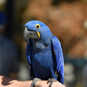
Birds most likely evolved from theropod dinosaurs, while mammals descended from carnivorous reptiles, the cynodonts. Their differing lineage possibly contributed to the differences observed in the avian cardiovascular structures when compared to mammalian ones. Similarities may represent the conservation of characteristics common to the two ancestral groups and/or convergence of evolution once the groups diverged.
The most striking features of birds is their ability to perform very rigorous functions in harsh environments, such as diving deeply in cold water, flying at high altitudes and running in hot deserts. This requires that their cardiovascular system (CVS) be able to meet the demands of providing adequate delivery of oxygen to vascular beds that are taxed by extreme metabolic demands. The CVS must also efficiently remove metabolic byproducts to maintain function and hence performance. As homeotherms, this CVS must maintain internal body temperature while conserving or removing excess heat.
Gross Anatomy & Function of the Avian Heart
The avian heart is four-chambered. The right side of the heart receives blood from the systemic circulation and pressurizes the pulmonary circulation. Blood returns to the left side of the heart, where the left ventricle then pressurizes the systemic circulation. Both the right and left ventricles receive blood at the central venous pressure before they enter their respective outflow tract. This resistance to blood flow (peripheral resistance) is less on the pulmonary side when compared to the systemic side. For this reason, the left ventricle has more muscle mass to overcome the increased resistance on the systemic side to produce the same flow rate as that on the pulmonary side.
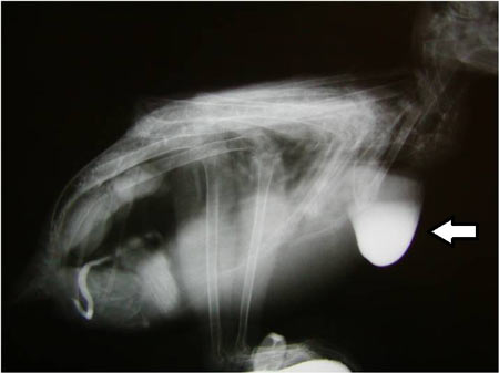
The heart is located in the cranial portion of the thoracoabdominal cavity with its long axis slightly to the right. Radiographically, the liver extends caudally from the apex of the heart, and this overlap results in an hourglass appearance of the two organs. The heart is enclosed in a tough, fibrous pericardial sac that contains a small amount of serous fluid for lubrication. The pericardial sac is loosely attached to the sternum, vertebral column, and adjacent air sacs and more firmly to the liver. By its peritoneal connections, it is attached to the hepatic peritoneal cavities. This arrangement makes the pericardial sac relatively noncompliant and thereby resistant to large increases in size from volume overload.
The relative size of the heart in birds is inversely related to a species’ body mass. This suggests that the larger species of birds have reduced heart mass in comparison to the smaller ones, which have proportionally larger hearts. This arrangement differs from mammals, as their body mass is directly proportional to the size of their hearts. This difference may result from the fact that the heart of larger birds, such as the barnacle goose, can hypertrophy (enlarge) prior to migration so that their hearts are proportional to their weight. Hummingbirds have proportionally larger hearts for their size, most likely as a consequence of the high aerobic demands of hovering flight.
The four chambers of the heart are completely divided into two atria and two ventricles. The right atrium tends to be larger than the left in most birds. The wall of these chambers consists of the same components as in mammals — the endocardium as the inside lining, the middle myocardium and the outer epicardium. The muscular myocardium of the atria is thinner than that of the ventricles. However, the myocardium is arranged in thick muscular bundles forming muscular arches. The atrial muscles contract to empty blood into the ventricles during ventricular diastole.
The muscular anatomy of the ventricles is more complex than the atria. The left ventricle is cone-shaped and extends to the apex of the heart, while the right ventricle is a crescent-shaped cavity that does not go to the apex. Its right wall forms the interventricular septum that separates the two chambers. The wall of the left ventricle is two to three times thicker than that of the right. The curvature of the wall of this left chamber is less than the right and results in a greater mechanical advantage for pressure generation. This allows it to generate systolic pressures that are four to five times greater than those produced by the right ventricle.
The atrioventricular (AV) valves of the heart of birds are similar in their anatomy to those of mammals; however, the cusps of the valves are poorly defined. The right AV valve is structurally distinct, however. It consists of a single spiral flap of myocardium attached to the free wall of the right ventricle. The AV valve of the left side is tricuspid, and not bicuspid, as it is in mammals. It appears that these valves are connected to the Purkinje system — a network of fibers that carry the cardiac impulse from the atrioventricular node to the ventricles of the heart and causes them to contract — and are electrically activated prior to activation of the myocardium to contract the valves and close the AV orifice at the start of ventricular systole. This arrangement differs from that of mammals, which allows the leaflets to float up into the atrial chambers.
The outflow valves of the ventricles are similar to those of mammals except for the fact that there is myocardial tissue that extends into the valves. Additionally, the myocardial extensions have connections to the Purkinje electrical system. This suggests that the valves act as a sphincter that is capable of constricting the outflow opening and changing flow dynamics.
Oxygenated blood from the left heart can enter the right and left aortic sinuses that become their respective coronary arteries. Most commonly, there are two coronary arteries in birds but there may be up to four in number. Often the superficial branches form a ring around the coronary groove to provide blood to the area. There are also deep branches that supply the ventricular walls along with the atria. The right branch is often the dominant vessel and supplies that majority of the blood to the heart. There are frequent anastomoses between branches of the coronary arteries particularly near the coronary groove.
There are often five groups of coronary veins with small tributaries that return blood to the right atrium and/or the right ventricles. There is often a coronary sinus where the blood is shunted before it enters the atrium.
Perfusion of the heart muscle is more active than in skeletal muscle and other avian tissues. As in mammals, perfusion (the passage of fluid through the lymphatic system or blood vessels to an organ or a tissue) occurs during diastole (the normal rhythmical dilatation of the heart during which the chambers fill with blood). The reduction in oxygen supply and increase in myocardial oxygen demand results in an increase in coronary blood flow. These factors come into play in birds that fly at high altitudes.
Coronary blood flow increases as a result of decreases in vascular resistance in response to hypoxia (inadequate oxygenation of the blood). As birds fly at even higher altitudes, the reduction of oxygen results in a compensatory increase in ventilation. Therefore, the arterial blood becomes alkalotic or the pH of the blood rises and the carbon dioxide levels decrease but the perfusion of the tissues with blood increases. This is very different from the dynamics of coronary flow in mammals where hypocapnia (abnormally low levels of carbon dioxide in blood) causes a decrease in blood flow.
Studies and dissections of avian hearts demonstrate that birds have larger hearts with bigger stroke volumes resulting in larger cardiac outputs than mammals.Birds have lower heart rates per unit size for greater perfusion of the tissues of the body. However, they have a greater mean arterial blood pressure than those mammals of comparable body mass. Values that compare birds of different sizes show that larger species have a greater increase in heart rate in absolute terms when transitioning from rest to flight. All of these factors are important to allow birds to have the oxygen capacity to flight and to handle these extreme conditions — something man has only been able to do with machines in the last 100 years of time!

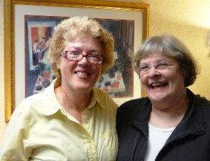 I really can’t remember when I first met Liz — it was so long ago. Avian medicine was in its infancy, and we would attend meetings and hang onto every word. Those words would provide clues that might help us save birds’ lives. There were few pieces of information written, so we relied on the experiences of others to help us move forward in the field of avian medicine.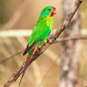
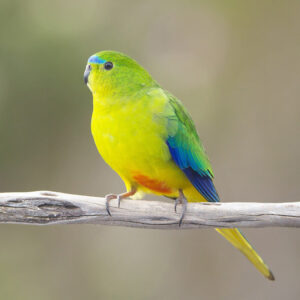

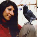
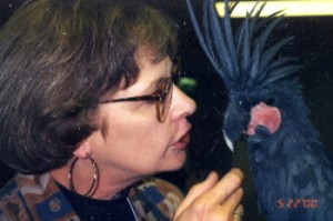 With prior training in elementary education and psychology, Liz began working with companion parrot behavior in 1989 and gained recognition internationally as a parrot behavior consultant thanks to her engaging lectures at avian veterinary and avicultural conferences, and at bird clubs both in the United States and Europe. She challenged bird owners to take a closer look at their own behavior as a catalyst for their birds’ behavior and offered a realistic glimpse at the benefits and challenges of sharing one’s life with a pet parrot. Liz wanted people to open their hearts and homes to a pet parrot for all the right reasons and didn’t shy away from talking someone out of getting a pet bird if it wasn’t in the bird’s best interest. In her articles, Liz often lovingly referred to her husband David Hearn, as well as her beloved 60-plus-year-old
 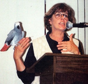 Liz’s passion to expand the avicultural community’s understanding of parrot behavior inspired her to establish the Association of Parrot Behavioral Professionals in 2001, which fostered collaboration among her colleagues, as well as created an invaluable source of information for bird owners. In 2005, she moved this organization into the International Association of Animal Behavior Consultants (IAABC) where it became its Parrot Division.
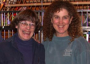 “Over the years I’ve met many people who had their views shaped by a simple truth spoken in raw honesty by Liz. Since she was dedicated to the life of the parrot, she was not patient with those who thought a situation was all about them, or that they did something right and the parrot did it wrong. That’s a good thing; parrots need this kind of advocate, someone who doesn’t dance around the edges of the hard issues. Parrots often pay the price for our stupidities, and Liz was bound and determined to help people get ahead of those, for the parrot’s sake. Liz, I really don’t know yet what we are going to do without you, as a friend and as a colleague. But I promise you, Phoenix Landing will always put the birds first, and we will do our best to promote education in your honor and good name. Love always to you and all the parrots now by your side.
— Ann Brooks, president & founder, Phoenix Landing Foundation (www.phoenixlanding.org)
“Over the years I’ve met many people who had their views shaped by a simple truth spoken in raw honesty by Liz. Since she was dedicated to the life of the parrot, she was not patient with those who thought a situation was all about them, or that they did something right and the parrot did it wrong. That’s a good thing; parrots need this kind of advocate, someone who doesn’t dance around the edges of the hard issues. Parrots often pay the price for our stupidities, and Liz was bound and determined to help people get ahead of those, for the parrot’s sake. Liz, I really don’t know yet what we are going to do without you, as a friend and as a colleague. But I promise you, Phoenix Landing will always put the birds first, and we will do our best to promote education in your honor and good name. Love always to you and all the parrots now by your side.
— Ann Brooks, president & founder, Phoenix Landing Foundation (www.phoenixlanding.org)

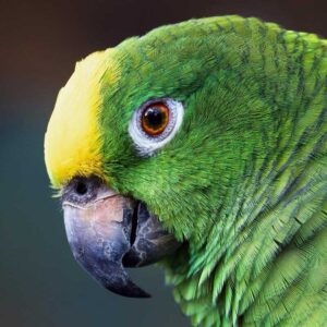
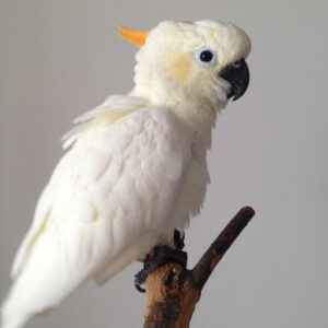

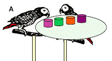 Clearly, if the birds figured out the system, they’d realize that if they cooperated and each chose green, they’d be rewarded all the time. We also added a little twist: We let Griffin, the dominant bird, choose first on half the trials, and let Arthur choose first on the other half to see if that made a difference. We ran the experiment for a long time — 500 choices for each bird — to see if their behavior might change over time and if they would learn to work together. The results were quite interesting.