Honoring Kevin Wright; What You Can Do

Kevin Wright

Dr. Kevin Michael Wright passed away at the age of 50 after a brief illness on Friday, September 27, 2013.

Remembering a distinguished career

Kevin Wright earned his Doctorate of Veterinary Medicine from the University of Florida. He worked in private practice before completing a residency in zoological medicine at the Philadelphia Zoo and the University of Pennsylvania’s School of Veterinary Medicine. Dr. Wright then served as the veterinarian and curator for the amphibian and reptile collection at the Philadelphia Zoo. In 1999, he went to work for The Phoenix Zoo, but returned to private practice in 2005 co-founding an exotic animal hospital. In 2012 he founded his own exotic pet mobile practice.

An original member of the Board of Directors of the Association of Reptilian and Amphibian Veterinarians, Dr. Wright was named the Exotic DVM of the Year in 2008. In both 2009 and 2012 he was awarded Exotics Speaker of the Year by the North American Veterinary Conference. In the first year this specialty was offered (2010), he became a Diplomate of the American Board of Veterinary Practitioners in Reptile and Amphibian Practice. Dr. Wright was the co-author of the highly respected text, Amphibian Medicine and Captive Husbandry, and the author of more than 150 articles related to zoological medicine for both professional and lay audiences.


Dr. Kevin Wright Memorial Fund

In a Facebook post dated October 5, 2013, his brother, Don Wright wrote:

As many of you know, my brother Kevin passed away after a brief illness on September 26, 2013. Our family is heartbroken and numb by the loss, but our attention is focused on helping Marlene through this extremely difficult time. Not only did she lose her beloved husband and life partner of 23 years, but she also lost her employer.

Although Kevin’s veterinary house call business was growing, many of you also know he was unable to acquire health or life insurance. In order to help Marlene with expenses, we have set up a memorial fund in Kevin’s honor to receive monetary gifts. All funds will be used to pay off final expenses and any excess will be used for living expenses to support Marlene until she can re-enter the workforce…We intend to donate a portion of the proceeds to organizations that represent Kevin’s varied passions including local animal rescues and others committed to preservation of native wildlife around the world.

The Fund can receive online payments or wire transfers using the following information:* Checks should be made payable to C. Marlene Wright
Desert Schools Federal Credit Union
Phoenix, AZ 85062
(602) 433-7000Name: C. Marlene Wright
Routing and transit: 122187238
Account: 6000201101
Account type: Savings
Note: Dr. Kevin Wright Memorial Fund
Note The Dr. Kevin Wright Memorial Fund in the memo field.Mail the check to:
2036 N. Gilbert Road
Suite 2-153
Mesa, AZ, USA 85203-2139

*Outside of the United States?
Desert Schools Federal Credit Union does NOT have IBAN or SWIFT numbers. If your bank requires more than a routing number, have your institution select a larger, local bank to serve as an intermediary for the wire transfer.


Obituary

Click here to visit an online obituary that includes a guest book where you can post your remembrances until November 2, 2013.

Administration of Medication in Reptiles

Reptile owners are routinely instructed on oral or intramuscular drug administration techniques for outpatient care. In many instances and in many species, parenteral injections are preferred over the oral route. Injectable medications can be delivered intramuscularly, subcutaneously, intracoelomically, intravenously, or . . .


To continue you need to be a LafeberVet.com member. (Français), (Español)

Pour continuer, vous devez être un membre LafeberVet.com

Para continuar, debe ser miembro de LafeberVet.com

Already a LafeberVet Member?

Please Login

Esophagostomy Tube Placement in the Ferret

Esophagostomy tube placement is an excellent choice for nutritional support of the debilitated small mammal patient requiring long-term feeding or for individuals that have suffered major orofacial trauma. Use this video clip or text with still images to review this important technique in the ferret . . .


To continue you need to be a LafeberVet.com member. (Français), (Español)

Pour continuer, vous devez être un membre LafeberVet.com

Para continuar, debe ser miembro de LafeberVet.com

Already a LafeberVet Member?

Please Login

Zoonotic concern: Rabies in Terrestrial Small Mammals

All mammals are considered to be susceptible to rabies although susceptibility appears to vary by species and viral variant. Rabies is exceedingly rare among small wild mammals and exotic pets. Among rodents in the United States, rabies is most commonly reported in . . .


To continue you need to be a LafeberVet.com member. (Français), (Español)

Pour continuer, vous devez être un membre LafeberVet.com

Para continuar, debe ser miembro de LafeberVet.com

Already a LafeberVet Member?

Please Login

World Lion Day Facts

World Lion Day is August 10 and LafeberVet celebrated on Twitter in 2014. Explore our collection of “Felidae-friendly” facts expanded for this post . . .


To continue you need to be a LafeberVet.com member. (Français), (Español)

Pour continuer, vous devez être un membre LafeberVet.com

Para continuar, debe ser miembro de LafeberVet.com

Already a LafeberVet Member?

Please Login

Presenting problem: Anticoagulant Rodenticide Toxicosis in Free-Living Birds of Prey

Why is this bird bleeding? Anticoagulant rodenticide exposure in birds of prey has been documented in the United States, Canada, and Europe. Anticoagulant rodenticides have been commonly used over the past decades for control of rodent populations and these rodenticides can cause toxicosis in birds of prey via consumption of poisoned prey.

Anticoagulant rodenticides are divided into two categories: first generation rodenticides like warfarin and second generation rodenticides, such as brodifacoum. Second generation anticoagulant rodenticides are more toxic and also have . . .


To continue you need to be a LafeberVet.com member. (Français), (Español)

Pour continuer, vous devez être un membre LafeberVet.com

Para continuar, debe ser miembro de LafeberVet.com

Already a LafeberVet Member?

Please Login

Yearly Checkup: Promote Exotic Pet Annual Exams

FREE video to promote exotic pet annual exams

Produced by exotic animal veterinarians for exotic animal veterinarians

We know that our patients need yearly exams to receive excellent medical care. Drs. Greg Rich, Scott Echols, Christal Pollock, and Ted Lafeber worked together to produce this video for the benefit of the exotic veterinary community.


Too many exotic pets miss their yearly exam

Lack of routine care often results in needless patient suffering, which can lead to expensive trips to the emergency clinic. This video serves to encourage clients to have yearly exams for their exotic animals.


Designed to easily work with your web page

This video can be posted on most websites and most web browsers. Simply cut and paste the embed code of your choice (size and title) below and place it on your site. This video is available with your choice of titles: Exotic 911: Help Your Vet Save Your Pet OR Keep Your Exotic Pets Healthy. Select the video title and size that best suits your practice and your website.


A free resource

We believe that veterinarians need to work together. This video is a result of veterinarians helping veterinarians.


Keep your exotic pets healthy with yearly checkups

700px wide

Courtesy of Drs Greg Rich, Scott Echols, Christal Pollock,Ted Lafeber, and Lafeber Company.

Copy the embed code for your site:

<iframe src="https://player.vimeo.com/video/71435826" width="700" height="381" frameborder="0" allowfullscreen="allowfullscreen"></iframe> <a href="https://lafeber.com">Courtesy of Drs Greg Rich, Scott Echols, Christal Pollock,Ted Lafeber and Lafeber Company</a>

500px wide

Copy the embed code for your site:

<iframe src="https://player.vimeo.com/video/71435826" width="500" height="281" frameborder="0" allowfullscreen="allowfullscreen"></iframe> <a href="https://lafeber.com"> Courtesy of Drs Greg Rich, Scott Echols, Christal Pollock, Ted Lafeber and Lafeber Company</a>

200px wide

Copy the embed code for your site:

<iframe src="https://player.vimeo.com/video/71435826" width="200" height="120" frameborder="0" allowfullscreen="allowfullscreen"></iframe> <a href="https://lafeber.com"> Courtesy of Drs Greg Rich, Scott Echols, Christal Pollock, Ted Lafeber and Lafeber Company</a>

Epidural Anesthesia in Small Mammals

Are you using epidurals in your practice? The epidural is a neuroaxial technique that provides preemptive analgesia by injecting drug into the epidural space surrounding the spine. Epidurals can be used for abdominal surgery, perineal surgery, and orthopedic procedures involving the pelvic limb or spine. Some opioids can also travel cranially to provide supplemental analgesia for chest and thoracic limb procedures. Epidurals serve as an adjunct to systemic analgesia, and epidural analgesia also reduces the amount of anesthetic drug needed. Epidurals can also shorten recovery time . . .


To continue you need to be a LafeberVet.com member. (Français), (Español)

Pour continuer, vous devez être un membre LafeberVet.com

Para continuar, debe ser miembro de LafeberVet.com

Already a LafeberVet Member?

Please Login

Laboratory Assessment of the Bleeding Exotic Animal Patient

Hemorrhage in the critical patient can occur from a number of reasons. Before a blood sample is collected, carefully weigh the risk to the exotic animal patient against the clinical value of the test results. What will you do with this information? How will it affect your clinical plan? EDTA is the most commonly used anticoagulant in small mammals; lithium heparin is commonly used in birds and reptiles. Whenever possible, make a blood film immediately after venipuncture using fresh blood free of anticoagulant. Most adult small mammal hematocrits range from . . .


To continue you need to be a LafeberVet.com member. (Français), (Español)

Pour continuer, vous devez être un membre LafeberVet.com

Para continuar, debe ser miembro de LafeberVet.com

Already a LafeberVet Member?

Please Login

Intravenous Catheter Placement in Small Mammals

Intravenous catheters are commonly placed in ferrets and rabbits to administer fluids and medications, induce anesthesia, and for delivery of analgesic drugs during and after surgery. Intravenous catheters are also placed with growing frequency in chinchillas, guinea pigs and other small exotic companion mammals. Use this video clip or text with still images to review patient selection, potential complications, equipment needed and step-by-step instructions for this technique, as well as daily fluid requirements, catheter maintenance, and patient monitoring . . .


To continue you need to be a LafeberVet.com member. (Français), (Español)

Pour continuer, vous devez être un membre LafeberVet.com

Para continuar, debe ser miembro de LafeberVet.com

Already a LafeberVet Member?

Please Login

Understanding the Illegal Parrot Trade

Introduction

America is a nation of pet keepers with approximately 63% estimated to own at least one pet. In 2008, Americans were estimated to own 16 million pet birds and many of these birds are parrots (APPMA 2012, AVMA 2002). Members of order Psittaciformes are highly sought as pets because of their brilliant coloring, social nature, intelligence, and ability to vocalize (Fig 1).

hyacinth macaw

Figure 1. Parrots are charismatic birds that are highly sought as pets. Shown here, hyacinth macaw (Anodorhynchus hyancinthinus).

While there are more parrots than ever before in captivity, there are fewer parrots in the wild now than at any time in recorded history. In fact, psittacine birds are the most threatened group of bird species in the world today (Wright 2001, Pires 2011). The situation is particularly dire in the neotropics where at least 46 out of 145 species (31%) are at risk of global extinction (Wright 2001).

Although the cause of declining parrot populations worldwide is complex (Box 1), the most important factors include habitat loss, culling, and capture of individuals for the pet trade (Snyder 1999,Wright 2001).

Box 1. Factors implicated in falling wild parrot numbers
  • Habitat loss
  • Culling by farmers
  • Capture for pet trade
  • Natural predators
  • Climate change
  • Inclement weather (e.g. hurricanes)
  • Disease or injury

Most parrots live in tropical rain forests, and destruction of their habitat is ongoing as land is cleared for cattle ranching, exotic tree plantations, settlement, and agriculture. As rain forest is turned into farmland, parrots can turn into crop pests, by eating or indiscriminately destroying plants. Farmers respond by culling bird populations, which can take a serious toll on bird numbers. The relatively low reproductive rate of parrots exacerbates their conservation status (Wright 2001) (Box 2).


Box 2. Features of parrot reproductive biology that worsen their conservation status
  • Small clutch size
  • One clutch or less per year (with rare exceptions)
  • Low survival rate of chicks and fledglings
  • Large proportions of nonbreeding adults
  • Restrictive tree cavity nest requirements

Although parrots are cavity nesters, they lack beaks that are capable of excavating holes within trees, so they must rely on cavities created by other species (Fig 2) (Pires 2012). If a parrot is unable to find a suitable nest cavity, it does not breed.

ring necked parakeets tree nest

Figure 2. Ring-necked parakeets (Psittacula krameri) nesting in a tree cavity. Image by Les Chatfield. Click image to enlarge.

While habitat loss is indiscriminate, affecting all species that claim the rain forest as their home, the pet bird trade only affects those species craved by humans (Fig 3) (Pires 2012). This fact should lead companion parrot owners, as well as professionals that dedicate their time and expertise to bird care, to some uncomfortable truths:

[L]et us…presume that you didn’t hop on a plane and pluck your bird out of a tree. We will also assume that none of us are directly responsible for the millions of parrots taken from their natural habitats. However, it is inescapable that we have directly benefitted from this exploitation. In essence, while we are only a gear in the machine that made parrots threatened, the machine wouldn’t work without us. We owe them.—Dicker 2000

smuggled parrots mom with parrots

Figure 3. Trapping for the legal pet bird trade & illegal smuggling have made a significant impact on wild bird populations. Image by Janice Waltzer from Flickr Creative Commons where the caption reads: [S]o sad…she tried to smuggle them into the US and they suffocated when customs kept them for hours going through everything… Click image to enlarge.

A dozen facts about the illegal bird trade

So if we “owe birds”, then the first, necessary step we can take is to learn more about the illegal bird trade, a complex and pressing issue.

  1. The illegal wildlife trade is the third largest black market industry in the world:  The wildlife trade is a massive global industry in which live animals are captured from their native habitats and sold as pets or for research, or animals are killed and their parts sold for medicines, food, clothing, or accessories (Gastañaga 2011). Interpol values the worldwide wildlife black market at (US) $10 billion annually, making it the third largest illegal trade in the world after guns and drug trafficking (Gastañaga 2011). The illegal parrot trade, which occurs largely in the neotropics, is a part of wildlife crime.
  2. Most parrot poachers are the quintessential “little guy”:  Although the role of professional poachers or members of organized crime groups is important in activities like tiger poaching in India or turtle poaching in Asia, this is not the dominant picture in the wild bird trade (Pires 2011). Local villagers opportunistically commit most poaching in underdeveloped regions of the neotropics, Africa, and Asia (Pires 2011). These individuals often subsist on very little income, and wildlife trade serves as a secondary, or sometimes primary, source of money (Pires 2011). Villagers often keep parrots like chickens until they can be sold to a trader (Pires 2012) The middleman then transports birds to open-air markets in major cities throughout the neotropics (Gonzalez 2003, Gastañaga 2011, Pires 2011, Pires 2012).
  3. Villagers usually target nestlings or juvenile birds during the breeding season:  Depending on the species of interest, anywhere from 30% to greater than 70% of nestlings are poached (Fig 4) (Pain 2006, Pires 2012).Species whose nestlings are most easily removed from the nest are captured in the highest numbers (Pires & Clarke 2012).
    juvenile macaw in nest

    Figure 4. Poaching of nestlings for the pet trade is believed to contribute to the decline of many parrot species. Image provided by LoraKim Joyner.

    The type of nest targeted determines the method used:

    • Only a few parrot species nest in cliff crevices, making them the most difficult to access and the least likely to be poached.
    • The few parrot species that nest closer to the ground or in arboreal termite mounds, like the canary-winged parakeet (Brotogeris versicolorus), are most likely to be poached. Villagers simply reach into nest holes and remove nestlings although they may need to bring along children whose smaller hands can more easily reach into the nest (Pires 2012).
    • Of course most psittacine bird nests are found high up in trees. The poacher climbs the tree using ropes and primitive ladders then reaches into the nest cavity. Sometimes a machete is used to enlarge the cavity. Alternatively cutting down the entire tree allows easy access to the nest. Of course machete use or demolition of an entire tree can cause bird death and also destroys a viable nest that could be used for future breeding purposes (Cantu 2007, Pires 2012).When smugglers plan to transport birds internationally, eggs can be less cumbersome to transport than live birds. Egg smugglers in Australia utilize specialized body vests to prevent egg damage while incubating and concealing their cargo (Alacs 2008, Coghlan 2012).Nest poaching requires no specialized gear, however professional poachers employ specific skills and equipment like cage traps, fishing line snares, or netting outside of the breeding season (Engebretson 2006, Cantu 2007, Wetson 2009, Pires 2012). Species targeted by mist nests, like the monk parakeet (Myiopsitta monachus) and yellow-chevroned parakeet (Brotogeris chiriri), spend much of their time in agricultural lands and may be easier to catch while feeding on crops (Pires 2012).


  4. Domestic trade rules the roost:  The large, international demand for birds fueled wildlife poaching during the 1960s to 1980s (Gastañaga 2011), but there are indications that a large segment of poached birds today are part of internal trade (Fig 5) (Desenne 1991, Best 1995, Wright 2001, Pires 2011). Throughout the neotropics, parrots are common household pets–like cats and dogs in the United States (US) (Drews 2002, Cantu 2007, Weston 2009, Pires 2012). A survey that interviewed many licensed trappers and environmental police inspectors in Mexico estimated that 86% to 96% of birds captured from the wild are sold within Mexico and are not exported abroad (Pires 2012).

    girl with-lorito

    Figure 5. Young girl with her pet parrot or “lorito” in Paraiso, Peru. Image by ‘Jagubal’.

  5. The most heavily poached species are usually the least expensive:  Although the media often focuses on the smuggling of species considered rare and valuable, the most heavily poached and traded species are actually the least expensive birds. Smaller species, like parrotlets and parakeets, generally lack the ability to mimic the human voice and demand the lowest prices. For instance the canary-winged parakeet, the most poached parrot in northern Peru, sells for only (US) $0.33. In Mexico, the Mexican parrotlet (Forpus cyanopygius) retails for (US) $5 and the orange-fronted conure (Aratinga canicularis) for (US) $18 (Fig 6). Amazon parrots (Amazona spp.) are higher priced since these fairly large birds are considered the best talkers. Macaws are generally the most expensive birds because of their large size, ability to talk, and longevity. For instance the military macaw (Ara militaris) sells for anywhere from (US) $600 to $1800 (Cantu 2007, Pires 2012).

    orange fronted parakeets

    Figure 6. A free-ranging orange-fronted conure (Aratinga canicularis). Image by Brian Ralphs.

  6. The illegal bird trade poses an immediate threat to species survival through loss of species from the wild:
    According to the International Union for Conservation of Nature (IUCN) Red List of Threatened Species, of the 330 existing parrot species, 100 psittacine birds are listed and 66 are directly threatened due to the illegal parrot trade (Gastañaga 2011, Pires 2012, Coghlan 2012, Fernandes 2013). Extensive poaching has the potential to eliminate species from the wild and the Spix’s macaw (Cyanopsitta spixii) is considered the latest victim of the wild bird trade. This species is now thought to be extinct in the wild (Pires 2012), and wild population estimates for the blue-throated macaw (Ara glaucogularis), red-fronted macaw (Ara rubrogenys), and Lear’s macaw (Anodorhynchus leari) hover around 150 individuals or less per species (Pires 2012). The hyacinth macaw (Anodorhynchus hyancinthinus) is also considered an endangered species because the population has undergone very rapid reductions in the past and the threat from illegal trapping and habitat loss remains (Fig 7) (Wright 2001, BirdLife International 2012).

    hyacinth macaws

    Figure 7. Free-ranging pair of hyacinth macaws (Anodorhynchus hyancinthinus) in southern Pantanal, Mato Grosso do Sul, Brazil. Image by Geoff Gallice.

  7. Poaching is associated with a very high mortality rates: Many birds do not survive the process of poaching. It is estimated that approximately 75% of birds taken from the wilds of Mexico (50,000-60,000 birds per year) die in transit. Researchers in Nicaragua estimate that in order to compensate for mortalities, up to four times as many parrots are captured than are needed at market (Engebretson 2006, Wetson 2009).Even parrots that do survive can become malnourished, ill, or injured. Feather plucking is also commonly observed in birds kept at illegal pet markets presumably due to the stress of capture, transport, and inadequate diet (Engebretson 2006, Wetson 2009, Pires 2012).
  8. The illegal wildlife trade is associated with potentially significant health risks: The global wildlife trade is considered an important source of emerging diseases (Gomez 2008). Smuggled birds can serve as a health risk to both humans and native bird species by exposing them to pathogens introduced from their region of origin or acquired during transit (Raso 2004, Karesh 2005, Gomez 2008). Pathogens diagnosed in illegally traded wild birds have included (but are not limited to) Chlamydophila psittaci, Newcastle’s disease, and avian influenza (Bruning-Fann 1992, De Schrijver 1995, Mase 2001, Raso 2004, Freitas 2004, Gomez 2008).
  9. The primary purpose of CITES is to ensure that international trade in wild animals and plants does not threaten their survival: Although its efficacy is greatly undermined by the illegal wildlife trade, the Convention on International Trade in Endangered Species of Wildlife Fauna and Flora or CITES is one of the most successful international environmental treaties ever established. Nations traditionally viewed environmental problems as domestic concerns, but by the 1960s the need for international cooperation had become clear. CITES originated as a 1963 resolution of the World Conservation Union and was entered into force on July 1, 1975 (Zimmerman 2003).CITES establishes a permit system for exportation and importation of regulated wildlife. The treaty divides regulated plant and animal species into three appendices (Box 3) (Zimmerman 2003). CITES has also banned trade in over 800 species of flora and fauna (Pires 2011).
    Box 3. Convention on International Trade in Endangered Species Appendices
    Appendix I Species in serious danger of extinction.
    Appendix II Species not necessarily threatened with extinction, but in which trade must be controlled in order to avoid use incompatible with their survival. Species that closely resemble Appendix I species (“look-alikes”) are also listed in Appendix II.
    Appendix III Species that are protected in at least one country that has requested assistance from other member states in controlling trade

    CITES covers all psittacine birds except budgerigar parakeets (Melopsittacus undulatus), cockatiels (Nymphicus hollandicus), ring-necked parakeets (Psittacula krameri), and peach-faced lovebirds (Agapornis roseicollis) (Coghlan 2012) because their wild populations are not threatened.

  10. ‘[P]eople and States are and should be the best protectors of their own wild fauna and flora’ (CITES 1973): Individual member states to CITES must take responsibility for protection of their native wildlife, and each country must also pass (and enforce) national legislation that regulates animal importation.
    • Australia elected to halt trade of its native wildlife in the 1950s.
    • In the 1980s and early 1990s, the US was the largest importer of neotropical parrot species, accounting for almost 50% of the international market for imported birds (Pires 2012, Wright 2001). In 1992 the US passed the Wild Bird Conservation Act (WBCA), which bans the importation of all endangered species listed by CITES (Wright 2001).
    • Although parrot-poaching rates were significantly lower in the years following the WBCA (Snyder 1999), over 75% of legal bird trade was redirected to Europe (Beissinger 2001, Wright 2001). In October 2005, the European Economic Union (EEU) temporarily banned wild bird trade after imported birds died from a H5N1 strain of avian influenza in the United Kingdom. In 2007 the EEU adopted a blanket ban on parrot trade that is irrespective of the species conservation status (Carrete 2008).


  11. A “top-down” approach to wildlife trade control appears ineffective in the neotropics: The “top-down” approach to trade control emphasizes state level concerns and policing. For instance, the Peruvian government has established legal trade quotas that allow parrot harvesters to operate under a state license (Weston 2009). Ideally, such quotas are based on population and habitat studies and are meant to remove ‘surplus’ individuals from the wild population (Munn 2006, Weston 2009).Unfortunately there is little that is done for violations of state wildlife laws. Law enforcement is generally indifferent to wildlife crimes given the perception that it is a relatively minor offense (Herrera 2007, Pires 2011). This mindset is particularly true in underdeveloped countries where an environmental police agency may not even exist (Pires 2011).Even when law enforcement takes the illegal parrot trade seriously, very little can be done to deter potential poachers (Pires 2011). Agents are faced with the daunting task of patrolling large areas of land despite inadequate resources. Law enforcement in Mexico, for instance, has been credited with halting only 1% to 2% of illegal parrot poaching on an annual basis (Cantu 2007, Pires 2011).
  12. The strongest solutions to the illegal parrot trade may come from the “bottom up”: Given that local villagers, and not professional poachers, are responsible for most wildlife poaching, it is not surprising that the typical policing model of catching offenders in the act has a negligible effect on reducing the illegal wildlife trade. Arresting and prosecuting individuals may even be counter-productive because it not only builds resentment against outside authorities but can also criminalize common village practices (Duffy 2010, Pires 2011). Viable solutions to the poaching problem must acknowledge that livelihoods sometimes depend on exploiting species for the illegal wildlife trade (Pires 2011).The best approach to the illegal wildlife trade in neotropical countries targets multiple issues by reducing the rewards associated with poaching by shutting down illegal markets in the cities, incentivizing locals, and increasing the risk to poachers (Pires 2011, Pires 2012).Increasing the risk to poachers is done by (Wright 2001, Pires 2011):
      • Increasing guardianship: Poaching rates at nest sites decrease significantly when breeding sites are actively monitored by local citizens or police through patrols or video, although this can be expensive and difficult to extend over large areas (Fig 8).
    parrot patrol

    Figure 8. Villagers patrolling local parrot nests in Honduras. Image provided by LoraKim Joyner.

    • Strengthening formal surveillance e.g. law enforcement stops on roads that middlemen use to transport species.
    • Making certain areas inaccessible e.g. remove make-shift ladders from trees
    • Eliminating the sale of mist-nets

    Research indicates that one of the best methods to preserve wildlife is to incentivize locals to not only abstain from poaching, but to actually protect parrots instead (Pires 2011). Locals can develop a vested economic interest in the conservation of their native wildlife when the existence of parrots provides jobs and money (Pires 2011). For example clay licks in Peru are a favorite destination of macaws year-round and draw many ecotourists internationally (Fig 9) (Pires 2011). Another tactic is parrot nest sponsorship programs, which pay an annual sum for protection of nest sites (Carrete 2008). Support of parrots by locals can also be encouraged through conservation education programs and awareness campaigns for national pride (Wright 2001).

    macaws at clay lick

    Figure 9. Clay licks attract large flocks and can potentially support ecotourism. Image by Brian Ralphs. Click image to enlarge.


What can you do?

Now that you have a better understanding of the illegal parrot trade, you are better equipped to help.

Conclusion

The illegal wildlife trade is a large and complex industry that has fed and clothed many of the poorest people in the world. Of course smuggling has also brought many species to the brink of extinction (Pires 2011). Although the topic can appear overwhelming, on Earth Day and in the days that follow, we owe it to parrots to learn more about the world around us—and to do what we can.

Nasogastric Tube Placement in the Rabbit

Nasogastric tube placement in the rabbit is an excellent choice for the weak, dehydrated patient that requires enteral nutritional support. Use of a nasogastric tube can be less stressful and more successful than syringe feeding. Nasogastric intubation is also indicated in rabbits that will undergo surgery involving the oral cavity, esophagus, stomach, or biliary tract. Use this video clip or text with still images to review contraindications and potential complications, equipment needed as well as the technique involved . . .


To continue you need to be a LafeberVet.com member. (Français), (Español)

Pour continuer, vous devez être un membre LafeberVet.com

Para continuar, debe ser miembro de LafeberVet.com

Already a LafeberVet Member?

Please Login

Presenting problem: Crop Burn in Birds

Crop burns are commonly caused by thermal injury in young birds and, in rare instances, by ingestion of caustic chemicals in adult birds. Crop burn is generally caused by feeding formula that is too hot (>110ºF or 43.3ºC) or less commonly, contact with a heat lamp or heating pad. Damage typically occurs in the gravity dependent right ventral region of the crop where the weight of the food bolus presses heated material against the skin. This increased thermal exposure can lead to necrosis of the crop wall and skin forming a fistula that can leak food . . .


To continue you need to be a LafeberVet.com member. (Français), (Español)

Pour continuer, vous devez être un membre LafeberVet.com

Para continuar, debe ser miembro de LafeberVet.com

Already a LafeberVet Member?

Please Login

Blood Collection in Ferrets

All but the weakest ferrets can be challenging to restrain for blood collection. Consider sedation or general anesthesia, particularly if the handler or phlebotomist is relatively inexperienced; however remember that anesthesia can affect ferret hematology.

Use this video clip or article with still images to review equipment needed, and potential venipuncture sites including the jugular vein, cranial vena cava, lateral saphenous vein, and cephalic vein . . .


To continue you need to be a LafeberVet.com member. (Français), (Español)

Pour continuer, vous devez être un membre LafeberVet.com

Para continuar, debe ser miembro de LafeberVet.com

Already a LafeberVet Member?

Please Login

Case Study Survey Radiographs: 5y Cockatoo With Open-Mouth Breathing, Poor Grip

Interpretations of radiographs from the article Veterinary Answers Case Study: 5-Year-Old Cockatoo With Open-Mouth Breathing and Poor Grip . . .


To continue you need to be a LafeberVet.com member. (Français), (Español)

Pour continuer, vous devez être un membre LafeberVet.com

Para continuar, debe ser miembro de LafeberVet.com

Already a LafeberVet Member?

Please Login

Veterinary Answers Case Study: 5-Year-Old Cockatoo With Open-Mouth Breathing and Poor Grip

This case study is based on a report prepared by Veterinary Answers Consultant, Gwen Flinchum, DVM, Dipl. ABVP (Avian). A 5-year old female umbrella cockatoo is presented for lethargy, open-mouthed breathing, and difficulty gripping of 2-3 days duration . . .


To continue you need to be a LafeberVet.com member. (Français), (Español)

Pour continuer, vous devez être un membre LafeberVet.com

Para continuar, debe ser miembro de LafeberVet.com

Already a LafeberVet Member?

Please Login

Reptile Physical Examination Form

Donated by Dr. Tom Tully of Louisiana State University School of Veterinary Medicine, use this reptile physical examination form to document evaluation of the reptile patient . . .


To continue you need to be a LafeberVet.com member. (Français), (Español)

Pour continuer, vous devez être un membre LafeberVet.com

Para continuar, debe ser miembro de LafeberVet.com

Already a LafeberVet Member?

Please Login

Oral Examination in Rabbits and Rodents

Evaluation of the oral cavity is considered an essential part of the complete physical examination in small exotic companion mammals, both symptomatic and clinically normal individuals alike. Use this video clip or article with still images to review equipment needed as well chemical and manual restraint techniques for extraoral and intraoral exams . . .


To continue you need to be a LafeberVet.com member. (Français), (Español)

Pour continuer, vous devez être un membre LafeberVet.com

Para continuar, debe ser miembro de LafeberVet.com

Already a LafeberVet Member?

Please Login

Waterfowl Housing Checklist

Waterfowl that require hospitalization may come from a variety of settings, however the basic approach to waterfowl patients remains the same. When hospitalizing any waterfowl patient, there are a number of basic facts to keep in mind . . .


To continue you need to be a LafeberVet.com member. (Français), (Español)

Pour continuer, vous devez être un membre LafeberVet.com

Para continuar, debe ser miembro de LafeberVet.com

Already a LafeberVet Member?

Please Login

Pet Food Labels: Unraveling the Fine Print

Introduction

A critical component for maintaining the health of all animals is proper nutrition. Every patient, healthy or ill, that visits the veterinary hospital should have an evaluation of their nutritional status and healthcare team members should be ready to make a nutritional recommendation based on this evaluation. Unfortunately the large number of pet foods available can make it difficult for healthcare team members to make a nutritional recommendation and effectively communicate this recommendation to their clients.

Nutrition can be defined as the relationship of food and nutrients to health. Proper nutrient intake is essential to normal development, overall health, and disease management in companion animals. Nutrients have many metabolic roles essential to normal physiologic function that can be compromised by insufficient or irregular intake. Minimum requirements for pets have been determined to achieve optimal nutrition, focusing on the key nutrients for physical development, mobility, immune response, cognitive functions, disease prevention, or target treatment.

Pet owners and healthcare team members today have access to a lot of information regarding pet nutrition via the Internet, news sources, and blogs. With this wealth of information comes sometimes confusing and incorrect nutritional information. Veterinary professionals need to understand companion animal nutrition and sort through the minutiae to educate well intentioned owners on what constitutes proper nutrition for their beloved pet. There is a lot of misinformation regarding pet food, so this paper will take a closer look at pet food regulation and the interpretation of pet food labels.


Pet food labels

The pet food label is the primary means by which product information is communicated from the manufacturer or distributor to pet owners, veterinary health care team members, and regulatory officials (Fig 1). Reading and interpreting pet food labels is one method that healthcare team members and pet owners can obtain information about pet foods. However, it is important to remember that pet food labels do not necessarily provide information about food quality such as digestibility and biological value. Contact pet food manufacturers or nutrition experts for additional information that can be used to evaluate pet food quality.

Pet food labels not only communicate information about the product, but also serve as a legal document. A number of agencies and organizations regulate production, marketing, and sales of pet foods in different countries. Each agency has different responsibilities with varying degrees of authority. Some of these agencies regulate information found on pet food labels whereas others influence the regulatory process. Pet foods are regulated at their point of sale, but foods sold outside the United States must also meet labeling requirements of the country in which the food is sold. Pet foods sold in the US must conform to Food and Drug Administration (FDA), Association of American Feed Control Officials (AAFCO), and state pet food labeling requirements.

Pet food labels include two main parts:

  1. The principal display panel and
  2. The information panel.

Certain pieces of information are required on each part of the label, whereas others are optional.


Principal display panel

The principal display panel is defined by the FDA as “the part of a label that is most likely to be displayed, presented, shown or examined under customary conditions of display for retail sale.” It is the primary means of attracting the customer’s attention and should immediately communicate the product identity. The product identity must include a designator such as “bird food”. The brand name is the name by which pet food products of a given company are identified. The product name is not essential and may be the same as the brand name; it is usually descriptive of the food and is subject to regulations dealing with composition of ingredients. The product vignette is a visual representation of the product and it must accurately depict the contents of the package. Food pictured on the label cannot appear better that the actual product.

Percentages of a specific ingredient can determine product names. For example, the term “chicken” requires that at least 70% of the product contain chicken. A “chicken dinner or entrée” must contain 10% chicken if moist and 25% chicken if dry. “With chicken” means that the product contains at least 3% chicken; “chicken flavor” means that chicken is recognizable by the pet (< 3% chicken).

A nutrition statement may be provided on the display panel; it is usually brief and can include such terms as “complete and nutritious,” “100% nutritious,” or “100% complete nutrition.” The use of these terms implies that the product contains ingredients in quantities sufficient to provide the estimated nutrient requirements of a pet or the product contains a combination of ingredients that when fed to a normal animal as the only source of nourishment will provide satisfactory results.


Information panel

The information panel is adjacent to the principal display panel and includes product information. The ingredient statement must be shown on the label and includes a list of ingredients, which must conform to AAFCO names, in descending order by weight. Ingredients are listed on an “as fed basis, which makes interpretation of ingredient lists difficult since many key ingredients are added with different moisture contents. Meats contain more moisture; therefore they may be listed first on the ingredient list however this is often very misleading to pet owners. In reality the primary component of the food is often a mixture of grains. Furthermore, the ingredient statement does not provide information about the quality of ingredients.

One limitation of the ingredient statement is that terms such as “meat by-products” are difficult to evaluate. Many owners are under the impression that foods containing “meat by-products” are inferior to foods containing whole meat.

  • The nutritive value of various meat by-products varies widely. For example, meat by-products such as liver, kidney, and lungs have excellent nutritional value, whereas udder, bone, and connective tissue have poor nutrient availability. Meat by-products do not include hair, horns, teeth, or hooves.
  • AAFCO defines meat on an ingredient label as any combination of skeletal, striated muscle or muscle found in the tongue, diaphragm, heart, and esophagus with or without the overlying fat and the skin, sinew, nerves, and blood vessels that normally accompany muscle. Meat must be suitable for use in animal foods and therefore excludes feathers, heads, feet, and entrails.

By-products are simply secondary products produced in addition to the principal product. Many human foods contain by-products, and the majority of pet foods contain by-products. For example, by-products of human milk production would be ice cream, cheese, and butter. Many by-products are excellent sources of nutrients for pets as well as people.

Food additives in pet foods added by the manufacturer, must be listed in the ingredient statement (Box 1).

Box 1. Food additives in pet foods

  • Vitamins

  • Minerals

  • Antioxidant preservatives

  • Antimicrobial preservatives

  • Humectants

  • Coloring agents

  • Flavors

  • Palatability enhancers

  • Emulsifying agents

Guaranteed analysis

The guaranteed analysis includes crude protein, crude fat, crude fiber, and moisture (Box 2). Additional guarantees are optional and may be included by the manufacturer.

Box 2. Guaranteed analysis includes:

  • Crude protein (minimum)

  • Crude fat (minimum)

  • Crude fiber (maximum)

  • Moisture (maximum)

The guaranteed analysis is a general idea of the nutrient content of a food but is of little value in comparing foods because specific nutrient contents are not given and values are listed on an “as fed” basis.

  • While crude protein is an accurate index of protein quantity, it is not indicative of the quality of the protein.
  • Crude fat may be used to estimate energy density of the food.
  • Crude fiber is an estimate of the indigestible portion of the food that usually underestimates the true level of fiber in the product. Foods that contain higher levels of fiber are generally lower in calories.
  • Moisture content represents the water content in the food. In the United States moisture levels cannot exceed 78%. Foods that exceed 78% moisture must use a different name such as “in gravy,” “in sauce,” or “in aspic.”
Box 3. Example of conversion to dry matter
Lafeber Nutri-Meals® contain 12% moisture; therefore each bar contains 88% dry matter. The label states that Breakfast Nutri-Meal Bars® contain 11% crude protein. Divide 11% crude protein by 88% dry matter to get 12.50% as the amount of protein present on a dry matter basis.

Now that we have this information, we can compare Nutri-Meals® with other products. Harrison’s Bird Food Adult Lifetime Coarse® contains 10% moisture and 15% crude protein. On a dry-matter basis, this product contains 16.67% protein. When comparing these products on a dry matter basis, Harrison’s product has more protein. The same calculations may be used for fat, fiber, and other nutrients on a dry matter basis.

Nutritional adequacy statement

The nutritional adequacy statement on the information panel is often more detailed than the brief statements found on the principal display. Examples include “complete and balanced nutrition for growth of kittens” and “meets…requirements for the entire life cycle of all dogs.” The nutrition statement will help you determine if the manufacturer is making claims for a specific purpose diet as opposed to an all purpose diet.

Pet foods with no statement of adequacy include snacks, treats, and some therapeutic foods. Therapeutic foods have a statement that they are to be used by or under the direction of a veterinarian. No further nutrition statement is required since the complete nutritional profile is usually available to the veterinarian.

The basis of the nutrition claim listed on the pet food label is based on the formulation method, and/or the feeding trial method.

  • Formulation method: The formulation method is simply a laboratory nutrient profile analysis and does not require any feeding or digestibility trials to prove availability of the nutrients in the profile analysis. This method is recognized on a label by a statement such as “meets or exceeds the minimal nutritional levels established by AAFCO” or “formulated to meet the AAFCO dog nutrient profile for…” AAFCO nutrient profiles are published for growth and reproduction as well as adult maintenance.
  • Feeding trial method: The feeding trial method is the preferred method for substantiating a claim and is considered the gold standard when determining nutritional claims. Feeding trials can results in adequacy claims for four categories including: gestation and lactation, growth, maintenance, and complete for all life stages. A food that has successfully completed a sequential growth and gestation/lactation trial can make a claim for all life stages. The required wording for labels that have passed these tests is: “Animal feeding tests using AAFCO procedures substantiate that (brand) provides complete and balanced nutrition for (life stage).” Pet foods that do not meet AAFCO requirements by either of the standard methods will have a nutritional statement as follows: “this product is intended for intermittent or supplemental feeding only.

Feeding directions

Dog and cat foods labeled as complete and nutritious for any or all life stages must list feeding directions on the product label. At minimum, feeding directions should include the instructions “feed (weight/unit) per (weight) of dog or cat” and frequency of feeding. However, it should be recognized that these feeding directions are general guidelines and should serve as a starting point. Adjustments may be needed to maintain optimal body condition. Therefore, as a veterinary professional the amount to feed should be calculated to attain a more accurate food dose dependent upon species, lifestyle, life stage, etc.


Natural vs. Organic vs. Holistic

Natural

The term “natural” has been defined by AAFCO as:

A food or ingredient derived solely from plant, animal, or mined sources, either in its unprocessed state or having been subject to physical processing, heat processing, rendering, purification, extraction, hydrolysis, enzymolysis, or fermentation, but not having been produced by or subject to a chemically synthetic process and not containing any additives or processing aids that are chemically synthetic except in amounts which might occur unavoidably in good manufacturing practices.”

This definition does not include the use of any synthetic preservatives, flavors, and colors in products labeled as “natural.” However, most added trace nutrients such as vitamins, minerals, and taurine are chemically synthetic, AAFCO guidelines do allow use of trace nutrients in “complete and balanced” pet foods. This is done by adding a disclaimer similar to the following: “natural ingredients with added vitamins and minerals”.


Organic

The term “organic” refers to the process by which ingredients are grown, harvested, and processed. Currently the term organic encompasses human foods and can only be used in pet foods, if the standards set for human foods are met. For example, “organic beef ” must come from cattle raised under certain conditions without the use of drugs such as hormones and antibiotics. Currently, pet foods meeting the human standard can display the USDA organic seal (Fig 2) if the contents of the package meet specific requirements (Table 1).

Table 1. USDA organic seal package categories
CategoryDefinitionProduct label can display USDA organic seal
100% organicLabeling term that indicates food has been produced through methods that foster cycling of resources, ecological balance, and biodiversityYes
Organic≥ 95% of content is organic by weight excluding water and saltYes
Made with organic≥ 70% of content is organicNo; the front product panel can display the phrase “made with organic” followed by no more than three specific ingredients or glasses of ingredients

When less than 70% of content is organic, the label may list only those ingredients that are organic on the ingredient panel with no mention of organic elsewhere on the label. These products may not display the USDA organic seal.

USDA organic label

Figure 1. Pet foods that meet specific human food requirements may display the USDA organic seal.

Holistic” is a term that has been used in a number of pet foods with a variety of ingredients and characteristics. Holistic is not legally defined or regulated by pet food regulations therefore this term is meaningless. The term is also considered misleading in that it may falsely imply therapeutic benefit.


Manufacturer information

In the United States the name and address of the pet food manufacturer, distributor, or dealer must be found on the label, generally on the information panel. The following phrases: “Distributed by…” or “Manufactured for…” or “Imported by…” are indicative of a company other than the one selling the product has manufactured the pet food. With private label brand pet foods, this is fairly common. In these instances the manufacturer is referred to as a co-packer.

Regulations in the US also require that the manufacturer’s information be accompanied by “Product of (country of origin)”, if the product is manufactured in a country other than where it is sold. Additionally, many manufacturers will include a universal product code (UPC) or bar code on the label, although this is not a legal requirement. Other important information frequently found on pet food containers or labels are batch numbers and date of manufacture. This type of information assists both the purchaser and the manufacturer when communicating about product in a specific container. Finally, a freshness date (e.g., “Best before [date]”) or a list of other guarantee policies may be found on the product’s packaging.


Summary

Pet owners at present want and expect the very best for their pets. However, the information that pet owners are given although well intentioned can be misleading. The veterinary healthcare team should focus on proper nutrition for every pet that presents to their hospital. To do this, the healthcare team must perform a complete nutritional history and patient assessment and be knowledgeable about the wide variety of foods that are on the market today. Not all foods are created equal and pet foods labels can be misleading and misinterpreted. The veterinary healthcare team must familiarize themselves with properly reading a pet food label and understanding how the pet food label can affect the nutritional recommendation being made.


References

Backyard Poultry Primer

Backyard or hobby flocks consist of meat and game birds; or ornamental or show birds. This review article, co-authored by poultry veterinarian, Teresa Morishita, offers tips on the basic clinical approach to backyard poultry as well as differential diagnosis lists for common clinical problems. Conditions commonly encountered in backyard chickens and turkeys often include endoparasites, like Eimeria spp., pasteurellosis or fowl cholera, mycoplasmosis, staphylococcosis, and colibacillosis. Diseases important for public health concerns, such as avian influenza and Newcastle disease, are also discussed . . .


To continue you need to be a LafeberVet.com member. (Français), (Español)

Pour continuer, vous devez être un membre LafeberVet.com

Para continuar, debe ser miembro de LafeberVet.com

Already a LafeberVet Member?

Please Login

Grooming Companion Birds: A Review

Grooming in the bird can refer to clipping wing feathers, trimming nails, and smoothing and/or trimming the beak. Grooming can be performed by the veterinarian or an astute, skilled veterinary technician, however before the procedure begins one must always ask should the bird be groomed and should the bird be groomed at my practice . . .


To continue you need to be a LafeberVet.com member. (Français), (Español)

Pour continuer, vous devez être un membre LafeberVet.com

Para continuar, debe ser miembro de LafeberVet.com

Already a LafeberVet Member?

Please Login

10 Tips for a Great Baby Bird Visit

For most veterinarians, chicks are seen only sporadically in clinical practice–which can shake your confidence. Use LafeberVet’s NEW Ten Tips for Evaluating Chicks in the Exam Room as a clinical aide to start your pediatric visit off right . . .


To continue you need to be a LafeberVet.com member. (Français), (Español)

Pour continuer, vous devez être un membre LafeberVet.com

Para continuar, debe ser miembro de LafeberVet.com

Already a LafeberVet Member?

Please Login

Lafeber in the Field: Stick-A-Roos at Zoo Atlanta

Lafeber Company’s work isn’t always tied to our family farm and production facility. Lafeber Company has worked with Atlanta Zoo veterinarian, Dr. Samuel Rivera, to create Stick-A-Roos (Stickaroos), a diet designed for use in interactive parakeet and cockatiel aviaries. Stick-A-Roos provide maintenance nutrition and foraging enrichment . . .


To continue you need to be a LafeberVet.com member. (Français), (Español)

Pour continuer, vous devez être un membre LafeberVet.com

Para continuar, debe ser miembro de LafeberVet.com

Already a LafeberVet Member?

Please Login

Basic Cytology of the Avian Gastrointestinal Tract

Historically, routine Gram’s stains were performed in apparently healthy birds. As our understanding of avian medicine has grown, avian veterinarians have questioned the reliability and validity of Gram’s stain cytology as a screening test. Cytology is indicated when specific problems are reported during a detailed medical history or when . . .


To continue you need to be a LafeberVet.com member. (Français), (Español)

Pour continuer, vous devez être un membre LafeberVet.com

Para continuar, debe ser miembro de LafeberVet.com

Already a LafeberVet Member?

Please Login

Basic Information Sheet: Parakeet

Parakeet

Parakeet

Species


“Parakeet” is a broad term for a small parrot with a long tail. There are many parakeet species, however the most popular pet parakeet is the budgerigar parakeet or “budgie”. Free-ranging “budgies” live in large flocks in a variety of habitats such as woodlands, open grassland, and dry scrub throughout non-coastal Australia and Tasmania. Another popular species is Bourke’s parakeet, which is from the southern and eastern regions of Australia.

Taxonomy


Class: Aves

Order: Psittaciformes

Family: Psittacidae

Melopsittacus undulates – budgerigar parakeet or “budgie”

Neopsephotus bourkii – Bourke’s parakeet

Conservation status


Bourke’s parakeet is on CITES Appendix II, which means they are not necessarily threatened with extinction, but may become so unless their trade is strictly regulated.

Six parakeet species are listed on Appendix I: blue-chested, Forbes’s, golden, horned, Mauritius, and red-fronted parakeets. Appendix I species are threatened with extinction, and commercial trade is prohibited and importation/exportation for scientific research requires special permits. The International Union for Conservation of Nature lists the budgerigar parakeet’s conservation status of “least concern”.


Physical description


  • The standard or wild-type budgerigar parakeet has yellow feathers barred with black on the back of its heads and body with plain yellow feathers on the forehead and throat. A row of black dots sit across the throat and greenish-yellow feathers are found on the underside.
  • The two basic types of pet budgerigar parakeet are the English “budgie”, a show bird with a bigger head and chest, and the American “budgie” a more active bird with a shorter tail.
  • Numerous mutant color varieties have been bred into the captive population.
  • Budgerigar parakeets are sexually dimorphic. The cere may be blue in males, pink or brown in females, and pearlescent in juveniles.


Diet


  • Budgerigar parakeets are granivores. Free-ranging birds are ground feeders foraging for grass seeds and chenopod seeds. Budgies have also been reported to eat grain crops.
  • Since psittacine birds hull seeds before ingestion, they do not require grit. In fact, some individuals will overeat grit when ill putting the bird at risk for impaction.
  • All-seed diets are deficient in protein, vitamins, and minerals including calcium and vitamin A.


Husbandry


  • Provide a cage that is a minimum of 18 in (46 cm) wide and long. Cage length is more important than height.
  • Cage bar spacing should be approximately 3/8 in (0.95 cm).
  • Perch diameter should be between 3/8 to 3/4 in (0.95-1.9 cm). Sand paper perch covers are very abrasive to the feet, and are not recommended.
  • Consider housing in pairs or groups.
  • Offer regular baths or misting with water to maintain healthy plumage.


Behavior


  • The English budgerigar parakeet may be slightly more docile than the American.
  • Parakeets are social and can become hand-tamed.
  • Parakeets are great whistlers and learn to mimic sounds and words.
  • Foraging is an important part of normal daily parrot activity. Teach and encourage pet birds to play and forage.


Normal physiologic values


Temperature (average)* 41.8 C 107.1 F
Resting heart rate (beats/min) Approximately 274
Respiration (breaths/min) 60-75
Body weight (g) 25-35
Mean life span (years) 7-15
Mean number of incubation days 18
Average number of eggs laid 4-6
Target environmental temperature Mimic natural environment. Household temperatures of 70-80°F (21-27°C) are generally acceptable, however healthy birds can tolerate hot and cold temperatures.
* Routine avian exam does not include measuring body temperature


Anatomy and physiology


  • The “budgie” heart rate is 7 times faster than in humans.
  • Anatomic traits of Order Psittaciformes include:
    • Communication of the right and left nasal sinus
    • The only avian tongue with intrinsic muscles
    • Simply syrinx
    • Craniofacial hinge of beak is a synovial joint
    • Ceca absent
    • Gall bladder often absent
    • Zygodactyl foot: two toes pointed backward and two pointed forward


Restraint


Budgies may be restrained by holding the head between index and middle fingers. Support the body with the palm of the hand as well as the thumb and little finger.

Venipuncture


Use a 27-gauge needle and 1 to 3-mL syringe to draw blood from the right jugular vein. Collection of up to 1% of body weight is acceptable in healthy patients.

Preventive medicine


Injections


Intramuscular (IM) Reasonably safe, most accurate.Inject middle of muscle mass. Ideal location –Pectoral muscle mass
Subcutaneous (SQ) Large volumes can be injected, poor absorption. Location:  Inguinal or precrural fold
Intravenous Effective, narrow safety range. Right jugular vein or brachial vein is most commonly used. Alternative option: superficial metatarsal vein.


Important medical conditions


Non-Infectious Diseases

  • Obesity and fatty tumors
  • Goiter
  • Neoplasia
    • Renal
    • Gonadal
    • Pituitary
  • Reproductive problem such as chronic egg laying and yolk peritonitis
  • Xanthomatosis

Infectious Diseases

  • Trichomoniasis
  • Avian polyomavirus or “Budgerigar fledgling disease”
  • Knemidokotpes (“Scaly face and leg”) mites

**Login to view references**

References

Basic Information Sheet: Macaw

Macaw

macaws

Blue and gold macaw (Ara ararauna) (left); green-winged macaw (Ara chloropterus )(right)

Natural history


Macaws are found in a variety of habitats throughout large areas of the Amazon Basin, such as gallery forests and mangrove swamps.  Most macaws are listed on Convention on International Trade in Endangered Species (CITES) Appendix I, however the blue and gold, green-winged, Hahn’s, noble, red-bellied, severe and yellow-collared macaws are listed on CITES Appendix II. Appendix I species are threatened with extinction globally, and commercial trade is prohibited and importation/exportation for scientific research requires special permits. Appendix II species are not necessarily threatened with extinction, but may become so unless their trade is strictly regulated.

Taxonomy


Class: Aves

Order: Psittaciformes

Family: Psittacidae

Anodorhynchus hyacinthinus – hyacinth macaw

Ara spp.

Ara ararauna – blue and gold macaw

Ara chloropterus – green-winged macaw

Ara macao – scarlet macaw

Diopsittaca nobilisD. n. cumanensis – noble macaw


There are 15 species of macaws in the genus Ara and 3 species in genus Anodorhynchus. Blue and gold, scarlet, and green-winged macaws are among the most popular pet macaw species. Visit LafeberVet’s Macaw ID Slideshow for additional information.


Physical description


  • Macaws are slender birds with long tapered tails; strong, heavy bills; and a bare facial patch.
  • Macaws are sexually monomorphic.
  • The green-winged macaw may be distinguished from the scarlet macaw by its larger size and the bands of small, red feathers around the eyes. Scarlet macaws also have yellow feathers on their wings.

    Scarlet macaws (Ara macao)

    Scarlet macaws (Ara macao). Image by Dunleavy. Click image to enlarge


Diet


  • Macaws are frugivores, granivores, and/or florivores. Free-ranging birds feed on seeds, fruit, figs, palm nuts, leaves, nectar, and/or flowers.
  • Some macaws need more fat in their diet. Hyacinth macaws consume only palm nuts in the wild.
  • Since psittacine birds hull seeds before ingestion, they do not require grit. In fact, some individuals will overeat grit when ill putting the bird at risk for impaction.
  • All-seed diets are deficient in protein, vitamins, and minerals including calcium and vitamin A.


Husbandry


  • Macaws require a large amount of space in a quality made cage or room with appropriate toys.
  • Cage bar spacing should be 0.75-1.5 in (1.9-3.8 cm).
  • Perch diameter should range from 5/8 in (1.6 cm) for Hahn’s macaw up to 2 in (5 cm) for hyacinth macaws.
  • Provide frequent baths or showers.


Behavior


  • Most of the large macaws are considered gentle birds, however all large macaws can be quite noisy and destructive.
  • The scarlet macaw has a reputation for a particularly strong personality making them a difficult pet for inexperienced owners.
  • All macaws are extremely intelligent that require a lot of stimulation.
  • Feather destructive behavior may be more common in some of the mini macaw species.
  • Most macaw species have a bare facial area, which can flush pink indicating a change in mood.
  • Foraging is an important part of normal daily parrot activity. Teach and encourage pet birds to play and forage.


Normal physiologic values


Temperature (average)* 42°C 107.7°F
Heart rate (beats/min) Small Macaws 389
Large Macaws 275
Respiration (breaths/min) 20-25
Average body weight (g) Hahn’s 120-160
Military 900
Scarlet 900-1000
Blue and Gold 900-1300
Green-Winged 1200-1600
Hyacinth 1250-1695
Mean life span (years) Small Macaws 20-25 (35 reported)
Large Macaws 30-45 (75-100 possible)
Sexual maturity Small Macaws 4-6 years
Large Macaws 5-7 years
Mean number of incubation days 23-28 Longer for larger birds
Average number of eggs laid 1-3
Weaning age (days) Small Macaws 90-120 – parent raised
Large Macaws 120-150 – parent raised
Target environmental temperature Mimic natural environment. Household temperatures of 70-80°F (21-27°C) are generally acceptable, however healthy birds can tolerate hot and cold temperatures.
*Routine avian exam does not include measuring body temperature


Anatomy and physiology


  • Macaws have a rolling or waddling gait due to walking on their feet as well as caudal tarsometatarsus.
  • The pressure exerted by a large macaw beak can be greater than 200 psi.
  • Macaws share the following anatomic traits with other members of Order Psittaciformes:
    • Communication of the right and left nasal sinus
    • The only avian tongue with intrinsic muscles
    • Simply syrinx
    • Craniofacial hinge of beak is a synovial joint
    • Ceca absent
    • Gall bladder often absent
    • Zygodactyl foot: two toes pointed backward and two pointed forward


Restraint


  • Most macaw species have a bare facial patch that can flush pink color indicating a change in mood.
  • Restrain large macaws by placing the thumb and forefinger beneath the mandible. Secure the outside wing with three fingers and use the other hand to hold the feet.
  • Avoid touching the bare facial patch as the skin can bruise after aggressive restraint.


Venipuncture


Use a 25-gauge needle and 1 to 3-mL syringe to draw blood from the right jugular vein. Collection of up to 1% of body weight is acceptable in healthy patients.

Preventive medicine


  • Obtain a complete history and perform a thorough annual physical examination.
  • Establish baseline data with regular clinical testing (complete blood count, protein electrophoresis, and plasma biochemistries.
  • Ensure proper nutrition and husbandry.
  • Recommend quarantine of newly acquired birds.
  • Perform additional testing for select diseases based on history and physical exam findings: avian polyoma virus and psittacosis.
  • Determine origin and history of newly acquired sick birds to contain and prevent further spread of disease.
  • Birds housed in large groups or aviaries are at higher risk of Pacheco’s disease virus and use of the vaccine may be indicated.
  • The avian polyoma virus vaccine is recommended for breeding populations.


Injections


Intramuscular (IM) Reasonably safe, most accurate.
Inject middle of muscle mass.
Ideal location –Pectoral muscle mass
Subcutaneous (SQ) Large volumes can be injected, poor absorption. Location:  Inguinal or precrural fold
Intravenous Effective, narrow safety range. Right jugular vein or brachial vein is most commonly used.
Alternative option: superficial metatarsal vein.


Important medical conditions


Non-Infectious Diseases

  • Aspergillosis
  • Atherosclerosis
  • Beak malocclusion
  • Degenerative cardiac disease
  • Feather destructive behavior in some mini macaw species
  • Goiter
  • Inflammatory skin disease
  • Internal papillomatosis
  • Respiratory disease including pulmonary hypersensitivity syndrome or allergic pneumonitis: The incidence of respiratory disease increases in birds exposed to low relative humidity and/or birds that produce lots of powder down such as cockatoos.
  • Yolk peritonitis

Infectious Diseases

**Login to view references**

References

Presenting problem: Trauma in Ferrets

Ferrets are small, flexible, mischievous, and curious. This combination means that traumatic injury is a common problem, particularly when owners do not supervise their pets or “ferret proof” their living quarters. For instance, reclining chairs have been implicated in the injury and even death of many ferrets. Ferrets may also be accidentally stepped on or become trapped within a confined space. Trauma can result in a constellation of injuries that may affect the head, thorax, abdomen, spine, and limbs . . .


To continue you need to be a LafeberVet.com member. (Français), (Español)

Pour continuer, vous devez être un membre LafeberVet.com

Para continuar, debe ser miembro de LafeberVet.com

Already a LafeberVet Member?

Please Login

Endotracheal Intubation in the Rabbit

Endotracheal intubation of the rabbit is challenging because it difficult to directly visualize the tracheal opening in all but the largest individuals. Rabbits have a long, narrow oral cavity and the larynx, which sits on a ventral slope, is prone to spasm.

Use this video clip or text with still images to review equipment needed, potential complications as well as the techniques involved for blind intubation and intubation using visualization . . .


To continue you need to be a LafeberVet.com member. (Français), (Español)

Pour continuer, vous devez être un membre LafeberVet.com

Para continuar, debe ser miembro de LafeberVet.com

Already a LafeberVet Member?

Please Login

Intravenous Catheter Placement in Rabbits

Intravenous catheters are commonly placed in rabbits to administer fluids, medications, induce anesthesia, and for delivery of analgesic drugs during the perioperative and postoperative periods. Catheterization techniques used in dogs and cats can also be used for rabbits. To reduce the time in handling and reduce stress, supplies needed for catheterization should be set out and ready to go, prior to removing the patient from the cage . . .


To continue you need to be a LafeberVet.com member. (Français), (Español)

Pour continuer, vous devez être un membre LafeberVet.com

Para continuar, debe ser miembro de LafeberVet.com

Already a LafeberVet Member?

Please Login

Blood Collection in Rabbits

The rabbit has a relatively short prothrombin time and whole blood quickly clots at room temperature. To minimize the risk of clot formation, it can be helpful to pre-heparinize the needle and syringe by drawing heparin into the needle and expelling the excess from the hub. The total volume of blood that can be safely collected typically ranges from 0.5% to 1.0% body weight. Collect smaller volumes from geriatric patients or those suspected to have anemia or hypoproteinemia.

Use this video clip or article with still images to review equipment needed, and potential venipuncture sites including the . . .


To continue you need to be a LafeberVet.com member. (Français), (Español)

Pour continuer, vous devez être un membre LafeberVet.com

Para continuar, debe ser miembro de LafeberVet.com

Already a LafeberVet Member?

Please Login

Mammary Tumors in Small Mammals

An important differential for lumps and bumps: Mammary gland tumors are relatively common in rats and mice, and are also seen in African pygmy hedgehogs and guinea pigs. Get the facts about mammary tumors in small mammals. Review diagnostics, management, prognosis and prevention of this important condition . . .


To continue you need to be a LafeberVet.com member. (Français), (Español)

Pour continuer, vous devez être un membre LafeberVet.com

Para continuar, debe ser miembro de LafeberVet.com

Already a LafeberVet Member?

Please Login

Referring Non-Traditional Species

Depending on the problem encountered, referral may be in the patient’s best interest as the general practitioner or emergency clinic may lack the necessary expertise, equipment, or medication. Referral may also be indicated when you have an inconclusive diagnosis or when the client requests a second opinion . . .


To continue you need to be a LafeberVet.com member. (Français), (Español)

Pour continuer, vous devez être un membre LafeberVet.com

Para continuar, debe ser miembro de LafeberVet.com

Already a LafeberVet Member?

Please Login

Thoracocentesis in Ferrets

Exotic animal care is frequently a balancing act between concepts true for all medicine and species-specific information. Management of pleural effusion in the ferret relies on the same tests and treatments used in dogs and cats, including chest taps when indicated . . .


To continue you need to be a LafeberVet.com member. (Français), (Español)

Pour continuer, vous devez être un membre LafeberVet.com

Para continuar, debe ser miembro de LafeberVet.com

Already a LafeberVet Member?

Please Login

Use of Nutri-An Cakes for Weight Reduction and Maintenance

Introduction

Weight reduction requires a multi-step plan that includes commitment from the pet owner, metabolically controlled food for weight maintenance or reduction, an appropriate feeding method, an exercise plan, communication with the pet owner, and patient monitoring.

Orosz taking history

Weight reduction requires a multi-step plan that includes commitment from the pet owner and careful communication with the pet owner.

In humans and small animals, the combination of feeding reduced-calorie foods, providing regular exercise, and using behavior modification to change eating patterns bring about the best chance of achieving and maintaining weight loss (Toll 2010). A key nutritional factor for weight loss and the prevention of weight gain is to feed calorie-restricted diets. These diets should reduce energy density (e.g., calories per gram) while providing normal levels of other nutrients required for balanced nutrition. If a maintenance diet is fed at a reduced rate, energy is reduced, but so too are all the other nutrients, so that the diet I no longer in balance. Nutri-An Cakes provide an energy-restricted diet for psittacine birds with a normal complement of the other nutrients to maintain a nutritional balance.


Commitment from the pet owner

The first problem is for the owner to understand that their beloved companion bird is overweight. Palpation of the pectoral muscle mass and showing the owner the fat over the muscle, having them feel the fat in the coelom and/or showing them the fat obstructing the view of the jugular vein are all helpful for them to understand that their bird is overweight. Information on why this is a serious medical problem should then be discussed. Birds that are overweight have been shown to have hepatic lipidosis, reduced immune function, cholesterol plaques in the great vessels of the heart, lipomas, greater problems with osteoarthritis, and a greater likelihood of seizures. Once owners understand and are willing to be a major player in controlling their bird’s weight, their next step is to commit to a weight loss program. One motivational factor is to resolve a problem caused or exacerbated by obesity. This should be part of the plan tailored for their bird.


Metabolically controlled food

Nutri-An Cakes for Foraging and Weight Maintenance (FW) is a therapeutic diet designed for avian patients that need nutritional enrichment and/or weight reduction. Nutri-An Cakes FW provide high quality protein from a variety of sources but at normal levels for companion parrot species, making it useful for a number of medical conditions:

  • Molting or other skin conditions
  • Egg laying
  • Renal disease
  • Problems affecting the liver where highly digestible protein may help function

In addition, Nutri-An Cakes FW contain balanced omega 3:6 polyunsaturated fatty acids (PUFA) that reduce pro-inflammatory mediators affecting the function of a number of organs. This diet may be helpful as a complement to conditions where non-steroidal drugs are used to reduce inflammation.

The lower energy content of Nutri-An Cakes FW makes it an excellent food for weight maintenance or reduction:

  • 2826 ME kcal/kg in the parakeet, cockatiel, lovebird and conure cakes
  • 2858 ME kcal/kg in the parrot, macaw and cockatoo cakes


Appropriate feeding methods/exercise plan

Snyder et al documented that Puerto Rican Amazon parrots (Amazona vittata) spend approximately 4-6 hours per day foraging and they routinely travel several miles between sites. It is presumed that other psittacine species spend similar amounts of time searching for and acquiring food as part of their daily behavioral repertoire. In contrast, companion birds such as orange-winged Amazons (Amazona amazonica) in our homes, spend approximately 30-72 minutes per day eating a pelleted diet (Oviatt 1997) without traveling, manipulating food items, or attempting to balance their own diet (Meehan 2003).

These data suggest that birds need to forage for their food items and expend additional energy in the food acquisition process or as an exercise routine to mimic their normal behavioral repertoire. This is certainly the goal in birds that are obese as increased physical and mental activity enhances weight loss. A foraging plan and exercise program should be part of the weight loss plan. It is important to start with basic steps and information that is tailored to the avian patient. For example, Amazon parrots are good paper shredders but often do not handle complex plastic toys that require manipulation that are easy for cockatoos to manipulate. Another problem is that complexity needs to be increased based on the learning curve of the patient as owners often stop at the first level.

Please refer to Table 1 below for appropriate amounts of Nutri-An Cakes to be fed per day. These cakes should be fed as part of the foraging program developed for this avian patient. Since birds spend a large portion of their day acquiring food, other food items that do not enhance weight gain can be provided. These items are listed in Table 2.

Table 1. Daily food intake for weight reduction* using Nutri-An Cakes
Body weight (grams) Small Nutri-An Cakes Large Nutri-An Cakes
20 0.66 0.22
40 1.1 0.33
50 1.5 0.44
80 1.8 0.54
100 2.2 0.62
120 2.5 0.73
140 2.7 0.81
160 3.0 0.90
180 3.3 0.98
200 3.6 1.0
250 4.2 1.2
300 4.8 1.4
350 5.4 1.6
400 5.9 1.8
450 6.7 1.9
500 7.0 2.0
550 7.4 2.2
600 7.9 2.4
650 8.4 2.5
700 8.9 2.6
750 9.3 2.8
800 9.8 2.9
850 10.2 3.0
900 10.7 3.2
950 11.0 3.3
1000 11.5 3.4
1100 12.3 3.7
1200 13.2 3.9
1300 13.9 4.1
1400 14.7 4.4
1500 15.5 4.6

* Food not consumed or waste risks the patient not receiving sufficient calories. Calculations assume that birds live in cages indoors and have an average amount of activity. Body weight should be monitored daily and food amounts adjusted accordingly.

Table 2. Some foods may be offered with Nutri-An Cakes (right), while others are best to avoid during weight loss (left)
Foods to avoid Foods that may be offered with Nutri-An Cakes
  • Rice
  • Potatoes
  • Corn
  • Peas
  • Lima beans
  • Banana
  • Apples
  • Grapes
  • Broccoli
  • Green beans
  • Summer squash
  • Pumpkin, cubes of
  • Spinach
  • Parsley
  • Walnuts
  • Flax seeds

The exercise plan may vary with the species and the physical condition of the bird. A cardiac work-up to include echocardiography may be indicated. Wing flapping on a swing or by hand with a gradual increase in time provides one of the best options for expenditure of calories. Controlled flying, if possible, is one of the best forms of exercise as it requires between 11-20 times the basal metabolic rate in birds.


Communication with the client/patient monitoring

Customizing the weight loss program is important for avian patients as there is great variation between species than with breeds of dogs and cats.

  • The plan should be in writing with an explanation of goals with dates included.
  • Monitoring is an important factor in weight loss. Owners should acquire a food scale and weigh their birds at the same time each day and put their weights on a provided chart.
  • It is great to have a check-off list for elements of the exercise program as well.
  • In dogs, the use of an appropriate feeding plan and monthly hospital weight rechecks with subsequent weight maintenance periods was sufficient to achieve good results in dogs (Toll 2010). It is important to compliment and encourage the owner at these visits to maintain their commitment to weight management.

References

Presenting problem: The Fluffed and Ruffled Bird

A relaxed, comfortable bird may briefly fluff out its feathers before its plumage again appears flat and sleek. The critically ill bird may exhibit persistently fluffed and ruffled plumage . . .


To continue you need to be a LafeberVet.com member. (Français), (Español)

Pour continuer, vous devez être un membre LafeberVet.com

Para continuar, debe ser miembro de LafeberVet.com

Already a LafeberVet Member?

Please Login

Presenting problem: Cloacal Prolapse in Birds

Understanding the cloaca and cloacal prolapse

The cloaca is a common receiving chamber for the reproductive, urinary and gastrointestinal systems. Each system empties into its own ‘compartment’, which is separated by folds. The vent is a transverse opening in the body wall through which droppings and reproductive products are expelled from the cloaca.

Prolapses can originate from the cloaca, oviduct or intestinal tract. The cloaca normally prolapses during egg laying or oviposition, and normal retraction of the cloaca may be slowed or absent in an obese hen or one with hypocalcemia. Excessive abdominal contractions caused by an abnormal egg, dystocia, cloacal disease, gastrointestinal disease or chronic mastubatory behavior can also promote prolapse (Fig 1).

cloacal prolapse

Figure 1. Cloacal prolapse in a parakeet. Photo credit:  Dr. Isabelle Langlois. Click image to enlarge.


Key points of urgent care

Cloacal prolapse is a serious and potentially life-threatening problem. When prolapse is identified during physical examination, liberally and gently apply water-soluble lubricant to keep all exposed tissues as moist as possible.

The prolapse should be reduced as soon as possible to prevent further trauma, infection, and necrosis of cloacal tissue.

  1. Induce general anesthesia.
  2. Place the patient in dorsal recumbency.
  3. Hypertonic saline, 50% dextrose or dimethyl sulfoxide (DMSO) may alleviate some of the edema in prolapsed tissues making reduction easier.
  4. If an egg is within the prolapsed tissue, perform ovocentesis. Insert an 18-gauge needle to aspirate egg contents, then apply pressure laterally to collapse the eggshell.
  5. Gently swab the tissue for bacterial culture and sensitivity testing.
  6. Gently clean exposed tissues by lavage with sterile saline and dilute antiseptic solution.
  7. Replace prolapsed tissues using an appropriately sized, lubricated, blunt probe or gloved finger.Do not merely stuff tissues back into the cloaca. Tissues should invert back inside in an anatomically correct orientation, like a sock being turned inside out.
  8. Place two (or three) simple interrupted or horizontal mattress sutures across the vent using 3-0 to 5-0 non-absorbable monofilament suture such as nylon or PDS on a fine-curve cutting edge needle.

Make small, shallow parallel bites. Purse string sutures are not recommended for birds due to anecdotal reports of cloacal atony. Pull both ends of suture material with equal tension and take care not to cinch sutures down tight to minimize the risk of post-operative swelling.

Leave a lubricated cotton-tipped applicator or red rubber catheter in place while placing sutures to ensure the opening is large enough for droppings to pass.


Case management

Stay sutures provide a temporary treatment and should be left in place at least 48 hours. Meanwhile the underlying cause of the prolapse must be identified and addressed.

Signalment

Reproductive disease is most commonly seen in small parrots:

History and examination

Obtain a complete history including information about mineral supplementation. Carefully evaluate droppings. Blood may be seen in the droppings with dystocia.

In addition to standard questions about husbandry and clinical disease, the reproductive history should include:

  • When was the last clutch (or collection of eggs) laid?
  • How many eggs were laid?
  • Were the eggs normal in size and shape?
  • Has broody (or reproductive) behavior been observed such as a increase in appetite, particularly for calcium-rich foods? Does the bird seek dark places or exhibit nest-building behavior like paper shredding? Some hens may become cage protective or aggressive?

A chronic egg layer produces a larger than normal clutch or it produces repeated clutches, regardless of the existence of a suitable mate or the season. Without special modifications to the diet, repeated egg production leads to a depletion of body calcium and protein stores, which may promote egg binding, dystocia, and weight loss.

Diagnosis of prolapse is based on physical examination findings. Exam reveals prolapsed tissue through the vent that may be intermittent or persistent. There may also be hematochezia, and if the cloaca is infected, there may be foul-smelling droppings.

With a uterine prolapse, the lumen of the oviduct gives the tissues a “donut-like appearance”. Although prolapsed tissue can become edematous, the longitudinal folds of the uterus are still easily visible in most cases. With a rectal prolapse, the colon appears as a tubular structure devoid of folds.

Complete examination of the bird with cloacal prolapse should include careful evaluation of the oropharynx and crop, coelomic palpation, and inspection of the vent.

Oropharynx
  • Evaluate the mucous membranes for pallor and look for dry, tacky mucus as evidence of dehydration.
Crop
  • Gently palpate the crop for evidence of distension with fluid or food. Crop stasis may be a secondary finding in any ill, stressed bird.
Coelomic palpation
  • A shelled egg is easily detected. Carefully and gently palpate the egg to get a general idea of its size, shape, and orientation.
Vent
  • Cloaca mucosa is normally smooth, pink and shiny, although prolapsed tissue will become edematous over time.
  • Papillomatous tissue may have a cobblestone appearance or the mucosa can be red and friable.
  • Infection of the cloaca or cloacitis is a common secondary finding in birds with a history of excessive straining due to chronic cloacal prolapse and masturbation and other reproductive related behaviors. Tissue appears red, smooth, and swollen and the entire cloaca is rounded–like a ball.


Diagnostics

After the prolapse has been reduced, work to identify the underlying cause of dystocia. Obtain a minimum database whenever possible including a complete blood count, biochemistry panel and survey whole body radiographs.

Look for the presence of osteomyelosclerosis on radiographs. Ossification of long bones or osteomyelosclerosis is a normal radiographic finding in the hen gearing up to lay eggs is. Bone marrow ossification occurs secondary to rising estrogen levels and provides a critical calcium reserve for the hen to shell and pass the egg through the reproductive tract (Fig 2). The absence of osteomyelosclerosis in a hen with a shelled egg is very significant because this suggests she lacks the calcium reserves needed for normal uterine contraction waves that will expel the egg.

Ossification of long bones or osteomyelosclerosis

Figure 2. Ossification of long bones or osteomyelosclerosis (yellow arrows) compared to normal radiographic appearance of bone in the bird (red arrows). Click image to enlarge.


Therapy

After the prolapse has been reduced, place the patient on systemic antibiotics, anti-inflammatories (Meloxicam 0.2-0.5 mg/kg PO, IM, SC q12-24h), and possibly a stool softener such as lactulose (200 mg/kg PO q8-12h).

If egg laying played a role in the prolapse, also administer a drug to inhibit egg laying such as human chorionic gonadotropin (HCG) (500 IU/kg IM on Day 1, 3 and 7), or more commonly, leuprolide acetate (Depo Lupron, TAP Pharmaceuticals) (100-200 µg/kg IM q 2-4 weeks). Additionally improve the bird’s plane of nutrition, particularly the calcium and protein content of the diet. Also recommend behavioral and environmental changes that can halt egg laying. See the Client Handout: Chronic Egg Laying.

Once all medical and behavioral issues have been corrected, if the prolapse persists or recurs, then an advanced surgical procedure can be considered such as ventplasty or cloacopexy. Salpingohysterectomy may also be recommended to prevent future episodes of egg retention or in cases of oviductal prolapse.


References

Basic Information Sheet: Red-Footed Tortoise

“Red-foots” are a medium-sized, hardy tortoise with energy and personality. The red-footed tortoise is native to a wide variety of habitats ranging from humid tropical forests to the dry savannah or semi-arid land of Central and South America. Generally, the species prefers a humid environment.

Red-footed tortoises make excellent pets. Hatchlings in the pet trade are usually captive-bred in the United States, whereas adults may be wild-caught imports . . .


To continue you need to be a LafeberVet.com member. (Français), (Español)

Pour continuer, vous devez être un membre LafeberVet.com

Para continuar, debe ser miembro de LafeberVet.com

Already a LafeberVet Member?

Please Login

Presenting problem: Hemorrhage in Birds

When a bird is presented for bleeding as with a broken blood feather, it is important to distinguish between frank hemorrhage and blood on the cage or bird without active bleeding. This brief presenting problem article on hemorrhage in birds reviews the key points of urgent care: Hemorrhage that has stopped is best left undisturbed initially, while active bleeding requires immediate intervention by applying firm, steady pressure . . .


To continue you need to be a LafeberVet.com member. (Français), (Español)

Pour continuer, vous devez être un membre LafeberVet.com

Para continuar, debe ser miembro de LafeberVet.com

Already a LafeberVet Member?

Please Login

Presenting problem: Broken Blood Feather

Why is a broken blood feather an emergency? When the blood feather breaks, the feather shaft acts like a straw making the vessels bleed much longer than they would otherwise due to capillary action. The degree of blood loss can be significant, particularly in small birds. Use this video clip or article with still images to review the basic structure of the blood feather, key points of urgent care as well as follow-up care . . .


To continue you need to be a LafeberVet.com member. (Français), (Español)

Pour continuer, vous devez être un membre LafeberVet.com

Para continuar, debe ser miembro de LafeberVet.com

Already a LafeberVet Member?

Please Login

Otitis in Rabbits

The pathogenesis of otitis is often multifactorial in the rabbit. Predisposing factors such as ear conformation increase the risk of otitis in certain breeds. All rabbits have a relatively narrow ear canal, however in Lop-eared rabbits the fold in the ear cartilage is such that the lumen is entirely closed off preventing normal drainage of cerumen from the ear . . .


To continue you need to be a LafeberVet.com member. (Français), (Español)

Pour continuer, vous devez être un membre LafeberVet.com

Para continuar, debe ser miembro de LafeberVet.com

Already a LafeberVet Member?

Please Login

People and Parrots of Honduras and Guatemala: Update 2010

Lafeber conservation


Honduras

Goals Accomplished 2010:

  1. Presentation at Universidad Pedagógica Nacional Francisco Morazan, (National Autonomous University of Honduras and Instituto para la Ciencia y la Conservción de la Biodiversidad en Honduras also invited and students attended): “Avian Conservation in Mesoamerica: The intersection of veterinary medicine, biology, and human rights.”
  2. Conducted a collaborative field investigation of the status of the Scarlet Macaw (Ara macao) in La Mosquitia, Honduras and preliminary evaluation of nest and chick health, including field instruction in conservation medicine. Collaborators include the three organizations listed in #1.
    For more details, see Status Update 2009
  3. Initiated long term working group to investigate and stabilize population of psittacines in La Mosquitia through teaching at universities, ongoing field investigation and teaching, and establishment of biological research station in Mabita, La Mosquita. Dr. Joyner was invited to be on the faculty of University Pedagogica to teach conservation medicine in the field.
  4. Presented 2-day course in avian conservation, rescue, rehabilitation, management, and liberation at the Tegucigalpa Metropolitan Zoo.
  5. Conducted a collaborative field investigation of the status of indigenous communities in La Mosquitia, including ecosystem health and human security issues.
  6. Conducted an ethno-ornithological research to develop approaches and techniques in sustainable compassionate conservation techniques, to be presented at the International Congress of Conservation Biology in Edmonton, Canada July 2010, “Flocking and Flying High Together: Avian Conservation as Lived Religion.”
  7. Documented status of parrots, ecosystems, and people through pictures, video, and internet reporting (Liberating Wings blog, Parrots International Magazine blog, Lafeber Conservation and Wildlife blog, Liberating Wings Twitter, and Lafeber Conservation and Wildlife Facebook page).
  8. Donated NutriStart and EmerAid Carnivore and Omnivore to psittacine rescue efforts of confiscated parrots at National Zoo.
  9. Advised university student psittacine rescue group for confiscated parrot and treat their group of sick white-fronted Amazon chicks.

Findings:

  1. Thirteen macaw nests were identified in the Rus Rus area. The majority had signs of poaching. Only 2 chicks were handled, both of which were slightly to moderately thin. Two other nests had chicks which could not be removed. Others had eggs or were empty. Honduran biologists suspected some kind of climate/environmental shifting due to the young age of nests late in the season.
  2. Significant human security issues in area hamper conservation practices, but a working group from the Universities , governmental agencies, NGO’s, and indigenous people are willing to work in the area to stabilize the people and parrot populations.
  3. Honduras does not currently have a significant parrot project and they desire collaboration to handle various aspects of parrot conservation, including field research and management, education and awareness, community development, law enforcement, avian medicine for confiscated parrots, and rehabilitation.

Next Steps:

  1. Lafeber Conservation and Wildlife will work with various agencies to build and maintain a biological research station in Mabita, La Mosquitia. This includes seeking international support and conducting and teaching avian conservation medicine in 2011.


Guatemala

Goals Accomplished 2010:

  1. Consulted with Wildlife Conservation Society’s Scarlet Macaw conservation program in areas of conservation medicine, especially psittacine pediatrics, pediatric nutrition and feeding, incubation, and disease surveillance of psittacines (wild and captive).
  2. Assist in setting up field incubation center in El Peru, Laguna del Tigre National Park, Mayan Biosphere Reserve.
  3. Donated NutriStart and EmerAid Carnivore and Omnivore and offered a presentation on the use of feeding supplements to conservation team members.
  4. Presented to San Carlos University Masters in Conservation and Wildlife Management: “Avian Conservation in Mesoamerica: The intersection of veterinary medicine, biology, and human rights,” and “Rescue, Rehabilitation, and Liberation of Psittacines.”
  5. Investigated field sites for promoting avian conservation in the Southcoast of Guatemala where the Yellow-naped Amazon is threatened.
  6. Taught avian conservation in the field to veterinary students at Facultad de Veterinaria and Zootechnica at San Carlos University.
  7. Arranged for and attended a meeting of interested parties to form working group for conservation of psittacines in the southcoast of Guatemala. This includes census of targeted areas, including the 1990 –1996 Proyecto Loro census areas, education and awareness, captive breeding, protection, reforestation, and avitourism.
  8. Donated to WCS conservation medical kit: 2500 gram psola, various syringes/needles, feeding syringes and needles, assorted supplies, vibromycin and digital caliper (given in part with Gainesville Bird Fanciers).
  9. Assisted WCS in working with BBC for program highlighting their conservation efforts.
  10. Met with officials to form collaborative group to sample psittacines for infectious diseases (conservation veterinarians, WCS biologists, and CONAP).
  11. Documented status of parrots, ecosystems, and people through pictures, video, and internet reporting (Liberating Wings blog, Parrots International Magazine blog, Lafeber Conservation).

Findings:

  1. The time is now for widespread collaboration in Guatemala for Southcoast psittacines:
    • Have support of CONAP (similar to USFWS)
    • Have support of San Carlos University
    • Have support of Mesa National and National Association of Private Reserves
    • Sugar cane monoculture continues to heavily impact ecosystems and Yellow-naped Amazon populations appear drastically reduced in the last 20 years.
    • Have support from Lafeber Conservation and Wildlife
    • A small working group formed in May 2010 and agreed to be a nucleus for a growing working group in the next year.
  2. The WCS Scarlet Macaw project benefited greatly from the consultation LCW offered last year and Dr. Joyner remains a member of this conservation team.

Next Steps:

  1. Continue to collaborate with newly formed south coast Yellow-naped Amazon Working Group. Assistance from Lafeber Conservation and Wildlife includes: energetic support and consultation, international networking and publicity, funds, in-country teaching and facilitation, and focus on research and management experimentation.
  2. Consult for and get permits for sample submission from Guatemala (and Honduras) to identify parasites and disease exposure in captive and wild populations.
  3. Arrange for census of prior parrot census areas (3 finca sanctuary system that is now mostly sugar cane fields).
  4. Attend/present at Mesoamerica Psittacine Conference, Costa Rica, November 2010.

Looking for more information on avian conservation? Go to Lafeber.com: Conservation.


Acknowledgement: Funded by Lafeber Company, LoraKim Joyner, Wildlife Conservation Society, Gainesville Bird Fanciers, Instituto para la Ciencia y la Conservción de la Biodiversidad en Honduras, Universidad Pedagógica Nacional Francisco Morazan

Intravenous Catheter Placement in the Bird

Introduction

Fluid therapy is a vital part of avian medicine, and appropriate administration of fluids is essential. Intravenous (IV) catheters are commonly used intraoperatively or in more stable hospitalized patients. Unfortunately intravenous catheter placement in the bird can be challenging. Bird veins can be difficult to access, and the vessels are also prone to hematoma formation. Intraosseous catheter placement is generally faster and easier in birds and should be used as a first choice in an emergency situation.


Video


Video produced by Dr. M. Scott Echols and narrated by Dr. Susan Orosz.


Equipment needed

  • Small, short indwelling catheter (Select a 24 or 26-ga catheter for most parrots, a 22-ga catheter may be used in a large bird).
  • T-port or PRN adapter with a plug-tip (Avoid screw-tip or luer lock adapters, as the weight of the screw tends to pull the catheter back out of the vein).
  • White porous tape
  • Bandage material
  • Suture
  • Surgical glue (optional)
  • Elizabethan collar (optional)

Potential complications

Significant bleeding can occur if an IV catheter is pulled out or slides out, particularly if a large bandage covers the catheter.


Accessible veins for IV catheter placement

  • Jugular catheters may be placed in birds as small as 75 grams (Fig 1).
    The right jugular vein tends to be larger.

    Figure 1. The right jugular vein tends to be larger. Photo credit: Dr. Lauren Powers. Click image to enlarge.


  • Catheters may be placed in the medial metatarsal vein of birds exceeding 300 grams (Fig 2).

    The medial metatarsal vein

    Figure 2. The medial metatarsal vein is selected in larger birds or in species that lack a featherless tract overlying the jugular vein such as pigeons. Click image to enlarge.

  • The basilic vein, also known as the cutaneous ulnar or “wing” vein, is very superficial and at particular risk for hematoma formation. Therefore this vessel is typically used only for surgical procedures (Fig 3).

    The basilic vein, also known as the cutaneous ulnar or wing vein

    Figure 3. The basilic vein, also known as the cutaneous ulnar or wing vein, is visible just underneath the skin as it passes over the medial surface of the elbow.  Photo credit: Dr. Lauren Powers. Click image to enlarge.


Step-by-step instruction for jugular catheter placement

  • General anesthesia is typically required unless the patient is extremely weak.
  • Identify the featherless tract or aptyerylae overlying the right jugular vein (Fig 4).

    The jugular vein

    Figure 4. The jugular vein of most birds lies within a featherless tract or apterylae. Click image to enlarge.

  • Prepare the skin aseptically.
  • Enter the vessel in the distal one-half to one-third of the neck. Select a catheter of sufficient length to reach the thoracic inlet to minimize the risk of kinking (Fig 5).

    Enter the vessel in the distal one-half to one-third of the neck

    Figure 5. Enter the vessel in the distal one-half to one-third of the neck. Click image to enlarge.

  • Secure the catheter using butterfly tape and stay sutures (Fig 6). Place the suture caudal to the catheter. If placed cranially, the catheter can easily come out with neck movement.

    Secure the catheter with a butterfly strip of tape and suture

    Figure 6. Secure the catheter with a butterfly strip of tape and suture. Click image to enlarge.

  • Loosely encircle the catheter with a padded wrap to improve stability.
  • If the catheter will be maintained in a conscious patient, tape the T-port to an area on the outside of the bandage that the bird is least likely to reach with its beak (Fig 7).

    Tape T-port to an area outside of the bandage

    Figure 7. Tape the T-port to an area outside of the bandage not easily reached by the beak. Click image to enlarge.

  • Monitor the bird closely for signs of chewing, and consider using an Elizabethan collar on birds with leg or wing catheters.


References

Syringe Feeding the Ferret

Loss of appetite is a common finding in the sick ferret and nutritional support is often required. Ferrets with insulinoma may also require regular assist feedings to help maintain normal blood glucose levels. Fortunately syringe feeding the ferrets is a relatively straightforward process. The short, simple gut of the ferret has only a limited ability to absorb nutrients. So even healthy ferrets require a highly digestible diet. Use this video or article to review the equipment needed and the technique involved . . .


To continue you need to be a LafeberVet.com member. (Français), (Español)

Pour continuer, vous devez être un membre LafeberVet.com

Para continuar, debe ser miembro de LafeberVet.com

Already a LafeberVet Member?

Please Login

Ferret Restraint

Pet ferrets are easily handled using minimal restraint and a little petting. And with the exception of nursing females, ferrets rarely bite although young ferrets or “kits” may nip. Manual restraint is required for these lively, active creatures during uncomfortable procedures like obtaining a rectal temperature or during procedures that call for the animal to remain still like abdominal palpation. Use this video clip or text with still images to review handling techniques such as scruffing and stretching . . .


To continue you need to be a LafeberVet.com member. (Français), (Español)

Pour continuer, vous devez être un membre LafeberVet.com

Para continuar, debe ser miembro de LafeberVet.com

Already a LafeberVet Member?

Please Login

Rabbit Restraint

Rabbits possess a relatively lightweight, delicate skeleton paired with extremely strong, well-developed back and leg muscles. If improper restraint allows the rabbit to struggle or kick powerfully, they are in danger of breaking their back or a leg. Use this video clip or text with still images to review the equipment needed and techniques involved in rabbit handling and restraint . . .


To continue you need to be a LafeberVet.com member. (Français), (Español)

Pour continuer, vous devez être un membre LafeberVet.com

Para continuar, debe ser miembro de LafeberVet.com

Already a LafeberVet Member?

Please Login

Syringe Feeding Herbivorous Small Mammals

Mild to moderately ill exotic companion mammals are often syringe fed, and proper syringe-feeding technique is an essential skill for critical care nutrition of ferrets, rabbits, and rodents. Gastrointestinal stasis is one of the most common medical problems seen in small herbivores. A cornerstone of treatment is delivery of food containing high dietary fiber. Aggressive fluid therapy, often in the form of oral and subcutaneous fluids, is also crucial for successful management. Always address dehydration before beginning nutritional support. Get specific tips to improve your clinical success with this video clip or read the article with still images . . .


To continue you need to be a LafeberVet.com member. (Français), (Español)

Pour continuer, vous devez être un membre LafeberVet.com

Para continuar, debe ser miembro de LafeberVet.com

Already a LafeberVet Member?

Please Login

Urethral Catheterization in Male Ferrets

Urethral catheterization of the male ferret is challenging due to the animal’s small size and J-shaped os penis, however the principles of catheterization as well as monitoring during catheter placement are essentially the same as in the domestic cat. Use this video or the article with still images to review equipment needed, potential complications and the steps involved in this critical care technique . . .


To continue you need to be a LafeberVet.com member. (Français), (Español)

Pour continuer, vous devez être un membre LafeberVet.com

Para continuar, debe ser miembro de LafeberVet.com

Already a LafeberVet Member?

Please Login

Subcutaneous Fluids in Birds

The subcutaneous route is the most common method of fluid administration in the avian patient. Subcutaneous fluids are an excellent way to provide maintenance fluids or to correct mild dehydration in birds. This video clip and article with still images describe the equipment needed and the technique involved in this supportive care procedure.

Fluids are most commonly given in the inguinal space (crural patagium). With the bird secured by an assistant, have them extend one of the bird’s legs out and to one side. Wet down the area on the inner thigh to see the skin better at a . . .


To continue you need to be a LafeberVet.com member. (Français), (Español)

Pour continuer, vous devez être un membre LafeberVet.com

Para continuar, debe ser miembro de LafeberVet.com

Already a LafeberVet Member?

Please Login

Passerine Handling and Restraint

Manual restraint is required for virtually any medical procedure in the songbird or passerine. Warn owners of the inherent risk of handling the critically ill bird. Minimize handling time so the bird does not overheat or become overly distressed, and monitor the bird closely for any change in strength, breathing, or attitude. Use this video clip or text with still images to review equipment needed as well as handling and restraint techniques . . .


To continue you need to be a LafeberVet.com member. (Français), (Español)

Pour continuer, vous devez être un membre LafeberVet.com

Para continuar, debe ser miembro de LafeberVet.com

Already a LafeberVet Member?

Please Login

Intraosseous Catheter Placement in the Bird

Introduction

Fluid therapy is indicated in critical patients including exotic animals. Vascular access can be extremely difficult or even impossible during shock, making intraosseous or IO catheterization necessary. Fortunately fluids and medications given through IO catheters are immediately taken up into the vasculature. The distal ulna is the site of choice for IO catheter placement in birds (Fig 1).

Distal ulna

Figure 1. The distal ulna is the site of choice for intraosseous catheter placement in birds. Click image to enlarge.

In some patients, such as those with respiratory distress, abdominal distension, or extreme debilitation, subcutaneous fluid therapy may be the safest route initially.


Video


Video produced by Dr. M. Scott Echols and narrated by Dr. Susan Orosz.


Equipment needed

  • Spinal needle or hypodermic needle of appropriate size
  • Local anesthetic
  • 3-6 ml syringe partially filled with heparinized saline
  • White porous tape
  • Suture
  • Needle holders
  • Bandage material
  • Catheter cap or infusion plug
  • Sterile gloves
  • Fluid or syringe pump


Potential complication

The patient can drown if the catheter is placed in a long bone with an air sac diverticulum like the humerus (Fig 2).

Humerus diagram

Figure 2. The patient can drown if the catheter is placed in a long bone with an air sac diverticulum like the humerus. Click image to enlarge.


Step-by-step instructions

  1. Intraosseous catheter placement is considered painful so provide pre-emptive analgesia.
  2. Flex the carpus and identify the needle insertion site on the dorsal surface of the ulna just distal to the condyle. The insertion site may be felt as a plateau-like depression just ventral to the dorsal condyle (Fig 3).

    Depression just ventral to the dorsal condyle

    Figure 3. The insertion site may be felt as a plateau-like depression just ventral to the dorsal condyle (arrow). Click image to enlarge.

  3. Infuse local anesthetic to anesthetize the periosteum and overlying structures. Be sure you are confident about the insertion site beforehand since the bony depression will now be obliterated by the infusion of a local anesthetic.
  4. Perform an aseptic surgical preparation of the skin using antiseptic solution and alcohol or sterile saline.
  5. Use the non-dominant hand to grasp the limb, place one finger on the long axis of the bone.
  6. Grasp the needle securely between the thumb and first finger of your dominant hand.
  7. Insert the needle in the mid portion of the condyle and in the same plane as the curve of the bone (Fig 4). The curve of the ulna varies among species so palpation of the bone will assist in placing the needle correctly.

    Palpation of the ulna

    Figure 4. The curve of the ulna varies among species so palpation of the bone will assist in placing the needle correctly. Click image to enlarge.

  8. Seat the tip of the needle into the periosteum. Then rotate the needle to advance the needle into the medullary cavity of the bone. A gentle “pop” may be felt as the needle passes through the cortex and into the medullary cavity.
  9. Flush the catheter immediately with a small volume of heparinized saline. If a hypodermic needle is used, bone may plug the catheter. If the needle is plugged, just remove and place a new needle in the same hole. It often helps to use the next size up (For instance, if a 22-gauge needle becomes plugged then next place a 20-ga needle).
  10. Secure the catheter using butterfly tape and stay sutures (Fig 5).

    Secure the catheter using butterfly tape and suture

    Figure 5. Secure the catheter using butterfly tape and suture. Click image to enlarge.

  11. Lightly bandage the limb, using a figure-of-eight wrap to immobilize the wing (Fig 6).

    Figure-of-eight wrap

    Figure 6. Figure-of-eight wrap. Click image to enlarge.

Like intravenous catheters, IO catheters may be left in place for up to 72 hours. Check catheter placement regularly.


Further reading

Antinoff N. Catheters in birds: When, where, and how? Proc Annu Conf Assoc Avian Veterinarians; 2009.

Bowles H, Lichtenberger M, Lennox A. Emergency and critical care of pet birds. Vet Clin North America Exotic Animal Practice 10(2):355, 2007.

Intraosseous Catheter Placement in Small Mammals

Fluid therapy is an important part of supportive care in the critical patient. When intravenous catheter placement fails or when veins are too small or too fragile, an intraosseous or IO catheter is an excellent option in exotic companion mammals. Use this video or text with still images to review equipment needed, potential complications, as well as the technique for intraosseous catheter placement in small mammals . . .


To continue you need to be a LafeberVet.com member. (Français), (Español)

Pour continuer, vous devez être un membre LafeberVet.com

Para continuar, debe ser miembro de LafeberVet.com

Already a LafeberVet Member?

Please Login

Parrot Handling and Restraint

Manual restraint and handling is required for most medical procedures in the companion parrot, also known as the psittacine or hookbill. Warn owners of the inherent risk of handling the critically ill bird and minimize handling time so the bird does not overheat or become overly distressed. Also monitor the bird closely for any change in strength, breathing, or attitude. Parrot handling also carries potential risk for veterinary medical staff since even friendly birds may bite if they feel threatened . . .


To continue you need to be a LafeberVet.com member. (Français), (Español)

Pour continuer, vous devez être un membre LafeberVet.com

Para continuar, debe ser miembro de LafeberVet.com

Already a LafeberVet Member?

Please Login

Tube Feeding Birds

Tube feeding, also known as gavage feeding, is an essential part of avian supportive care. Sick birds are often presented with a history of anorexia and glycogen stores may be depleted within hours in small species with relatively high metabolic rates. Another important indication for gavage feeding is a documented drop in body weight of 5% to 10 . . .


To continue you need to be a LafeberVet.com member. (Français), (Español)

Pour continuer, vous devez être un membre LafeberVet.com

Para continuar, debe ser miembro de LafeberVet.com

Already a LafeberVet Member?

Please Login

Oral Drug Administration in Birds

Oral drug administration in birds can be a fairly straightforward process that works quite well for small doses. Use this video and text with still images to review the equipment needed as well as the technique involved in per os dosing for the veterinary avian patient . . .


To continue you need to be a LafeberVet.com member. (Français), (Español)

Pour continuer, vous devez être un membre LafeberVet.com

Para continuar, debe ser miembro de LafeberVet.com

Already a LafeberVet Member?

Please Login

EmerAid Omnivore and the EmerAid Critical Care System: Hand and Gavage Feeding

Emeraid Omnivore can be hand fed or gavage fed to companion birds like parrots and songbirds and to omnivorous rodents like rats and mice. Emeraid Omnivore may be fed in combination with Emeraid Herbivore, to species like gerbils, hamsters, and the African pygmy hedgehog (Atelerix albiventris). Combinations of Emeraid Omnivore with Emeraid Carnivore can be used to meet the dietary requirements of these species . . .


To continue you need to be a LafeberVet.com member. (Français), (Español)

Pour continuer, vous devez être un membre LafeberVet.com

Para continuar, debe ser miembro de LafeberVet.com

Already a LafeberVet Member?

Please Login

Intramuscular Injections in Birds

Intramuscular injections in birds are given into the pectoral muscle mass, which consists of superficial and deep pectoral muscles with a prominent fascial plane in between. View the brief video illustrating this clinical technique or review text with image . . .


To continue you need to be a LafeberVet.com member. (Français), (Español)

Pour continuer, vous devez être un membre LafeberVet.com

Para continuar, debe ser miembro de LafeberVet.com

Already a LafeberVet Member?

Please Login

Nutritional Support in Reptiles

Nutritional support is indicated in reptiles with a 10% drop in body weight, and force-feeding is sometimes indicated with a history of anorexia. Interpretation of anorexia can be difficult in some reptiles, particularly snakes and chelonians. Never rush to feed a reptile. The patient must first be warm, housed at its preferred optimal temperature zone, and must be adequately hydrated . . .


To continue you need to be a LafeberVet.com member. (Français), (Español)

Pour continuer, vous devez être un membre LafeberVet.com

Para continuar, debe ser miembro de LafeberVet.com

Already a LafeberVet Member?

Please Login

Chinchilla Restraint

Chinchillas, like many small exotic mammals, are prey species that can become easily stressed in a hospital setting. Approach these patients calmly and quietly. Fortunately most pet chinchillas are relatively docile. They are typically used to being handled and will often come out of their cage voluntarily. Use this video and text with still images to review the cautions or potential complications of chinchilla restraint and handling well as the technique involved . . .


To continue you need to be a LafeberVet.com member. (Français), (Español)

Pour continuer, vous devez être un membre LafeberVet.com

Para continuar, debe ser miembro de LafeberVet.com

Already a LafeberVet Member?

Please Login

Foraging as a Means of Behavior Modification

Foraging as a natural behavior

Foraging is the act of searching for and finding food. Many wild birds spend more than 50% of their day foraging and feeding, particularly in the morning and evening. Because foraging occupies a significant portion of a bird’s daily activity, it likely has social and behavioral importance.


How lack of foraging may affect behavior

Bird behaviors can be divided into four categories: foraging, socialization, grooming or self-preening, and sleeping or resting. In a captive situation, normal behaviors are likely disrupted including foraging. If the ability to forage is removed, that leaves socializing, grooming, and rest. If birds are isolated and have limited contact with humans, this may leave preening and sleep as the only natural behaviors conducted.

Captive orange-winged Amazon parrots (Amazona amazonica) monitored remotely using video camera demonstrated grooming behaviors primarily in the morning and evening. These birds had a complete diet nearby and ingested food 3-4 minutes per hour for a total of 30-72 minutes daily. Birds were inactive for a significant amount of time. This is in contrast to wild parrots such as the Puerto Rican Amazon parrot (Amazona vittata) that forages for 4 to 6 hours daily.

When one behavior is altered or abolished, other behaviors may become emphasized. Such behavior displacement does not mean lack of foraging will lead to overzealous feather grooming, FDB, or inactivity, but it may serve as a risk factor. Although many species differences exist and direct conclusions cannot be made, feather picking may actually be a redirected foraging behavior.

Enriching the environment with appropriate foraging substrates and increasing physical complexity prevented or reduced psychogenic feather picking in young orange-winged Amazon parrots. Physical enrichments included alternate perching sites and moveable, climbing, and swing objects intended to increase the physical complexity of the cage. Foraging enrichments required parrots to chew and sort through, manipulate, and/or open objects to get food and were intended to provide parrots with an opportunity to perform some amount of work to retrieve food. Foraging was associated with significantly improved feather scores, and investigators strongly recommended a varied enrichment protocol for all captive parrots.

Psittacine species are not the only birds reported with FDB. In chickens (Gallus domesticus), inability to access substrates appropriate for dust bathing or foraging is highly correlated with feather picking.

Additional support for foraging may come from studies in other avian species. While it may be assumed that a foraging bird would attempt to obtain food as efficiently as possible to reduce the risk of predation and maximize energy stores, both pigeons (Columba livia) and domestic fowl preferred to peck at a key to find grain rather than eat the same freely available food. Starlings (Sturnus vulgaris) chose to obtain a high percentage of mealworms by searching through covered holes rather than freely from a dish.


Stereotypic behavior

Stereotypies are abnormal, repetitive, functionless behaviors that most commonly develop in animals kept in barren environments. While the cause is not completely understood, stereotypies may develop in captive environments where highly motivated behaviors are thwarted, functional goals are unattainable, or behavioral competition is low.
Four distinct phases of behavioral change characterize the development of stereotypic behaviors.

  1. Ritualization: Behaviors become less variable over time.
  2. Emancipation: Behaviors are elicited by a greater variety of environmental stimuli.
  3. Establishment: The behavior becomes fixed in routine actions even when the environment is modified.
  4. Escalation: Stereotypies become more frequent and occupy a greater proportion of time.

Oral and locomotor stereotypic behaviors have been described in pet birds. Locomotor stereotypies involve repetition of an identical pattern of movement such as pacing (walking back and forth across the perch), perch circles (in which the parrot walks the length of the perch, climbs the side wall of the cage, climbs across the top of the cage, and down the opposite wall), corner flips (small circles in a top cage corner), or repeating an identical route around the cage over and over again. Oral stereotypies involve repetition of identical oral movements, possibly within an identical location in the cage. Oral stereotypies include wire chewing, chewing movements but with nothing in the mouth, manipulating food items in the mouth over and over again, and dribbling (dropping and picking up an object repeatedly).

In a group of orange-winged Amazon parrots, 96% of birds performed locomotor and/or oral stereotypies. Certain individuals spent up to 85% of their active time performing these abnormal behaviors. Birds introduced to enrichment performed significantly less stereotypy. Also, behaviors were primarily limited to locomotor stereotypies in enriched birds, suggesting that foraging may have significantly reduced the incidence of oral stereotypies. Foraging enrichments were also used more frequently than physical enrichments, and physical enrichments were often used to gain access to foraging enrichments.

Declines in stereotypies are gradual with the introduction of enrichment, and resolution follows distinct phases. First there is a silent reversal phase that precedes significant behavioral changes or attenuation.


Practical applications of foraging

While feather destructive behaviors (FDB) are common, their causes are often complex. Many approaches to management of FDB have been proposed, and management concerns that should be addressed include:

  • Socialization with “bird confident” people
  • Dietary modification as needed
  • Underlying disease
  • Environmental stressors
  • Increasing availability of toys and encouraging play activity
  • Keeping the bird below shoulder level, and
  • Making a conscious effort not to encourage feather picking.

Foraging is another tool to manage abnormal behaviors in birds. In addition to the basic management changes listed above, ask owners to incorporate foraging strategies into bird daily activities (Video clip 1). Gradually introduce foraging until it is used as the main food source by the bird.

Video clip 1. A “treasure chest” toy is used to hide dry food items from a Quaker parrot (Myiopsitta monachus). Video clip provided by Dr. M. Scott Echols.


  • When away from home, instruct owners to provide a small amount, if any, food in the cage.
  • When home, instruct owners to place the bird on a “foraging tree” away from the cage. The foraging tree is offered as a separate structure that provides multiple areas to climb and suspend toys and foraging devices. The tallest section or branch should be shorter in height than the level of the owner’s shoulder. Construct foraging trees from scratch or modify bird tree stands, perches, or cage materials.
  • Use foraging toys that require the bird do some action to retrieve food. There are multiple levels of difficulty. Begin with simple toys and gradually increase complexity.
    • Simple toys include crumpled paper with food inside, paper covering a food bowl, and holes drilled through wood with food inserted.
    • A piece of food tied to a rope and hung from a branch, food hidden in opaque containers with a simple cover, and food hidden in see-through containers with a simple opening mechanism are all examples of intermediate level toys.
    • Advanced foraging toys include puzzle toys filled with food, toys that require the bird to untie knots or open latches to get food, and toys consisting of relatively durable materials such as untreated wood that require chewing apart to get to food.

If the owner’s work schedule requires the bird be kept in its cage throughout the day, hide food in foraging toys within the cage (Video clip 2).


Video clip 2. Foraging can be an active part of a pet bird’s day as shown here in an African Grey parrot (Psittacus erithacus). Video clip provided by M. Scott Echols.


To more completely understand how to build a foraging tree for captive birds and create other foraging toys, check out the Captive Foraging DVD (Fig. 1).

Captive Foraging DVD by M. Scott Echols

Captive Foraging DVD by M. Scott Echols.

References

The Nutritional Content of Pet Bird Diets

Introduction

Little is known about the nutritional requirements of companion bird species. Dietary recommendations for pet birds are extrapolated from domestic poultry nutritional requirements; however these parameters are generally calculated to minimize cost while maximizing meat and egg production. There are also anatomical and physiological between gallinaceous birds and pet birds, which undoubtedly affect nutritional needs. Anecdotal reports suggest that the use of formulated diets in avian nutrition reduce the prevalence of nutritionally based disease.


Abstract

A published study (Hess L, Mauldin G, Rosenthal K. Estimated nutrient content of diets commonly fed to pet birds. Vet Rec 150:399-404, 2002) directly compared the ability of seed-based diets and commercial diets to meet the current recommended nutritional requirements of pet psittacine and passerine birds. The owners of 135 pet birds were surveyed by questionnaire to determine their birds’ food consumption during one week. Birds were divided into six food groups, according to the amounts of seeds, human food, and formulated products fed.

Group 1 1-25% formulated diet
Group 2 25-50% formulated diet
Group 3 50-75% formulated diet
Group 4 75-100% formulated diet
Group 5 100% seed
Group 6 Seeds and human food only

Formulated products and seed were analyzed for their nutrient content by two independent laboratories. The nutrient content of human foods was obtained from a standard nutrition reference, and each bird’s nutrient intake was estimated. The dietary content of individual nutrients was then compared with the estimated maintenance requirements for pet birds.

  • Seventy-eight of the birds (57.8%) consumed less vitamin A than recommended for maintenance. (The diet of birds in Group 5 contained significantly lower levels of vitamin A than birds in all other groups).
  • One hundred and thirty-three of birds (98.5%) consumed less vitamin D3 than recommended for maintenance.
  • Only 29 birds (21.4%) consumed less vitamin E than recommended for maintenance.
  • One hundred and twenty-nine of birds (95.6%) consumed diets containing less calcium than recommended for maintenance; 125 (92.6%) consumed diets with less phosphorus than recommended for maintenance.
  • Eighty percent (80%) consumed less crude protein than recommended for maintenance. Although the total protein content of seeds may appear to be almost sufficient, they are generally deficient in several amino acids, including tryptophan, methionine, arginine, and lysine. Only birds in Group 3 consumed sufficient crude protein levels.
  • Diets containing 25-50% formulated diet (Group 2) contained significantly higher levels of vitamin A than diets containing large amounts of human food (Group 1 and 6).
  • Diets high in human food were low in protein, energy, vitamins, and minerals. Feeding a large proportion of human food apparently reduces the intake of dietary protein and available vitamins and minerals.
  • Seventy-seven percent (77%) of birds consumed at least 1.5 to 2 times of the maintenance dietary fat requirement (Groups 2,3,4). Eighty percent consumed three times maintenance fat requirements (Groups 1 and 5)


References

Venipuncture in Small Mammals

Hematological and serum chemistry tests are considered part of the minimum database, yet collecting blood samples from small mammals can be extremely challenging. This review article reviews the recommended venipuncture site in popular exotic companion mammals including many rodents, rabbits, ferrets, hedgehogs, and sugar gliders. Sample collection from peripheral vessels including the cephalic, saphenous, tail, jugular, ear, and submandibular vein is discussed.
Blind venipuncture sites such as the cranial vena cava and femoral vessels are also described. Veterinary health professionals are also acquainted with the potential risks associated with blood collection from these small species, especially those presenting in advanced . . .


To continue you need to be a LafeberVet.com member. (Français), (Español)

Pour continuer, vous devez être un membre LafeberVet.com

Para continuar, debe ser miembro de LafeberVet.com

Already a LafeberVet Member?

Please Login

Online Resources: Small Mammals

Although we’d like to believe you need look no further than LafeberVet, exotic animal medicine is a diverse and varied topic. View LafeberVet’s ever-growing list of additional online resources on small mammal medicine.

Small Mammals American Association of Laboratory Animal Science
aalas.org/index.aspxAmerican Association of Zoo Veterinarians
aazv.org/

American Board of Veterinary Practitioners
abvp.com/

American College of Laboratory Animal Medicine
aclam.org/

American Society of Laboratory Animal Practitioners
aslap.org/

ASPCA: Small Pet Care
aspca.org/pet-care/small-pet-care/

Assessing the Health and Welfare of Laboratory Animals (AHWLA)
ahwla.org.uk/index.html

AHWLA Practical Animal Handling in Small Mammals
ahwla.org.uk/site/tutorials/BVA/BVA02-Introduction.html

Association of Exotic Mammal Veterinarians
aemv.org/

Centers for Disease Control & Prevention
cdc.gov/

Charles River Infectious Agent Technical Sheets
http://www.criver.com/customer-service/resources/infectious-agent-information

Duke University Medical Center. Blood collection techniques-General concerns
vetmed.duhs.duke.edu/GuidelinesforBloodCollection

Florida Atlantic University Veterinary Services
http://www.fau.edu/research/comparative-medicine/veterinary-care.php

The Humane Society of the United States
humanesociety.org/animals/

Lab Animal
labanimal.com/laban/index.html

Laboratory Animals Limited
lal.org.uk/

Laboratory Animal Welfare Training Exchange
lawte.org/

Merck Veterinary Manual
merckvetmanual.com/mvm/index.jsp

Napa Nectar
selabgroup.com/napa_nectar.htm

National Centre for the Replacement, Refinement, and Reduction of Animals in Research. Blood sampling microsite
nc3rs.org.uk/bloodsamplingmicrosite/page.asp?id=313

Nutrient requirements of laboratory animals, Fourth revised edition, 1995
nap.edu/catalog.php?record_id=4758

Nutrition Advisory Group: Feeding Captive Insectivorous Animals-Nutritional Aspects of Insects as Food
nagonline.net/801/feeding-captive-insectivorous-animals-nutritional-aspects-insects-food/

Oxbow Animal Health
oxbowanimalhealth.com/

Research Animal Diagnostic Laboratory
radil.missouri.edu/index-2.html

Small Mammal Hematology: Leukocyte Identification in Rabbits and Guinea Pigs
vet.uga.edu/vpp/clerk/lester/index.php

Sound Diagnostics, Inc.
sounddiagnosticsinc.com/

Tufts Open Courseware
ocw.tufts.edu/Schools/4

Tufts Open Courseware: Wildlife and Exotic Animal Nutrition
ocw.tufts.edu/Content/60/supplementarymaterial/784154

United States Department of Agriculture, National Agriculture Library Animal Welfare Information Center
awic.nal.usda.gov

University of Miami Leonard M. Miller School of Medicine: Comparative pathology; Avian/exotic test listing
cpl.med.miami.edu/avian-and-wildlife/price-list

University of Missouri Diseases of Research Animals
radil.missouri.edu/info/dora/Dora.htm

Vetdent.EU
http://www.vetdent.eu/

Veterinary Information Network
vin.com/

VeterinaryPartner.com Small Mammal Health Series
veterinarypartner.com/Content.plx?P=SRC&S=5&SourceID=43

Veterinary Technicians and Assistants Resource Center
vtarc.com/

Worms & Germs Blog
wormsandgermsblog.com/

Chinchilla Chinchilla Planet
chinchillaplanet.com/Chinchillas.org
chinchillas.org/Lowcountry chinchillas: Is it a boy or a girl?
huggablepets.com/huggablepets/sexing.shtml

Sexing your chinchilla
chin-chillas.com/sexing_your_chin.html

TJ’s Chinchillas (includes helpful Herb Information)
tjschinchillasupplies.com/index.html

Ferret American Ferret Association
ferret.org/Ferret Central
ferretcentral.org/Ferrets Anonymous
ferretsanonymous.com/International Ferret Congress
ferretcongress.org/Pathology of the Domestic Ferret
afip.org/consultation/vetpath/ferrets/
Gerbil American Gerbil Societyagsgerbils.org/Learn/Gerbil_Care_Handbook/index.phpMongolian Gerbils: Care, Diseases, and Use in Research

UK National Gerbil Society
gerbils.co.uk/

Guinea pig American Cavy Breeder’s Association
acbaonline.com/Dystocia in cavies
acbaonline.com/articles/ACBA%20Health%20Dystocia.pdfGuinea Lynx: A medical care guide for guinea pigs
guinealynx.info/index.html
Hedgehog Hedgehog Central
hedgehogcentral.com/International Hedgehog Association
hedgehogclub.com/index.html
Mouse See Rat & Mouse below
Rabbit The Busy Bunny
busybunny.com/DebMark Rabbit Education Resource. Sexing rabbits
debmark.com/rabbits/sexing.htmHouse Rabbit Network. How to sex your rabbits
rabbitnetwork.org/articles/sexing.shtml

House Rabbit Society
rabbit.org/

Information Resources on the Care and Welfare of Rabbits

Omlet
omlet.co.uk/homepage/homepage.php

Pan-American Rabbit Science Site
easterplanet.ca

Rabbit Anesthesia and Analgesia
marquette.edu/researchcompliance/research/documents/RabbitAnesthesia-Analgesia.pdf

Successful management of rabbit anesthesia through the use of nasotracheal intubation.
ncbi.nlm.nih.gov/pmc/articles/PMC2679669/

Rat & Mouse Diseases of the Rat
aquavet.i12.com/Rats.htmLoyola Medicine. Mouse breeding informationRat & Mouse Club of America
rmca.org/

Rat & Mouse Gazette
rmca.org/Articles/respir.htm

Rat Behavior and Biology
ratbehavior.org/rats.html

Rat Guide
ratguide.com

Sugar glider Association of Sugar Glider Veterinarians
asgv.org/

Omega-3 Fatty Acids and Atherosclerosis in Birds

What is atherosclerosis?

Atherosclerosis is characterized by fibrous plaques between the tunica intima and the internal elastic lamina of the vasculature. The heart, great vessels, and peripheral vessels of all sizes can be affected. Atherosclerosis begins with the formation of fatty streaks, which can eventually progress into fibrous plaques and complicated lesions . . .


To continue you need to be a LafeberVet.com member. (Français), (Español)

Pour continuer, vous devez être un membre LafeberVet.com

Para continuar, debe ser miembro de LafeberVet.com

Already a LafeberVet Member?

Please Login

Raising Orphaned Raptors

Imprinting is an important, natural part of a young animal’s development where it learns to recognize its own species. Imprinting utilizes the senses of sight, touch, and sound. Imprinting via sound probably begins in the egg during the pip-to-hatch stage when the parent and chick vocalize back and forth. After hatching, sight becomes an important factor in imprinting as the chick’s visual ability improves. The chick associates the images it sees with the sounds and tactile sensations with which it is already familiar.

It is not enough to prevent imprinting on humans . . .


To continue you need to be a LafeberVet.com member. (Français), (Español)

Pour continuer, vous devez être un membre LafeberVet.com

Para continuar, debe ser miembro de LafeberVet.com

Already a LafeberVet Member?

Please Login

Mange in Exotic Small Mammals

Most species of mites are host-specific, however take special precautions, such as wearing exam gloves, to minimize the spread of potentially zoonotic pathogens. Humans that become infested with Sarcoptes scabei may develop wheals, vesicles, papules, and intense pruritus. Pet owners, especially children, may become infected with . . .


To continue you need to be a LafeberVet.com member. (Français), (Español)

Pour continuer, vous devez être un membre LafeberVet.com

Para continuar, debe ser miembro de LafeberVet.com

Already a LafeberVet Member?

Please Login

Clinical Pathology for Exotic Small Mammals

Small mammals, such as rabbits and rodents, are stoic by nature and have evolved to mask their illness to avoid predation. This behavior can create a false sense of security in owners and a clinical challenge for veterinarians. In some cases, an animal that appears clinically normal may in fact have a terminal illness. Use hematology and biochemistry analysis to characterize the true physiological status of these species and aid in disease diagnosis . . .


To continue you need to be a LafeberVet.com member. (Français), (Español)

Pour continuer, vous devez être un membre LafeberVet.com

Para continuar, debe ser miembro de LafeberVet.com

Already a LafeberVet Member?

Please Login

Physical Examination of Small Exotic Mammals

Introduction

Physical examination in exotic small mammals is performed similarly to examinations in dogs and cats, however many small mammals can easily become stressed. Approach these patients calmly, gently, and quietly. Gather all items that may be needed during the physical exam beforehand since it is essential to keep handling time to a minimum. Ideally schedule examination of nocturnal species such as sugar gliders (Petaurus breviceps), rats (Rattus norvegicus), and mice (Mus musculus) during the evening hours (Fig 1). It can also be helpful to dim the lights while examining these species.

rat exam Nugent

Figure 1. Physical examination of a pet rat (Rattus norvegicus). Photo credit: Jody Nugent-Deal. Click image to enlarge.

Visual examination

Perform a visual examination before beginning the hands-on portion of your exam. Gather visual information about the pet’s caging: note the bedding and the degree of cleanliness, the diet offered, as well as the presence of any toys or cage furniture.

The visual examination is particularly important in small, pugnacious rodents like the hamster that will allow only a quick physical exam. Gather information about the appearance and mentation of the pet. What is the appearance of the stool? Does the skin and hair coat appear healthy? Or is there evidence of fur or quill loss? Are there signs of respiratory disease such as increased respiratory effort, increased respiratory rate, sneezing, coughing, chattering, or congestion? As obligate nasal breathers, open-mouth breathing is a sign of respiratory distress in rabbits (Oryctolagus cuniculus) and many rodents.

Many small mammals, like rabbits and rodents, tend to mask pain and discomfort, especially when frightened. Signs of fear in the rabbit may include the body flattened in a crouched, motionless position with the feet tucked underneath and the head extended. The ears are often tucked tightly against the head, and the eyes may bulge. Signs of pain may include teeth grinding in a slow, loud crunching fashion in rabbits and ferrets (Mustela putorius furo), reluctance to move, decreased interest in the environment, and a hunched posture.


Physical examination

Obtain heart rate and respiratory rate as soon as the animal is removed from the cage (Table 1). If you suspect respiratory problems, supplemental oxygen may be warranted. Since many exotic mammals are very small, obtain weight using a scale that weighs to the nearest gram (Fig 2). Obtain body temperature using a small, quick-acting, flexible digital thermometer when appropriate. If the patient appears overly stressed at any time during the examination, halt the exam and allow the animal to relax.

Syrian hamster

Figure 2. Syrian hamster (Mesocricetus auratus) in a weight bucket. Photo credit: Dr. Lauren Thielen. Click image to enlarge.


Table 1: Reported Normal Vital Signs for Exotic Small Mammals.
Temperature °C (°F) Pulse (bpm) Respiration (bpm)
Chinchilla 37-38 (98.5-100.4) 150-350 40-80
Degu 38.3 (100.9)
Ferret 37.8-39.4 (100-103) 180-250 30-40
Gerbil 37.4-38.2 (99.3-100.8) 260-600 85-160
Guinea pig 37.2-38.6 (99-101.5) 230-380 40-100
Hamster 36.1-38.9 (97-102.3) 250-500 34-114
Hedgehog 35.4-37 (95.7-98.6) 180-280 25-50
Mouse 37.1 (98.8) 400-700 90-200
Rabbit 38.0-39.6 (101.3-103) 180-325 30-60
Rat 37.7 (99.9) 300-500 70-150
Sugar glider 32 (89.6) 200-2300 16-40

Select a link above for additional species-specific information in our Basic Information Sheets

A general rule of thumb is to begin the physical examination at the head and work your way down to the tail to ensure nothing is overlooked. The primary deviation from the “head to tail” method is the oral exam in rabbits and rodents. Since this procedure can be very stressful for small prey species, it is generally performed last. In predator species, such as dogs, cats, and ferrets, the mouth is usually evaluated at the same time the head is examined. Broken canines are a common finding in adult ferrets. Gingivitis and periodontitis are frequently seen in African pygmy hedgehogs (Atelerix albiventris).

Continue the examination by looking at the nares, eyes, and ears. Ferrets often have dark, waxy debris in the ears, however they should be evaluated if there is excessive debris or a history of scratching. Most ferrets in the United States have two small round tattoos on the inside of the pinna. These tattoos indicate the ferret is from a facility called Marshall Farms. One dot indicates the ferret has been descented and the second dot indicates neutering (Fig 3).

Ferret with two small round tattoos on the inside of the pinna.

Figure 3. Most ferrets (Mustela putorius fur) in the United States are from Marshall Farms, a facility that places two small round tattoos on the inside of the pinna. One dot indicates the ferret has been descented and the second dot indicates neutering. Photo provided by Nugent-Deal. Click image to enlarge.

Auscult the heart and lungs using an infant or pediatric stethoscope (Figs 4A and 4B), noting the presence of any potential heart murmurs, arrhythmias, harsh lung sounds, or any other abnormalities. When compared to the cat or dog, the ferret heart is located more caudally (Fig 5). Also a prominent sinus arrhythmia is frequently ausculted in the normal ferret.

Physical exam of a hamster with an infant stethoscope.

Figure 4A. Auscultation of small mammals with an infant stethoscope. A, Golden hamster (Mesocricetus auratus). Photo provided by J. Nugent-Deal. Click image to enlarge.

Auscultation of a rat

Fig 4B. Auscultation of a rat (Rattus norvegicus) Click image to enlarge.

Physical exam auscult ferret

Figure 5. Auscultation in a ferret (Mustela putorious furo) illustrating the more caudal location of the heart when compared to the cat or dog. Photograph provided by J. Nugent-Deal. Click image to enlarge.

Palpate all peripheral lymph nodes. Normal ferrets may have a significant amount of fat around the peripheral lymph nodes that may be confused with lymphadenopathy.

Evaluate the hair coat and skin for evidence of skin lesions, hair loss, or ectoparasites. Progressive hair losson the tail, tail base, and trunk is the most common sign of hyperadrenocorticism in ferrets (Fig 6). Normal hedgehog skin is relatively dry and flaky but excess crust or scale, quill loss, or redness is associated with skin disease such as dermatophytosis or mange. Guinea pigs often have mild to moderate dark sebaceous debris on the skin overlying the back. Older boars may develop a collection of debris at the base of the spine.

Physical exam of bald ferret tail

Figure 6. Alopecia on the tail, tail base, and trunk is the most common sign of hyperadrenocorticism in the ferret (Mustela putorius furo). Photograph by C. Pollock. Click image to enlarge.

Also check for any masses, especially in rats and mice. Be aware of normal glandular structures. The male sugar glider has a ventral chest gland as well as a scent gland on top of the head that may resemble a “bald spot”. There is also a dark brown, fur-covered flank gland in male and female hamsters. This gland tends to be larger and more prominent in males.

Palpate the abdomen. In many small mammals, the stomach, kidneys, cecum (if present) and bladder (if full) can be easily palpated. The normal rabbit cecum should be soft and compressible. A mildly enlarged spleen is often palpable in the normal ferret. Even marked splenomegaly may merely indicate normal extramedullary hematopoiesis when associated with a normal texture. Auscult the abdomen in rabbits, guinea pigs (Cavia porcellus), and chinchillas (Chinchilla laniger) (Figs 7A and 7B). Healthy herbivores typically have one or two gut sounds or borborygmi per minute. Disease or stress can reduce gut movement.

Auscultation of the gastrointestinal tract in an herbivore

Figure 7A. Auscultation of the gastrointestinal tract in an herbivore. Photo by J. Nugent-Deal. Click image to enlarge.

Auscultation of the gastrointestinal tract in a rabbit

Fig 7B. Auscultation of the gastrointestinal tract in a rabbit (Oryctolagus cuniculus). Photo by J. Nugent-Deal. Click image to enlarge.

Palpate the limbs and joints and examine the feet. Hedgehogs may suffer from threads wrapped around digits or ingrown toenails. Check the bottoms of the feet for evidence of pododermatitis (Fig 8). Be sure to note the fur overlying the forelimbs since matted fur may build up when rabbits fastidiously groom oculonasal discharge.

Pododermatitis in a guinea pig

Figure 8. Pododermatitis in a guinea pig (Cavia porcellus); note the swelling and redness. Photograph by J. Nugent-Deal. Click image to enlarge.

Examine the genitalia, the perineal region, the prepuce, and the mammary glands. Assign a body condition to the patient using a system similar to that used in dogs and cats, with 1 out of 9 being emaciated and 9 out of 9 being grossly obese.

Examine the mouth last. Palpate the cheek and jaws for swellings or asymmetry, and look for evidence of drooling, which may also be seen with dental disease. Carefully evaluating the gums, tongue, and all of the teeth including the incisors. The oral examination can be difficult in a conscious patient, and light sedation or general anesthesia may be required in uncooperative or anxious individuals. Use a mouth speculum with a bright light source to examine the oral cavity of rodents and rabbits (Oryctolagus cuniculus). One of the most effective instruments is an illuminated bivalve nasal speculum (Fig 9 and Fig 10). A rigid endoscope along with a mouth speculum may also be used to examine the mouth. Less effective instruments include an otoscope with a long otoscope cone or a small vaginal speculum and a penlight. Look for dental abnormalities such as malocclusion, tongue entrapment (generally seen in guinea pigs with severe dental disease), incisor overgrowth, fractured teeth, or points on the lingual or buccal surfaces of the cheek teeth, common in guinea pigs, rabbits, and chinchillas.

Oral exam of a guinea pig using an illuminated bivalve nasal speculum.

Figure 9. Oral exam of a guinea pig (Cavia porcellus) using an illuminated bivalve nasal speculum. Photograph provided by J. Nugent-Deal. Click image to enlarge.

Intraoral view of a guinea pig using an illuminated bivalve nasal speculum.

Figure 10. Intraoral view of a guinea pig (Cavia porcellus) using an illuminated bivalve nasal speculum. Photograph provided by J. Nugent-Deal. Click image to enlarge.

Evaluate patient hydration status in a manner similar to that used in cats or dogs. Mucous membranes should be moist and pink, and capillary refill time should be between 1 to 2 seconds. A common sign of dehydration is dry or tacky mucous membranes. Severe dehydration may be associated with sunken eyes. Also tent or pull upward on the skin overlying the back to assess hydration since another sign of dehydration is a reduction in skin elasticity.

If the initial respiratory rate seemed exaggerated, recount the rate upon completion of the examination after the animal has been placed back into the cage.


Conclusion

Before you begin the hands-on portion of your examination, perform a visual examination to obtain information about the cage as well as the appearance and mentation of the animal. The primary deviation from the typical “head to tail” method of physical examination is the oral examination. The oral examination can be very stressful for small mammals and therefore is generally performed last. Once a complete physical exam has been performed and the animal is stable, diagnostic may be performed when indicated.

References

Basic Information Sheet: African Pygmy Hedgehog

African Pygmy Hedgehog (Atelerix albiventris)

juv hedgehog

Natural history


The African pygmy hedgehog is a native of West and Central Africa.

The African pygmy hedgehog (Atelerix albiventris), also known as the central African, white-bellied, or four-toed hedgehog 1, is native to the savannah and steppe regions of Central Africa, extending from Somalia in the East and Senegal in the West 1,4,20.

The pet African pygmy hedgehog is probably descended from hybrids of the North African or Algerian hedgehog (A. algirus) and the African hedgehog 14. All pet hedgehogs in the United State are captive-bred.

In 1991, it became illegal to import hedgehogs from Africa into the United States because African hedgehogs can carry foot and mouth disease 17,21. The potential transmission of foot and mouth disease to cattle 17,21 has also made hedgehogs illegal in some states and municipalities. A permit is required in some states and a permit from the United States Department of Agriculture is required for persons who breed, transport, sell, exhibit or use HH for research or teaching purposes. 17

Taxonomy


Class: Mammalia

OrderEulipotyphla (moles, shrews, solenodons) 4

FamilyErinaceidae (gymnures, hedgehogs)

Conservation status


The International Union for Conservation of Nature Red List lists Atelerix albiventris as a species of least concern. The population is stable 4.

Diet


Hedgehogs are omnivores however they mainly consume a variety of invertebrates. Hedgehogs also feed on, frogs, lizards, snakes, small mammals,, carrion, as well as vegetables, and fruit in the wild 9.

Captive diets typically consist of a moderate level of protein (30%-50%, dry basis) and fat (10%-20%)9.:

  1. Protein sources such as high-quality, reduced-calorie cat food (2-3 tsp), live insects (5-6 mealworms or 1-2 crickets 3-4 times weekly), avoid waxworms; bird of prey diet, insectivore diet , or hedgehog diet.
  2. Produce such as chopped mixed vegetables and/or fruits (1-2 tsp)
  3. Avoid dairy products and eggs in non-breeding pets.

Fresh water should be available at all times.

Husbandry

Cage design House “hedgies” in an enclosure with smooth walls and flooring such as 15-20 gallon aquarium with a secure, wire mesh lid.

Hedgehogs are nocturnal and they prefer a quiet, dim environment.

Substrate Provide about 10 cm of soft, absorbent bedding (recycled paper product, aspen shavings) to allow burrowing.
Cage furniture Provide visual security in the form of a cardboard box with one open end, a plastic flower pot, hollow tubes, such as PVC piping, plants, or logs. If multiple hedgehogs are housed together, provide one hide box per animal 17.

Some hedgehogs may also be litter box trained 17. Alfalfa pellets or recycled paper product generally work best.

Solid exercise wheels provide exercise and enrichment opportunities (1,17
Other enrichment items can include swimming tubs, climbing structures, straw or hay, and cardboard tubes (1,,17

Target environmental humidity Low humidity < 40% is preferred 1 17
Target environmental temperature 17 75-85°F 24-30°C
In a hospital setting, maintain weak or debilitated hedgehogs between 80-85°F (27-29°C). 17

To prevent torpor, provide supplemental heating if temperature falls below 65°F (18°C). Below this temperature, hedgehogs becomes inactive and the immune system is compromised. 16

Normal physiologic values

Body temperature (see Metabolism below)17 95.7-98.6°F 35.4-37.0°C
Pulse 180-280 bpm
Respiration 25-50 bpm
Body weight Adult male 400-600 g 17
Adult female 250-400 g 1
Mean life span 3-5 years 4-6 years 17
Up to 10y recorded
Sexual maturity Male 2-6 months 16
Female 6-8 months 16
Females sexually mature at 2 months but should not be bred before 6 months of age 1
First breeding at 6-8 months
Gestation 2 17 34-37 days 1
Litter size 2 1-9 pups 17 (average 3-4)1,17
Birth weight 10-18 g 17
Eyes open 13-2414-18 days 17
Weaning age 2 4-6 weeks Begin eating solids at 3 weeks 17

Anatomy ⁄Physiology

Dermatology The legs and ventral surface of the hedgehog are covered with light-colored fur.

The mantle is the dorsum of the hedgehog, which is covered with thousands of smooth, hollow spines made of keratin 17,24.

  • The spines serve to protect hedgehogs from predators, they buffer falls and help the thermoregulation (in winter) 22. Beneath the spines sits a large, fat-filled subcutaneous layer and a powerful muscle layer (orbicularis muscle) 17.

Juvenile hedgehogs are born with “nest spines” that sit just beneath the skin and emerge within hours after birth 1. “Nest spines” are shed at 1 month of age and are replaced with permanent spines, which last up to 18 months and are replaced one at a time 17.

Dental formula I (3/2) C (1/1) PM (3/2) M (3/3) = 36 teeth 1,17

  • Hedgehog teeth are more blunt than in most insectivores.
  • The incisors are sharp, canines are small, and the molars and premolars are flat and broad 1

Deciduous teeth erupt between 18-23 days permanent teeth erupt 7-9 weeks 1

Gastrointestinal tract Hedgehogs possess a simple stomach and lack a cecum. Gastrointestinal transit time is 12-16 hours 17
Musculoskeletal When threatened, the hedgehog curls into a ball by contracting the panniculus muscle. (It also extends its spines, puffs up, and hisses). Hedgehogs can remain rolled up for hours with relatively little muscular effort 17.

Most hedgehogs have five toes, but the African pygmy hedgehog only has four toes on the rear foot.1

The tibia and fibula are fused distally 17. The radius and ulna are also fused.1, 15

Reproduction The penis is external and located on the mid-ventral abdomen 1. The testicles are often abdominal, and are not easily palpable. (D’Agostino). Pressure on the abdomen can push the testicles into the inguinal space.

Hedgehogs are polygamous.

Female hedgehogs possess a bicornuate uterus and a single cervix. 1

  • Spontaneous ovulators 17
  • Capable of breeding at a young age (2 months) 1

Polyestrous, breed throughout the year in captivity 17

Milk composition 17
Protein 16g/100g
Fat 25.5g/100g
CHO trace

See additional information in physiologic values above.

Special senses Hedgehogs have a keen sense of smell and hearing and poor eyesight 1,17.
Metabolism Like most insectivores, hedgehog body temperature is relatively low

All hedgehogs are capable of entering torpor during periods of cool, dry weather (temp <65°F or 18°C) for up to 6 weeks 17. Body temperature can drop as low as 1°C (D’Agostino). Excessively high environmental temperatures can also induce a torpid state. 17

Physical activity


Hedgehogs are adept at climbing, digging, swimming and jogging 17.
Normal HH gait is slow and steady waddle but they are capable of bursts of speed 17.

HH can be placed into a large escape-proof enclosure for exercise on a daily basis 1,17. Solid exercise wheels provide exercise and enrichment opportunities 1,17. Other enrichment items can include swimming tubs, climbing structures, straw or hay, and cardboard tubes 1,17


Behavior

Nocturnal Hedgehogs are nocturnal and they prefer a quiet, dim environment.
Social structure Hedgehogs are solitary creatures that are generally housed alone. Males are particularly likely to fight.23

If multiple hedgehogs are housed together, provide one hide box per animal 17.

Defensive behavior When threatened, the hedgehog curls into a ball by contracting the panniculus muscle, to protect the vulnerable ventrum and feet 1. Hedgehogs can remain rolled up for hours with relatively little muscular effort 17.

The hedgehog’s spines, which extend along its head and back, are also a deterrent to predators. Threatened hedgehogs will hiss, puff up, and extend the spines (found along the dorsum).

Self anointing When exposed to a new object, hedgehogs may exhibit a unique “self anointing” or “anting” behavior where they place their thick, frothy saliva onto their spines 3,8. Encounter a new or irritating substance > lick substance until saliva is produced > then vigorously groom its quills. The reason for this behavior is unknown. (D’Agostino)

Restraint

  • African hedgehogs rarely bite but adult males may hiss 1.
  • Even the tamest hedgehog tends to roll into a ball when touched 1, therefore a careful visual exam should always proceed hands-on care.
  • Inspection of the stomach may be impossible 11.
  • Hedgehogs are tame for their owners if handled consistently from an early age 8,17, however light leather gloves are required to handle all but the tamest hedgehogs (D’Agostino, 17.
  • Gently extend the ear legs to “wheel-barrow” the hedgehog and place a finger underneath the chin to prevent the hedgehog from rolling up 17 If rolled up, a gently stroke of the back spines may trigger a relaxation and the hedgehog stretches out 11.
  • Use sedation or general anesthesia for complete examination or procedures.


Diagnostics


Venipuncture

Use the jugular vein for larger volumes, and use the cephalic, lateral saphenous, or femoral veins for small volumes. 5,6, Rossi et al. 2013)

Radiography

Spines greatly reduce radiographic detail. Plastic clips or tape can be used to gently pull dorsal skin away from the chest and abdomen to improve detail. (D’Agostino)

Anesthesia is required for proper positioning 8,17.

Preventive medicine


  • Biannual or annual physical examination 17
  • Routinely monitor body weight and body condition
    An African hedgehog with a normal body condition score should be able to roll up completely without any evidence of protruding fat deposits. 17. Overweight hedgehogs typically develop large axillary fat depots 17.
  • Regular dental cleanings (tartar control cat treats can be helpful
  • Routine toe nail trims (often anesthesia is necessary)
  • Microchip implantation for collections
  • Surgical sterilization is generally not necessary since most hedgehogs are housed alone

Therapeutics


Hospitalization

Maintain weak or debilitated hedgehogs between 80-85°F (27-29°C). 17
Provide familiar food items whenever possible. Live invertebrates may also stimulate feeding.

In hospital: for weak or debilitated HH

Oral

Oral medications can be difficult to impossible to administer to hedgehogs. Some patients will accept fruit-flavored medications via syringe 17. Mealworms injected with medication can also be offer per os 1.

Parenteral injections

Common intramuscular injection sites are the thigh and mantle (orbicularis muscle) 1.

The flanks are preferred for subcutaneous (SC) injections, however these furred areas are less accessible in a balled hedgehog. The dermis beneath the spiny skin is poorly vascularized, therefore absorption of fluids or medications injected SC under spiny skin may be poorly absorbed 1. SC injections beneath the mantel, should not be placed too laterally or ventrally; if the animal begins to roll up, the injection can inadvertently enter the thoracic cavity or peritoneal cavity. Injections in the shoulders or neck should also be avoided. as large fat depotscan impair the absorption 11,17,18.

Fluid therapy

Administer fluids intravenously via the cephalic vein or through intraosseous catheters placed in the proximal or distal tibia or the proximal femur 1,15.

Important medical conditions

  • Corneal ulceration 17
  • Dental disease, including periodontal disease 8,17
  • Dermatophytosis 17
  • Dilated cardiomyopathy, commonly affects males over 1 year of age 8,17
  • Leg and foot injury (wire cages and running wheels) 19
  • Mange
  • Neoplasia, including oral neoplasia, skin neoplasia, and uterine tumors (8,15<sup,17
  • Obesity, hepatic lipidosis 8,17
  • Ocular proptosis 17,25
  • Wobbly hedgehog syndromeWobbly hedgehog syndrome is a progressive, demyelinating paralysis condition first described in the 1990s in captive African hedgehogs 1,13,14,17. The incidence of disease is approximately 10% in North America 14,17. Onset typically occurs between 1-36 months of age. Clinical signs include falling to one side, hunched posture, seizure activity, tremors, exophthalmos, muscle atrophy, dysphagia, and paresis which leads to ascending paralysis. One of the earliest signs is an inability to roll up 17.Intervertebral disc disease has been reported in hedgehogs and is an important differential diagnosis for neurologic signs8.


Zoonotic potential


Hedgehogs are often asymptomatic carriers of several strains of Salmonella, particularly
Salmonella tilene, S. typhimurium, and S. enteritiditis 17.

Hedgehogs carry a variety of dermatophytes and therefore protection with gloves while handling is recommended21.

**Login to view references**

References

Dental Caries in Herbivores

Rabbits and rodents may suffer from a wide range of dental problems. Although the vast majority of cases are related injury or to lack of wear and tooth elongation, dental caries, a bacterial plaque-associated disease, may also be seen . . .


To continue you need to be a LafeberVet.com member. (Français), (Español)

Pour continuer, vous devez être un membre LafeberVet.com

Para continuar, debe ser miembro de LafeberVet.com

Already a LafeberVet Member?

Please Login

Dental Disease in Rabbits and Rodents

Dental problems in rabbits and rodents are often related to either trauma or lack of normal wear and tooth elongation. When herbivores like rabbits, guinea pigs, and chinchillas receive concentrates, in the form of grain or pellets, with only limited access to hay and natural vegetation this diet provides too little tooth wear to compensate for the natural growth of the teeth . . .


To continue you need to be a LafeberVet.com member. (Français), (Español)

Pour continuer, vous devez être un membre LafeberVet.com

Para continuar, debe ser miembro de LafeberVet.com

Already a LafeberVet Member?

Please Login

Dental Anatomy of Rabbits and Rodents

More hay please...Prolonged chewing of tough, abrasive foods such as hay causes rapid tooth wear in rabbits and herbivorous rodents. To compensate for this, these species have permanent teeth that grow and erupt continuously, never producing anatomical roots. Learn more in Dental Anatomy of Rabbits and Rodents by Dr. David Crossley . . .


To continue you need to be a LafeberVet.com member. (Français), (Español)

Pour continuer, vous devez être un membre LafeberVet.com

Para continuar, debe ser miembro de LafeberVet.com

Already a LafeberVet Member?

Please Login

Austral Peregrine Falcons in Patagonia, Argentina

Introduction

The Austral or Patagonian peregrine falcon (Falco peregrinus cassini) is one of the less known subspecies of this falcon worldwide, found along the Andes Mountains, Patagonian steppes and seacoasts of southern South America (Fig 1 A, Fig 1 B).

The Austral or Patagonian peregrine falcon

Figure 1A. The Austral or Patagonian peregrine falcon (Falco peregrinus cassini) shown A, close-up. Photograph provided by Miguel Saggese.

Peregrine falcon in flight

Figure 1. B The Austral or Patagonian peregrine falcon (Falco peregrinus cassini) shown B, in flight. Photograph provided by Miguel Saggese.

Why investigate the health status of Austral peregrine falcons?

The current conservation status of this falcon in Argentina is completely unknown and studies about its health status are lacking. Many species of raptors, including peregrine falcons, are globally threatened by human persecution, reduction in the availability of prey, use of pesticides, collisions with power lines and illegal commerce. Recently, the role of parasites and infections has been recognized as an additional cause of demographic changes in some wild raptors populations, including peregrine falcons. Understanding the role that diseases may play in this species’ decline in the wild may contribute to its management and conservation.

During the months of November and December 2009 we conducted field research on this species to investigate its health status in southern Patagonia, Argentina. As veterinarians interested in the role of diseases on wildlife populations, our specific goals were to:

  1. Establish baseline physiological reference values on hematology, serum biochemistry and plasma cholinesterases of free-ranging nestling Austral peregrine falcons;
  2. Investigate the prevalence of selected infectious and parasitic diseases in these birds, and
  3. Train Argentine veterinarians, biologists and students in biomedical sampling of birds of prey.

Hematological and clinical biochemistry reference values have not been reported for Austral peregrine falcons. These reference values are considered useful for monitoring bird’s health status, establishing disease prognosis of diseases, and evaluating responses to therapy. Toxic exposure to pesticides such as organophosphates, organochlorines, carbamates and pyrethrins is well known for contributing to the death, decreased reproduction and population decline of several species of birds of prey. Peregrine falcons worldwide have been severely affected by organochlorine use in the past century and exposure to these pesticides have been previously reported in Austral peregrine falcons. Peregrine falcons are also particularly susceptible to the use of organophosphates and strychnine. They are widely used by Patagonian ranchers as baits to kill foxes, pumas and other carnivores, including birds of prey.


The field trip

Our field trip begun in Mid-November 2009 when raptor biologists, David Ellis and Wayne Nelson, conducted surveys for active eyries (nesting sites) of Austral peregrine falcons. Our trip took us to remote locations in the Patagonian steppes (Fig 2), where we were exposed not only to the rigors of Patagonia where winds can reach 100 miles per hour and temperature can reach up to 48ºC (120º), but also to its beauties. Breathtaking landscapes and amazing wildlife are common sightings for those visiting this remote and wild area of the world. Guañacos (Lama guanicoe), Culpeo foxes (Lycalopex culpaeus), Maras or Patagonian hares (Dolichotis patagonum), Choiques or Darwin’s rheas (Rhea pennata), Magellanic geese (Chloephaga picta), Andean condors (Vultur gryphus), and buzzard eagles are all common inhabitants of the steppe.

Peregrine falcon nesting

Figure 2 Austral peregrine falcon (Falco peregrinus cassini) nesting sites or eyries are found on cliffs. Photograph provided by Miguel Saggese.


Austral peregrine falcon eyries are localized in cliffs (Fig 2) so they only could be accessed using rock-climbing gear or by rappelling from the top of the cliffs (Fig 3). Many of these nest sites were previously surveyed by David Ellis and collaborators 25 years ago, but have not been revisited since then. The main goals of this survey were to evaluate nest site fidelity, count the number of active nests and collect samples for population genetic studies. All birds underwent a complete physical examination and biomedical samples including blood were collected.

Rock climbing the Patagonian steppes

Figure 3 Our trip took us to remote locations in the Patagonian steppes where nest sites could only be accessed using rock-climbing gear or by rappelling from the top of the cliffs. Shown here are members of the research team David Ellis (right) and Dr. Wayne Nelson (left). Photograph provided by Miguel Saggese.

What are the white or pallid Austral peregrine falcons?

Austral falcons are the only subspecies of peregrine falcon worldwide that present a white or pallid color morph (Fig 4 A, Fig 4 B). This color variation was considered a different species for a long time. It was originally called Kleinschmidt’s falcon (Falco kreyemborgii) until 1980, when detailed studies by David Ellis and Cesar Garat concluded it was just a color morph of the Austral peregrine falcon. We were lucky enough to investigate several nesting sites occupied with this little known color morph. Nevertheless we still have so much to learn about Patagonian white falcons. The biology, distribution and conservation status of these birds is still mostly unknown.


Side view white austral peregrine falcon

Figure 4 A Side (A) view of the white Austral peregrine falcon (Falco peregrinus cassini). Photograph by Miguel Saggese.

Front view white austral peregrine falcon

Figure 4 B Frontal (B) view of the white Austral peregrine falcon (Falco peregrinus cassini). Photograph by Miguel Saggese.

Conclusion

Our study has provided the first ever data available on baseline health parameters and reference values for the Austral peregrine falcon in South America. By combining biological data with population status the results will allow us to better assess the health status of Austral peregrine falcons in southern Argentina. Furthermore, these results will set the grounds for future monitoring of these populations. In late 2010 the whole team will head down south again to visit more nesting sites and continue our work.


Acknowledgements

This project was supported in part by generous funding provided by Lafeber Company and The Schubot Exotic Bird Health Center. Special thanks to Dr. LoraKim Joyner for all of her support and encouragement.

References

Cardiac Disease in Ferrets

Cardiac disease is common in middle-aged and older domestic ferrets. Dilated cardiomyopathy is the most common heart disorder in older ferrets, however hypertrophic cardiomyopathy and valve conditions also occur in the ferret. Clinical signs range from asymptomatic disease to fulminate heart failure with problems such as anorexia, weakness, and dyspnea . . .


To continue you need to be a LafeberVet.com member. (Français), (Español)

Pour continuer, vous devez être un membre LafeberVet.com

Para continuar, debe ser miembro de LafeberVet.com

Already a LafeberVet Member?

Please Login

Encephalitozoonosis in Pet Rabbits

Neurologic diseases are relatively common in companion rabbits, and recognition of these conditions by the exotic animal clinician is very important. Encephalitozoonosis is endemic in the pet rabbit population worldwide, and is an important cause of neurologic disease in this species . . .


To continue you need to be a LafeberVet.com member. (Français), (Español)

Pour continuer, vous devez être un membre LafeberVet.com

Para continuar, debe ser miembro de LafeberVet.com

Already a LafeberVet Member?

Please Login

Respiratory Disease in Rats

One of the more common illnesses in rats is respiratory disease. Although most cases of respiratory disease in the rat are multifactorial, the most significant and serious bacterial pathogen is Mycoplasma pulmonis . . .


To continue you need to be a LafeberVet.com member. (Français), (Español)

Pour continuer, vous devez être un membre LafeberVet.com

Para continuar, debe ser miembro de LafeberVet.com

Already a LafeberVet Member?

Please Login

People and Parrots of Guatemala: Update 2009

Introduction

Guatemala is a land of wonder and possibility. The more than 720 species of birds living in Guatemala interweave in beauty and grace with the natural and cultural history of this land. The ancient peoples of Guatemala, the Maya, revered the scarlet macaw (Ara macao cyanoptera) that represented the sun and daylight, as well as the quetzal (Pharomachrus spp.), that represented freedom and wealth, and is now on the national currency (Fig. 1). Be it the mountains, the rain forests, the pacific plains, or the arid valleys of Guatemala, one encounters wonder in the birds and people.

Scarlet macaw in Guatemala

Figure 1. The quetzal is considered emblematic of light, freedom, and wealth in Guatemala. Shown here, the pavonine quetzal (Pharomachrus pavoninus). Click on image to enlarge.

Unfortunately, the future for the parrots and people remains uncertain. A complex interaction of neocolonialism, extraction economies, corruption, extreme income gap between the wealthiest and the poor, widespread poverty, and racism has led to widespread and continuous human right violations, genocide in the 1980’s, rampant poaching, and tremendous habitat loss and degradation. To combat this, conservation efforts need tremendous support to secure sustainable efforts and favorable outcomes. In order to obtain more support for the birds and people, I returned to Guatemala with a twenty-two year span of experience to explore the status of the people and parrots and to contribute directly to ongoing conservation efforts as both an avian veterinarian and a researcher in socioscience and ethno-ornithology. Besides visiting multiple conservation groups, I participated in a yellow-naped Amazon (Amazona auropalliata) roost census, and consulted with the Wildlife Conservation Society’s biologists and veterinarians during a three-week study of wild Scarlet macaw chick health (Fig. 2). (For more information see Lafeber Conservation, Liberating Wings, and the Wildlife Conservation Society.

Dr. LoraKim Joyner (center) with Wildlife Conservation Society (WCS) members

Figure 2. Dr. LoraKim Joyner (center) with Wildlife Conservation Society (WCS) members in Guatemala. Click on image to enlarge.


Dr. Melvin Merida of WCS working with a Scarlet macaw

Figure 2b. Dr. Melvin Merida of WCS working with a Scarlet macaw (Ara macao cyanoptera) chick. Photographs provided by Dr. LoraKim Joyner. Click on image to enlarge.

Current status

Despite the Peace Accords in 1996 and the US Wild Bird Conservation Act in 1992, poaching, violence, economic oppression, poverty and habitat destruction continue. Nearly 50% of Guatemalan’s children under 5 years of age suffer from malnutrition and are stunted, and poverty strikes up to 75% of the indigenous population. A recent overlay includes the increasing power of the drug cartels, which results in killings, disappearances, corruption, colonization, road and air strip building, and slash and burn techniques, especially in the Mayan Biosphere Reserve, the second largest tropical forest after the Amazon.

This Maya Biosphere Reserve (15,000-16,000 km²) occupies the northern 40% of the Petén forest, encompassing nearly 10% of Guatemala’s land area. In the last 10-15 years, the human population in the Petén has increased dramatically and the forested areas reduced substantially, threatening the remaining population of less than 200 Central American Scarlet macaws that used to fly over most of Guatemala. In April 2009, limiting factors for these macaws included poaching, fires, habitat loss, predation, inadequate government control, and external parasites and malnutrition for chicks. Initial impressions of fledge rates for chicks indicate that without focused and sustained intervention, these populations face a significant probability of extinction.

In the rich agricultural areas of the Pacific Plains for the last 10-15 years the expansion of monoculture sugar cane plantations has led to massive ecosystem destruction. A return to the site of the Guatemala Psittacine Research Project (Proyecto de Loros) southeast of Escuintla in March 2009 revealed that much of the habitat had been converted to sugar cane production. One yellow-naped Amazon roost site, once surrounded by pastures and thick fence lines of large trees, now experiences a constant roar of sugar cane trucks that travel through fields leveled down to dust and debris, and coated with the ashes that come from the burning of the sugar cane for harvest. Only 12 adults roosted in this area, representing 5% of previous counts for this time of year 14 years earlier.

There is much work to be done under difficult situations. Though no one can discern exact outcomes, hope arises as many groups continue to work with humans and non-humans for a better future for all.


Organizations

Many organizations are involved with avian conservation in Guatemala. Only a fraction were visited in 2009. Below is a brief summary of the wonderful work these organizations are doing. Lafeber Conservation gives thanks to the people of these organizations for their work and their graciousness in involving Lafeber Conservation in their work and lives.

  1. Mariana Aviaries
    • This longstanding aviary no longer breeds birds but serves as a substantial reservoir of knowledge and experience in psittacine captive propagation. They will relocate in the next year and will convert to a “bird park” that offers environmental and conservation education.
  2. Proyecto Loro – Guatemala Psittacine Conservation Project
    • This project is no longer active but past participants reunite to count birds in the Pacific Plains and offer experience and knowledge from conservation attempts and research that spanned 1990 – 1995.
  3. Associación de Rescate y Conservación de Vida Silvestre (ARCAS)
    • Provides ecotourism opportunities, environmental education, psittacine rehabilitation consulting, rehabilitation and release of confiscated parrots, and increasing production of captive-raised Scarlet macaws.
  4. Wildlife Conservation Society, Guatemala
    • This organization is a major presence in the Petén researching, protecting habitats, and managing the wild scarlet macaw.
  5. San Carlos University, Veterinary Medicine and Wildlife Management
    • Faculty at San Carlos, such as Dr. Dennis Guerra Centeno, teach avian medicine to veterinary students. There are plans to complete a captive psittacine breeding facility this year, and the program also hopes to open an avian clinical pathology laboratory.
  6. Mesa Nacional de Aviturismo
    • Members actively promote avitourism in Guatemala and have a broad understanding of the status, problems, and possible solutions to increased pressures on avian populations.


The Future

Success may not save every habitat and species, nor significantly improve the lives of the next generation of Guatemala’s children, but there is change in the wind that might, just might, clear the air long enough for new tools and strategies to emerge and take effect. New tools include ethno-ornithology, socioscience, and multidisciplinary conservation teams. By bringing disciplines together “we may be able to forge synergistic conservation programs capable of protecting the vivid splendor of life on earth”5 and to see “people and things not only as objects of scientific and technocratic interest, but as ‘matters of concern” (Bonta 2003).

This hopeful synergism exists today. Conservation projects in Guatemala work as teams more than ever before and are increasingly open to the wide array of gifts and experiences, including not just veterinary and wildlife management skills, but also economics, publicity, information science, socioscience, fund raising, and local knowledge, experiences, and structures of meaning. There are plenty of opportunities for every one to become involved, contribute, and discover the beauty of the birds and people of Guatemala.


What you can do

There are a variety of things you can do for the birds and people of Guatemala. You might choose to contact any of the organizations listed above or Lafeber Conservation to see how you might get involved. Also see Ten Things You Can Do to Promote Avian Conservation.

Upcoming projects that need concrete and immediate support include:

  1. Visit Guatemala to discover the flights, plights, and rights of the people and parrots of Guatemala so as to discern what you might do in avian conservation.
  2. Contribute to a hematology course at San Carlos University and set up of avian clinical pathology laboratory. They need equipment, funds, and expertise.
  3. 3. Fund scholarships for Masters in Wildlife Management, San Carlos University. Cost approximates $1200 per year for a two-year program for students focusing on avian conservation.

May you, like the Maya, hear the quetzal calling to you, inviting you on the journey of a lifetime as you discover the wealth of Guatemala and the richness of the gifts you have to offer. Let the Scarlet macaw’s bright rainbow colors keep you steadfast in commitment to this earth and the goal that all people and birds may flourish in freedom upon an eternity of the sun’s rising.

Free-flying Scarlet macaw

Figure 3. Free-flying Scarlet macaw (Ara macao cyanoptera) in Guatemala. Photograph by Dr. LoraKim Joyner. Click on image to enlarge.


Acknowledgement:  Funded by Lafeber Company, LoraKim Joyner, the Wildlife Conservation Society, and private donors.


Suggested reading

Barcott B. The Last Flight of the Scarlet Macaw: One Woman’s Fight to Save the World’s Most Beautiful Bird. New York: Random House; 2008.

Bonta M. Seven Names for the Bellbird: Conservation Geography in Honduras. College Station: Texas A&M University Press; 2003.

Boyd JD, McNab RB (eds). The Scarlet Macaw in Guatemala and El Salvador: 2008 Status and Future Possibilities. Findings and Recommendations from a Species Recovery Workshop 9-15 March 2008, Guatemala City and Flores, Petén, Guatemala. Wildlife Conservation Society Guatemala Program 2008. Available at:http://hatchedtoflyfree.homestead.com/WorkshopMarch08/WorkshopMarch08.html. Accessed Mar 17, 2010.

Grandin G. The Blood of Guatemala: A History of Race and Nation. Durham: Duke University Press; 2000.

Drew JA, Henne AP. Conservation biology and traditional ecological knowledge: integrating academic disciplines for better conservation practice. Ecology and Society. 2006; 11(2). Available at  http://www.ecologyandsociety.org/vol11/iss2/art34/. Accessed Mar 17, 2010.

Joyner L. Conservation and human rights in Guatemala: a twenty-two year perspective. Proc Annu Conf Assoc Avian Vet. 2009. Pp. 429-430.

Joyner L. The socioscientific arts of avian medicine. Proc Annu Conf Assoc Avian Vet. 2009. Pp. 239-250.

Joyner KL. Psittacine conservation techniques. Proc Annu Conf Assoc Avian Vet. 1995. Pp. 81-93.

Kraus W. Migratory birds and migratory scientists: multi-sited ethnography of a contested coastal landscape. National Centre for Research Methods and ESRC Symposium on Small and Large-N Comparative Solutions, University of Sussex, Brighton. 2005. Available at http://eprints.ncrm.ac.uk/54/. Accessed March 17, 2010.

LaBastille A. Mama Poc: An Ecologist’s Account of the Extinction of a Species. New York: WW Norton and Comp; 1990.

Lovell WG. A Beauty That Hurts: Life and Death in Guatemala. Austin: University of Texas Press; 2000.

Maslow J. Bird of Life, Bird of Death: A Political Ornithology of Central America. NY, NY: Laurel Trade Paperback; 1986.

Schwartz NB. Forest Society: A Social History of Peten, Guatemala. Philadelphia, PA: University of Pennsylvania Press; 1990.

Clinical Perspectives on the Principles of Avian Nutrition

Introduction

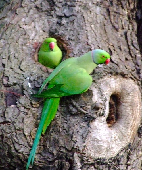
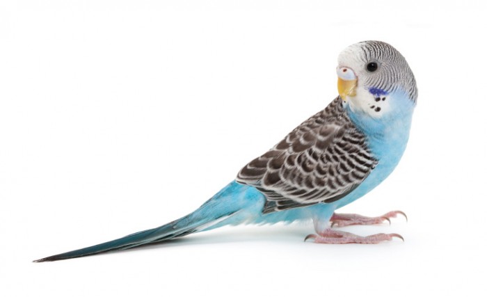
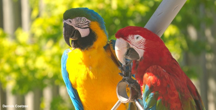
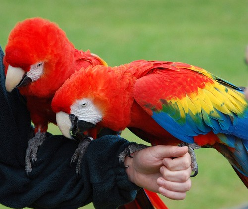
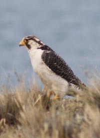
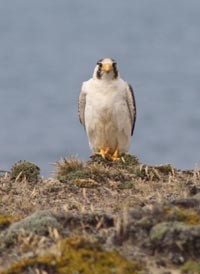
The types of foodstuffs consumed in the wild are often used to classify the nutritional requirements for groups of animals. Usually birds within the Order Psittaciformes are considered to consume plant-based foodstuffs and are classified as florivores. Subdivisions within this category include granivores (budgies and cockatiels), frugivores (many of the macaws, e.g. green winged macaw), and nectarivores (lorikeets and lories). Yet these artificial lines are sometimes too simplistic, as many psittacine birds cross over a category to consume a larger variety of foodstuffs. An example is the scarlet macaw (Ara macao), which is classified as a frugivorous-granivorous psittacine.

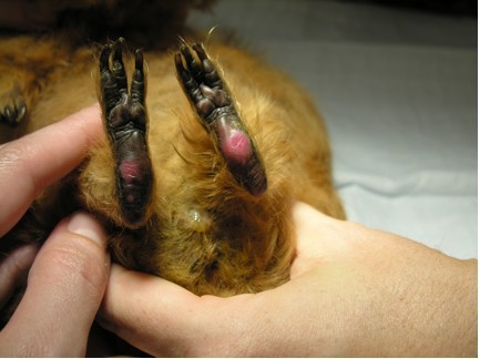
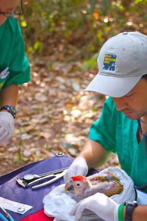
Psittacine birds generally consume plant-based foodstuffs and are classified as florivores. Photo credits:  Florida Center for Instructional Technology (upper left), Steve Bittinger via Flickr Creative Commons (upper right), Peter via FCC (lower left), Derrick Coatzee (lower right)


The availability of certain foods, the sex of the bird, and its age also influence what it eats. However, some species have a very limited diet, such as the glossy backed cockatoo (Calyptorhynchus lathami), which feeds almost exclusively on seeds from a single species of tree in their native environment.


Table 1. Feeding strategies and common diet ingredient of wild psittacine birds.
(Table adapted from Koutsos, et al. Nutrition of birds in the order Psittaciformes: a review. J Avian Med Surg. 2001;15:257–275.)
Species name Feeding strategy Common diet ingredients Time spent feeding*
Blue and gold macaw
(Ara araraunda)
Florivore Seeds, fruits, nuts NR
Military macaw
(Ara militaris)
Florivore Seeds, nuts, berries, fruits NR
Green-winged macaw
(Ara chloroptera)
Frugivore Fruits (hymenaea), palm nuts, seeds NR
Orange-winged Amazon      (Amazona amazonica) Frugivore Fruit (85% from palm fruit) NR
Scarlet macaw
(Ara macao)
Frugivore-granivore Fruits, nuts, bark, leaves, shoots NR
Budgerigar parakeet
(Melopsittacus undulatus)
Granivore Seeds NR
Cockatiel
(Nymphicus hollandicus)
Granivore Seeds (prefers soft, young overmature, hard seeds 3 h/d
Hyacinth macaw
(Anodorhynchus hyacinthinus)
Granivore Palm nuts (50% lipid content) NR
Sulphur-crested cockatoo
(Cacatua galerita)
Omnivore Seeds (primarily sunflower), grubs, rhizomes NR

Nutrient requirements

Nutrients from foods provide the energy to maintain life and the building blocks (precursors) needed to synthesize the structural and functional macromolecules. The macromolecules provide the majority of the diet and include lipids, proteins, carbohydrates, and water. Micronutrients from the diet include vitamins and minerals. Optimal health requires essential nutrients. The quantity of nutrient needed is described as the requirement for that particular nutrient. Some nutrients may not be synthesized in sufficient amounts to meet metabolic demands.

The qualitative and quantitative components of the nutrient requirements are well known in some of the domestic galliforms. Essential nutrients are required in similar proportions with these species and for this reason have been used as a model for psittacines. A variety of nutrients can be oxidized during metabolism of foodstuffs to produce energy.

The physiology of a particular species determines its nutrient requirements. Requirements are determined for three physiologic states: basal, maintenance, and total. The basal requirements are those needed to maintain basic life functions. The maintenance requirement is the amount of nutrients needed for basal functions, plus that needed to find and consume food, interact with other animals, and maintain body temperature. The total energy requirement is the combination of all requirements for life and its stages including growth, reproduction, and molt.

Few nutrient requirements have been evaluated scientifically in the various psittacine birds, so nutrient requirements are often based on the “best guess” from those derived from galliforms. Two methods have been used to determine requirements: 1) empirical calculations and 2) calculations based on factorial summation of specific needs. With empirically based recommendations, experimental diets with graded nutrient levels are fed to a particular species. The minimal level that optimizes the birds’ health and performance is considered the requirement for that nutrient.

One example of an empirical calculation involved feeding groups of galliform chicks increasing levels of methionine. With this nutrient, their growth rate increased linearly until a point was reached where the growth rate did not increase with increasing levels of methionine; this point was empirically determined to be the requirement level of this nutrient. However, the line between levels that are deficient and those needed is not often sharp and instead follows the law of diminishing returns.

Factorial calculations involve adding together various requirements to determine the needs for a particular situation. For example, the requirement of methionine during the egg laying stage has been determined by adding the maintenance requirement of the egg (as determined from the amounts of this nutrient found in the egg by chemical analysis) with the amount needed by the reproductive tract during the egg laying period. This later method is often used for calculating energy requirements, amino acid requirements, and necessary calcium levels. This technique can be very accurate as long as the information that it is based on is also accurate.

One problem with companion birds is that these requirements are often not well established. Another problem is that the efficiency of absorption of nutrients, especially for life stages, may not be accurate. Varying absorption efficiencies may thus throw off the calculations. Both empirical methods and calculations based on factorial summation of specific needs have been used to formulate the requirements for galliforms and ducks  published in the US National Academy of Sciences report. These values represent the nutrient requirements under optimal conditions for these species. Particularly in companion birds, a margin of safety is added to these values in galliforms and provides the “best educated guess” for adequate nutrition. The Association of Avian Veterinarians worked with a panel of experts in diverse areas of avian nutrition to provide general recommendations for psittacines and passerines. These values were the best collective guess to start the discussion on nutrient requirements in these groups of birds.

As avian clinicians, we realize that individual species have differing needs based on our clinical impression over time and the natural behaviors of their wild cousins. Additional concerns about feeding whole food diets in mammals and providing foods that allow our companion birds to exhibit their natural foraging behaviors is adding to a better understanding of their nutritional needs.

Birds in the wild do not necessarily select adequate diets nutritionally, although they appear to be able to balance their energy needs, amino acids, and calcium, but not their needs for other requirements. Birds in captivity do not appear to select appropriately either. A recent dietary approach has been to provide a pelleted or extruded mixture that provides all of the required nutrients above the estimated requirements. This approach has greatly improved the health of companion birds over an all-seed diet but has some shortfalls. The major criticisms are that seed-eating companion birds are difficult to switch to pelleted diets; there is concern about the lack of variety of food stuffs compared with their experience in the wild; various species may have nutritional needs not met with a pelleted diet and these diets offer no opportunity for birds to display their innate foraging behavior.

As we learn more about ways to provide foraging opportunities, changes will emerge in how companion birds are fed in captivity. Presently, the addition of vegetables and fruits to the diet for variety and enrichment suggests that nutrient dilution may occur. Theoretically, that would require that the diet be reformulated. However, this only becomes more of an issue when the bird eats a smaller portion of the formulated diet than the table foods offered. The reason a nutritional deficiency may not occur is that most of the domestic fruits and vegetables are predominately water.


Energy

Birds eat to meet their daily metabolic requirement. For example, a bird confronted with food items that have reduced energy will adjust the amount consumed to meet its energy needs. However, birds may continue to eat out of boredom in captive environments or may become obese when the foods that make up the diet are high in energy but are unbalanced from an amino acid perspective.

Daily energy requirements have only been documented in budgerigars, so calculations for other psittacine species are based on extrapolation, using body weight and a multiplication factor based on the housing condition. The amount of food required to meet those requirements is dependent on the density of the food items consumed, the digestibility of the items, and the ability of the digestive tract of that species to digest and absorb nutrients. Granivorous and omnivorous species are comparatively more efficient than nectivorous species in their ability to digest and absorb nutrients.

The maintenance energy requirement is the amount of dietary metabolizable energy (ME) needed to support basal metabolism. It can be expressed as the sum of the basal metabolic rate (BMR) plus the energy required for supporting maintenance activities and thermoregulation.

Understanding the energy requirements of the bird in its life stage is important for determining the amount of diet that can be fed and/or consumed. It appears that the BMR of psittacine birds depends on the thermal climate of the species of origin and is unrelated to food habits and water availability. Tropical psittacine species have BMRs similar to other tropical nonpasserines and can be expressed as BMR (kcal/d) = 73.6 X body weight (kg)0.73. Species from temperate climates have a BMR that is 21% higher than that found in tropical species. However, the BMR actually reflects the energy expended while sleeping, because the act of alertly and actively perching in budgerigars requires a two-fold increase in energy. The energy required in free-living Port Lincoln parrots (Barnardius zonardius) and galahs is about 3.0&3.23 times the BMR respectively because of their increased expenditures through foraging, thermoregulation, and defense.


Protein and amino acids

The essential amino acids for birds are arginine, isoleucine, leucine, lysine, methionine, phenylalanine, valine, tryptophan, and threonine. Research with chickens suggests that glycine, histidine, and proline are also considered essential as their rate of synthesis is less than their metabolic needs. Research suggests that glycine is also an essential amino acid for budgerigars. Protein levels consumed must meet the nitrogen requirements for that species in its housing condition and life stage. Growing chicks require greater amounts of protein, especially hatchlings. Hens laying large clutches of eggs also require larger amounts of protein than those at maintenance. Birds within a dietary strategy (e.g., granivorous) that have an increased body size also have higher protein requirements than smaller species.

Feathers compose the largest portion of the protein mass of birds. In budgerigars, they represent 5.7% of the protein mass, which is 28% of the total body protein. Molt results in increased protein needs. Feathers are enriched with cysteine and many of the nonessential amino acids. These amino acids are incorporated into the feather during its formation on a continual basis, while uptake from the GI tract occurs only after consumption of a meal, requiring that they be manufactured from tissue protein sources. Molt is also expensive energetically as the bird loses insulation during that period, consequently requiring increased energy. There are also increased expenditures to acquire more protein to synthesize feather proteins.

Protein digestibility is assumed to be similar in the granivorous psittacines, when compared to chickens. This assumption is based on the fact that assimilation efficiencies of complete diets are similar in psittacines, when compared with chickens. The quality of the protein in the diet reflects the digestibility of the components and the amino acid balance. Rate-limiting amino acids require that more food be consumed to meet that need, which may lead to obesity. If more food is not consumed, a clinically observed deficiency may result. For example, methionine deficiency during chick growth results in dark stress bars on feathers.

High protein diets have been assumed to lead to gout in birds, but this supposition has not been supported by research. Adult male cockatiels, when fed diets with up to 70% crude protein for 11 months, maintained their body weight and general body condition without evidence of renal dysfunction. There was evidence of liver damage because birds developed sinusoidal and periportal lipogranulomas at the 70% level, but not at the 20% or 35% level. It has been suggested that sudden changes to high protein diets might lead to hyperuricacidemia with possible nephritis or gout. However, there is no research at present that indicates that this hypothesis is valid. However, sudden changes are not recommended.


Vitamins

Vitamins play an important role in avian nutrition. Vitamins A, D, E, and K are fat-soluble vitamins and their excretion is more problematic than the water-soluble ones. Vitamin A is the vitamin that is more likely to be deficient in both captive and wild birds as the amount consumed in foodstuffs can be quite variable. Vitamin A represents all noncarotenoid derivatives that have a biological activity similar to all trans-retinol. Most appear as retinol and retinol esters that are absorbed from the gastrointestinal tract and transported by portomicrons for storage in the liver. There are two basic functions of vitamin A in cells&the hormone-like regulatory actions of retinoic acid and the photoreceptor actions of retinal. The hormone-like action involves binding to nuclear and cytoplasmic receptors to induce the regulation of cellular replication, differentiation, and preprogrammed cell death.

The exact requirement of vitamin A is unknown in psittacine birds. Feeding trials in female cockatiels suggest that the range may be between 2000 and 10,000 IU/kg in this species as there were no clinical signs that the birds experienced signs of a deficiency or toxicity. However, less grain-dependent species may have greater requirements than the low levels required of cockatiels. Birds deficient in vitamin A exhibit signs of hyperkeratinization of their mucous membranes (appearing as immature basophilic cells on gram stains), anorexia, poor conditioning, and increased susceptibility to infection. Parrots also exhibit squamous metaplasia of the salivary glands, including their excretory ducts and glandular epithelium. Vocalizations in cockatiels may be affected by vitamin A status, as cockatiels fed a diet high in vitamin A (100,000 IU vitamin A/kg diet) had increased numbers of vocalizations compared with birds fed 2000 IU/kg. Cockatiels fed a diet entirely deficient in vitamin A had reduced peak amplitude and total power of vocalization.

Vitamin D activity is found in a group of related sterols including cholecalciferol (vitamin D3), ergosterol (vitamin D2), and other metabolites. Birds are able to synthesize cholecalciferol from cholesterol but require an adequate amount of sunlight to do so. Because most companion birds do not have sufficient UV exposure for endogenous conversion, they need to have a dietary source of vitamin D. Vitamin D has hormonal actions as it regulates calcium and phosphorus metabolism including bone mineralization and eggshell formation. Vitamin D-deficient hens lay eggs with thin shells and develop osteomalacia with pathologic fractures. Vitamin D toxicity is associated with increased mobilization of calcium with soft tissue mineralization. Recent unpublished data suggest that increased levels of vitamin D can result in renal failure in chicks (D. Phalen, unpublished data, October 2003).

Vitamin E actually consists of 2 groups of compounds with antioxidant activity: the alpha-tocopherols and the alpha-tocotrienols. The most biologically active form of vitamin E is the D-α tocopherol, which is used as the dietary standard. In birds, it appears that only the alpha-tocopherols are incorporated into tissues. Vitamin E is transported by plasma lipoproteins and incorporated into the lipid bilayer of the cell’s membranes. It acts to stabilize these membranes and to quench reactions of oxygen intermediates and polyunsaturated fatty acids. These alpha-tocopherols can compete for free radicals faster than polyunsaturated fatty acids. They can also work in concert with a number of enzymes including superoxide dismutase, glutathione peroxidase, and catalase to protect cell membranes. These enzymes require some of the trace minerals including zinc, manganese, selenium, and iron as cofactors for proper function. When deficient, the absolute requirement of vitamin E increases, demonstrating that many of the nutrients are linked.

Vitamin E has other physiologic effects primarily through modulation of eicosanoid metabolism. Conversion of arachidonic acid into prostaglandins and thromboxanes can be modulated by vitamin E levels. This would result in alteration of the immune responses to a challenge.

The levels of vitamin E required in the diet varies, depending on a number of factors including the levels of polyunsaturated fatty acids, levels of vitamin A and/or beta carotene, the presence and quantity of other dietary antioxidants, rancidity of the fats in the diet, and the content of selenium. A selenium deficiency results in an impairment of the glutathione peroxidase antioxidant system, increasing the need for vitamin E.

Deficiencies of vitamin E result in a number of symptoms that relate to cell membrane dysfunctions. These commonly observed symptoms include encephalomalacia, exudative diathesis, muscular dystrophy, myopathy of the ventriculus, and increased fragility of red blood cells. Encephalomalacia in chicks results in crazy chick disease characterized by torticollis, crying out, and excessive wing flapping while maintaining abnormal postures. Higher than normal levels of vitamin E can result in the symptoms associated with deficiencies of the other fat soluble vitamins. The bioactivity of vitamin E is drastically reduced during processing due to heat and moisture, along with increased levels of unsaturated fatty acids and trace minerals.


Minerals

Calcium is necessary for bone mineralization and eggshell calcification; in its ionic form, it is needed for nerve conduction and for myofibril contraction. The calcium requirement(s) for psittacine species have not been determined but the maintenance requirement for chickens is 0.1% of the diet. Many of the seeds consumed by companion birds are less than 0.03% of the diet, suggesting that the requirement is larger than 0.05%.  Grey parrots are particularly prone to hypocalcemic seizures. The pathophysiology is unknown but may result from an inability to mobilize bone acutely. This problem seems less apparent clinically when birds eat at least half of their diet as pellets because vitamin D3 is provided. African greys are one species that have a uropygial gland and, consequently, they may have a higher requirement or sensitivity for cholecalciferol when not exposed to sunlight. Sunlight is needed to convert the inactive form of vitamin D to the active form or it needs to be supplied in some food on a regular basis.

The requirement for growing psittacine chicks in unknown, but they would be expected to require a similar ratio of calcium to phosphorous ratio (between 1.4:1 and 4:1), assuming the vitamin D levels are adequate. The calcium requirement for altricial species of birds is less than precocial species because the number and size per egg is greater in the latter birds. The calcium requirement in egg-laying chicken hens is 3.3% of the diet, while budgerigars and cockatiels require as little as 0.85% and 0.35%, respectively, for normal calcification. Calcium deficiency occurs when there is too little calcium or the active form of vitamin D or too much phosphorus in the diet. The lack of calcium and the active form of vitamin D in all-seed diets is the underlying problem with egg binding in cockatiels.


Conclusion

Avian nutrition has multiple components and dietary requirements are just a starting point for determining the needs of a diverse group of birds classified as psittacines. Many aviculturists know, through trial and error, that some species are not successful in captivity without certain food items. These nuances need to be added to the body of knowledge to provide improved health and well being for these magnificent creatures.

Disclaimer: Dr. Orosz consults with the Lafeber Company.


References

Microchip Placement For Identification of Birds

Microchip implantation provides permanent and unique identification. This article describes the equipment needed and the technique involved for this procedure. During microchip implantation, the avian patient may be awake and manually restrained or placed under general anesthesia. The battery-free transponder or microchip is placed superficially into the pectoral muscle mass. Potential complications are rare but can include evidence of pain in the bird, hemorrhage, infection, or accidental subcutaneous placement of the chip . . .


To continue you need to be a LafeberVet.com member. (Français), (Español)

Pour continuer, vous devez être un membre LafeberVet.com

Para continuar, debe ser miembro de LafeberVet.com

Already a LafeberVet Member?

Please Login

Psittacine Beak and Feather Disease

Shared by Dr. Eric Klaphake, this client education handout describes psittacine beak and feather disease (PBFD), an important disease of parrots. This handout discusses the cause of PBFD, bird species at greatest risk, transmission, as well as diagnosis or testing. Recommendations for prevention and control in an aviary or collection are also described.

Download the PDF version of this client education handout, or modify the DOCX version  for your veterinary hospital.

Cardiopulmonary-Cerebral Resuscitation in Birds

The basic principles of cardiopulmonary-cerebral resuscitation may be applied to birds. The prognosis for respiratory arrest, especially when caused by isoflurane anesthesia overdose, is good. Cardiac arrest in birds carries a poor prognosis, because direct compression of the heart is not possible due to the overlying sternum. Also, because birds lack a diaphragm, closed-chest compressions cannot utilize the thoracic pump mechanism to increase overall negative intrathoracic pressure. Therefore early recognition of cardiovascular instability is particularly important in avian species . . .


To continue you need to be a LafeberVet.com member. (Français), (Español)

Pour continuer, vous devez être un membre LafeberVet.com

Para continuar, debe ser miembro de LafeberVet.com

Already a LafeberVet Member?

Please Login

Avian Analgesia

Pain assessment in birds is very complex because it requires consideration of differences in age, gender, species, individual behaviors and environmental factors. Birds may exhibit different behaviors or may hide painful behaviors when outside of their home cage. Predatory species may exhibit painful behaviors more readily than prey species. Many clinical signs may be associated with pain in birds including . . .


To continue you need to be a LafeberVet.com member. (Français), (Español)

Pour continuer, vous devez être un membre LafeberVet.com

Para continuar, debe ser miembro de LafeberVet.com

Already a LafeberVet Member?

Please Login