As part of the Lafeber Company Student Program, Dr. David Scott of the Carolina Raptor Center presented this distance-learning event for the Cornell University College of Veterinary Medicine Zoo & Wildlife Society. View this 61-minute presentation, RACE-approved for 1 hour of continuing education. Dr. Scott explores proper triage, prognosis, and repair options for various fractures as well as post-operative care and protocols, including physical therapy . . .
Pour les vétérinaires. Par les vétérinaires.
Le site Lafervet.com est conçu pour une utilisation par les vétérinaires. Il est ouvert aux vétérinaires diplômés, aux techniciens vétérinaires diplômés, aux animaliers et aux étudiants dans ces domaines.
Créer un compte pour accéder à des articles et des ressources du site.
L'inscription est gratuite.
Para Profesionales Veterinarios. Por Profesionales Veterinarios.
El sitio Lafervet.com es para uso de los profesionales veterinarios. Está abierto a los veterinarios licenciados, técnicos veterinarios licenciados, rehabilitadores licenciados y estudiantes en estos campos.
Cree una cuenta para acceder a los artículos y recursos del sitio.
La registro es gratis.
Already a LafeberVet Member?
Please Login
David Scott, DVM
![]()
Dr. David Scott has served as Staff Veterinarian at the Carolina Raptor Center in Charlotte, North Carolina since 2008. Dr. Scott is the author of Raptor Medicine, Surgery, and Rehabilitation and The Red Tailed Hawk: A Surgical Dissection. He is also a software engineer and has developed the RaptorMed medical records software system, specifically designed for the medical management and husbandry of all types of animals. This software has been used at rehabilitation centers, aquariums, and for permanent collections all over the world. David earned a Bachelor of Science in Electrical Engineering in 1988 from the University of Illinois and a Doctorate of Veterinary Medicine from the University of Illinois in 1997.
Anesthetic Depth in Exotic Animals: Monitoring the Degree of Central Nervous System Depression
A dedicated anesthetist should be assigned to monitor every patient during the perianesthetic period. The anesthetist is fundamental to patient safety because she assures the patient is not aware, not moving, and not in pain, all while maintaining stable anesthetic depth. A deep plane of anesthesia can lead to hypoventilation and hypoxemia, reduced cardiac output, hypotension, inadequate tissue perfusion, central nervous system (CNS) depression, and prolonged recovery. This review article first explores the stages of anesthesia and then discusses assessment of anesthetic depth in exotic companion mammals, birds, and reptiles . . .
Pour les vétérinaires. Par les vétérinaires.
Le site Lafervet.com est conçu pour une utilisation par les vétérinaires. Il est ouvert aux vétérinaires diplômés, aux techniciens vétérinaires diplômés, aux animaliers et aux étudiants dans ces domaines.
Créer un compte pour accéder à des articles et des ressources du site.
L'inscription est gratuite.
Para Profesionales Veterinarios. Por Profesionales Veterinarios.
El sitio Lafervet.com es para uso de los profesionales veterinarios. Está abierto a los veterinarios licenciados, técnicos veterinarios licenciados, rehabilitadores licenciados y estudiantes en estos campos.
Cree una cuenta para acceder a los artículos y recursos del sitio.
La registro es gratis.
Already a LafeberVet Member?
Please Login
Anesthetic Monitoring Teaching Module
Upon completion of this RACE-approved learning aid, the participant will have a basic clinical understanding of anesthetic monitoring of exotic animal patients: birds, exotic companion mammals, and reptiles . . .
Pour les vétérinaires. Par les vétérinaires.
Le site Lafervet.com est conçu pour une utilisation par les vétérinaires. Il est ouvert aux vétérinaires diplômés, aux techniciens vétérinaires diplômés, aux animaliers et aux étudiants dans ces domaines.
Créer un compte pour accéder à des articles et des ressources du site.
L'inscription est gratuite.
Para Profesionales Veterinarios. Por Profesionales Veterinarios.
El sitio Lafervet.com es para uso de los profesionales veterinarios. Está abierto a los veterinarios licenciados, técnicos veterinarios licenciados, rehabilitadores licenciados y estudiantes en estos campos.
Cree una cuenta para acceder a los artículos y recursos del sitio.
La registro es gratis.
Already a LafeberVet Member?
Please Login
Veterinary Nursing Resources
Wow! Many LafeberVet resources can serve as a useful clinical refresher for veterinary technicians or as a learning aid for students in veterinary technology, including educational videos, RACE-approved webinar recordings, teaching modules, and a variety of articles.

After you’ve explored the content listed below, review LafeberVet’s forms/questionnaires and client education handouts, as well as basic information sheets for additional information.
Rabbit Basics Teaching Module Quiz
The Rabbit Basics Teaching Module was reviewed and approved by the American Association of Veterinary State Boards (AAVSB) Registry of Approved Continuing Education (RACE) program for 1 hour of continuing education, in jurisdictions which recognize AAVSB RACE approval . . .
Pour les vétérinaires. Par les vétérinaires.
Le site Lafervet.com est conçu pour une utilisation par les vétérinaires. Il est ouvert aux vétérinaires diplômés, aux techniciens vétérinaires diplômés, aux animaliers et aux étudiants dans ces domaines.
Créer un compte pour accéder à des articles et des ressources du site.
L'inscription est gratuite.
Para Profesionales Veterinarios. Por Profesionales Veterinarios.
El sitio Lafervet.com es para uso de los profesionales veterinarios. Está abierto a los veterinarios licenciados, técnicos veterinarios licenciados, rehabilitadores licenciados y estudiantes en estos campos.
Cree una cuenta para acceder a los artículos y recursos del sitio.
La registro es gratis.
Already a LafeberVet Member?
Please Login
Anesthetic Monitoring Quiz
The Anesthetic Monitoring Teaching Module was reviewed and approved by the American Association of Veterinary State Boards (AAVSB) Registry of Approved Continuing Education (RACE) program for 1 hour of continuing education, in jurisdictions which recognize AAVSB RACE approval . . .
Pour les vétérinaires. Par les vétérinaires.
Le site Lafervet.com est conçu pour une utilisation par les vétérinaires. Il est ouvert aux vétérinaires diplômés, aux techniciens vétérinaires diplômés, aux animaliers et aux étudiants dans ces domaines.
Créer un compte pour accéder à des articles et des ressources du site.
L'inscription est gratuite.
Para Profesionales Veterinarios. Por Profesionales Veterinarios.
El sitio Lafervet.com es para uso de los profesionales veterinarios. Está abierto a los veterinarios licenciados, técnicos veterinarios licenciados, rehabilitadores licenciados y estudiantes en estos campos.
Cree una cuenta para acceder a los artículos y recursos del sitio.
La registro es gratis.
Already a LafeberVet Member?
Please Login
James Haberfield, BSc, BVMS, PGCBM, MANZCVS (Unusual Pets, Avian Health)

James Haberfield is the founder, director and a current veterinarian at Unusual Pet Vets in Australia with clinics in Perth, Melbourne and Brisbane. Dr. Haberfield earned a Bachelor of Science (BSc) and Bachelor of Veterinary Medicine and Surgery (BVMS) as well as a post-graduate certificate in business management (PGCBM) from Murdoch University. He is a member of the Australian and New Zealand College of Veterinary Scientists in the fields of the Medicine and Surgery of Unusual Pets MANZCVS (Unusual Pets) and Caged and Aviary birds MANZCVS (Avian Health). James has also been involved with a range of field work activities, from microchipping Western spiny tailed skinks to filming with king cobras in India. He has also authored numerous scientific papers and contributed to a range of books.
Dacryocystitis and Nasolacrimal Flush in Rabbits
Ocular problems are common in both laboratory and pet rabbits ( Oryctolagus cuniculus), and disease of the nasolacrimal duct is one of the most frequently reported ocular diseases in rabbits. This review article features a brief video illustrating this clinical technique plus step-by-step guidance as well as clinically relevant anatomy and recommendations for diagnosis and treatment of dacryocystitis . . .
Pour les vétérinaires. Par les vétérinaires.
Le site Lafervet.com est conçu pour une utilisation par les vétérinaires. Il est ouvert aux vétérinaires diplômés, aux techniciens vétérinaires diplômés, aux animaliers et aux étudiants dans ces domaines.
Créer un compte pour accéder à des articles et des ressources du site.
L'inscription est gratuite.
Para Profesionales Veterinarios. Por Profesionales Veterinarios.
El sitio Lafervet.com es para uso de los profesionales veterinarios. Está abierto a los veterinarios licenciados, técnicos veterinarios licenciados, rehabilitadores licenciados y estudiantes en estos campos.
Cree una cuenta para acceder a los artículos y recursos del sitio.
La registro es gratis.
Already a LafeberVet Member?
Please Login
Joanne Sheen, BVM&S CertZooMed DABVP (Exotic Companion Mammals)
 Jo Sheen is an associate veterinarian at Sydney Exotics and Rabbit Vets and she consults as a primary accession and referral exotic animal medicine veterinarian at Veterinary Specialist & Emergency Centre North Shore in Sydney, Australia. Dr. Sheen graduated from the University of Edinburgh in 2004, and gained her postgraduate qualification in zoological and exotic medicine in 2008. In 2017, she became a Diplomate of the American Board of Veterinary Practitioners in Exotic Companion Mammal Practice. She is only the second Australian veterinarian to achieve these credentials.
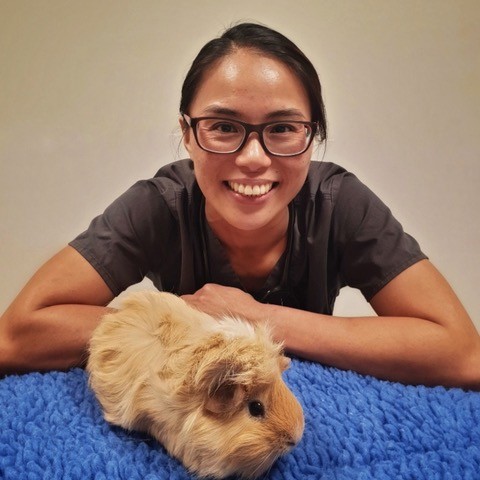
Katie Lennox-Phillibeck
 Katie Lennox-Phillibeck is a freelance video editor, videographer, and photographer. Katie graduated from Purdue University with a degree in Film and Video Studies. Katie is also a veterinary assistant at The Avian and Exotic Animal Clinic of Indianapolis, and she has worked for the clinic in some capacity for the last 17 years. She creates all of their educational videos and website. Some of her other regular clients include Oxbow Animal Health, Purdue University, Eli Lilly & Company, Indianapolis Motor Speedway Productions, The Caring Center, and Play Ball Indiana. Previously, Katie was a full-time video editor for Hall of Music Productions at Purdue University, and an intern at 4th Row Productions in New York City. As the daughter of a veterinarian, Katie has grown up around animals and she has a huge passion for them!
Angela M. Lennox, DVM, DABVP (Avian Practice), DABVP (Exotic Companion Mammal Practice), DECZM (Small Mammal Medicine)
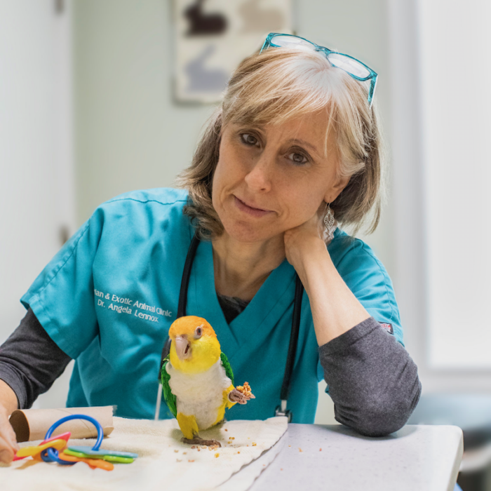 Angela Lennox is a graduate of Purdue University College of Veterinary Medicine. Dr. Lennox has exclusively practiced exotic animal medicine since 1991 and she is the owner of the Avian and Exotic Animal Clinic of Indianapolis. Angela is board certified through the American Board of Veterinary Practitioners in both Avian and Exotic Companion Mammal Practice, and through the European College of Zoological Medicine in Small Mammal Medicine. Dr. Lennox is also an Adjunct Professor at Purdue University College of Veterinary Medicine Department of Clinical Sciences, where she teaches various exotic animal medicine topics to both veterinary medical and veterinary technology students. Dr. Lennox is a Past President of the Association of Exotic Mammal Veterinarians. She also has many publications to her credit, including editor of the Handbook of Rabbit and Rodent Dentistry and co-author of Clinical Radiology of Exotic Companion Mammals.
Deborah Monks, BVSc (Hons) CertZooMed, FACVSc (Avian Health) DECZM
 Deborah Monks is the owner of Brisbane Bird and Exotics Service in Queensland, Australia. After graduating from the University of Queensland, Dr. Monks worked in a variety of practices (general, emergency, 50% avian/50% small animal practice), before finally moving to the United Kingdom to start an avian residency at Great Western Referrals. While in England, she obtained her Certificate of Zoological Medicine (CertZooMed). There are only 3 people in Australia with this qualification. Deborah is also a Diplomate of the European College of Zoological Medicine and Surgery (Avian). She earned her Diplomate status in 2006 and is the only Australian veterinarian with this qualification. Deborah is also a member of the Australian College of Veterinary Scientists in Avian Health since 1999 (MACVSc [Avian Health]). She improved on this initial qualification by achieving her Fellowship (Specialist) status in July 2006 (FACVSc [Avian Health]). Although there are now a number of Members of the Australian College of Veterinary Scientists in Avian Health, Deborah is only the fifth person to have attained Fellowship level and is one of only two in Queensland. Dr. Monk is President of the Avian Chapter of the Australian and New Zealand College of Veterinary Scientists, she serves on the Board of Directors, for the Australasian Chapter of the Association of Avian Veterinarians, and is a Policy Councillor, for the Unusual Pets and Avian Veterinarians, Special Interest Group of the Australian Veterinary Association. She is also a member of the International Committee of the Association of Avian Veterinarians, the European College of Zoological Medicine Examination Committee, and she serves as an online consultant for the Veterinary Information Network.
Tusk Trims in Miniature Pigs
Both males and female pigs possess modified upper and lower canine teeth or tusks, however, the tusks of the male retain an open root that allows these teeth to grow throughout life. Tusks can become long and extremely sharp and trims may be necessary to prevent injury to humans, other animals, household furniture, flooring, or even the pig itself. This brief article discusses relevant anatomy, equipment needed, potential complications, sedation, and step-by-step advice for successfully completing this clinical technique . . .
Pour les vétérinaires. Par les vétérinaires.
Le site Lafervet.com est conçu pour une utilisation par les vétérinaires. Il est ouvert aux vétérinaires diplômés, aux techniciens vétérinaires diplômés, aux animaliers et aux étudiants dans ces domaines.
Créer un compte pour accéder à des articles et des ressources du site.
L'inscription est gratuite.
Para Profesionales Veterinarios. Por Profesionales Veterinarios.
El sitio Lafervet.com es para uso de los profesionales veterinarios. Está abierto a los veterinarios licenciados, técnicos veterinarios licenciados, rehabilitadores licenciados y estudiantes en estos campos.
Cree una cuenta para acceder a los artículos y recursos del sitio.
La registro es gratis.
Already a LafeberVet Member?
Please Login
Hoof Trims in Miniature Pigs
Hooves that are not maintained can overgrow and curl, resulting in pain, difficulty walking, and damage to the soft tissue structures of the foot. The medial and lateral digits, that do not contact the ground much, will grow long and require trimming in all pet pigs. Therefore most pigs require hoof trims every 6-12 months. This brief article discusses relevant anatomy, equipment needed, potential complications, sedation, and step-by-step advice for successfully completing this clinical technique . . .
Pour les vétérinaires. Par les vétérinaires.
Le site Lafervet.com est conçu pour une utilisation par les vétérinaires. Il est ouvert aux vétérinaires diplômés, aux techniciens vétérinaires diplômés, aux animaliers et aux étudiants dans ces domaines.
Créer un compte pour accéder à des articles et des ressources du site.
L'inscription est gratuite.
Para Profesionales Veterinarios. Por Profesionales Veterinarios.
El sitio Lafervet.com es para uso de los profesionales veterinarios. Está abierto a los veterinarios licenciados, técnicos veterinarios licenciados, rehabilitadores licenciados y estudiantes en estos campos.
Cree una cuenta para acceder a los artículos y recursos del sitio.
La registro es gratis.
Already a LafeberVet Member?
Please Login
2020 AEMV Student Case Report Contest

Introduction
Lafeber Company was proud to sponsor the Association of Exotic Mammal Veterinarians (AEMV) Student Case Report Contest. Veterinary students from all over the world were encouraged to write a 2-page case report (1500 words or less) about an exotic companion mammal seen at their college of veterinary medicine or during a clinical experience.
Submissions closed March 27, 2020. Fifteen cases reports were received from eight countries, including the Czech Republic, India, Switzerland, Australia, USA, Canada, Portugal, and Romania. Judges from the Research Committee evaluating the case reports were blinded to the students, mentors, co-authors, and institutions at which the cases were seen.
Posted below are brief summaries of each winning case report. Each student has also been encouraged to submit their paper for peer-reviewed publication.
First place
Shanna Wong (Student – Oklahoma State University, USA): Intra-abdominal torsion of a neoplastic testicle in a rabbit (Oryctolagus cuniculus) with cryptorchidism
A 7-year-old Dutch rabbit was examined for sudden anorexia and lethargy. The rabbit was previously diagnosed with unilateral cryptorchidism 5 years earlier; however, the owner declined surgical treatment at that time. Transabdominal ultrasonography showed a structure consistent with an enlarged testis, with decreased echogenicity, and absent blood flow upon color Doppler ultrasonography. After induction of general anesthesia and during preparation for surgery, the patient had an episode of cardiac arrest. After successful resuscitation, bilateral cryptorchidectomy continued as planned. A ventral midline laparotomy was performed and confirmed presence of an enlarged, dark red left testicle torsed on its spermatic cord. The contralateral testicle was atrophic. Both testicles were removed after standard hemostasis. Recovery was uncomplicated. Histopathologic examination revealed a diagnosis of Sertoli cell tumor and extensive hemorrhage and necrosis in the torsed testicle. Based on literature search, this is the first reported case of intra-abdominal torsion of a neoplastic testicle in a cryptorchid rabbit. Early elective cryptorchidectomy before the potential development of life-threatening complications may be beneficial in rabbits as in other species.
Second place
Nicole Pauli (Student – University of Zürich, Switzerland):
Diagnosis and successful treatment of discospondylitis in a rabbit (Oryctolagus cuniculus) using MRI
A 2.5-year-old 5.3kg male neutered rabbit (Oryctolagus cuniculus) was presented for lameness and progressive exercise intolerance. In the clinical and neurological examinations, the rabbit showed an unphysiological hindlimb position and a non-ambulatory paraparesis with absent proprioception and increased spinal reflexes. Radiographs revealed an aggressive lesion of the endplates of the lumbar vertebrae L4-L5. Under general anesthesia (hydromorphone (0.3mg/kg IM), midazolam (1 mg/kg IM), ketamine (2.5 mg/kg IV), isoflurane in oxygen after intubation, flumazenil (0.05 mg/kg IM) as a reversal), an MRI scan was performed. The MRI study revealed typical signs for discospondylitis at the vertebrae L4 and L5, including T2-hypointense endplates, a T2-hypointense intervertebral disc and contrast enhancement of the surrounding soft tissue and vertebral bodies. Initially, the patient was not able to pass urine. Ultrasound of the urinary system and urine analysis were indicative of a cystitis. Blood analysis revealed an increased CRP (392 mg/l) and other unspecific signs of inflammation. The patient was treated with injectable penicillin (60’000 IU/kg; BID for 7 days, afterwards q2d) and marbofloxacin (5 mg/kg; SID) orally for a total of eight weeks, as well as meloxicam (1 mg/kg PO, SID) for the first three weeks. During treatment, the patient regained normal function of the hindlimbs and was able to pass urine again. On recheck exam after eight weeks of treatment, radiographs revealed signs of ongoing healing and the CRP returned to a normal level (10 mg/l).
Third place
Joana Soares (Student – Lisbon University, Portugal):
Polyostotic lymphoma with vertebral involvement and spinal extradural compression in a ferret (Mustela putorius furo)
A 4-year-old implanted male domestic ferret (Mustela putorius furo) was presented with a sudden onset of hind limb paralysis and anorexia. Physical examination findings included dehydration, paraplegia with both limbs lacking proprioceptive and withdrawal reflexes, urine retention resulting in a distended urinary bladder, absence of perineal reflex and poor body condition. Radiographs revealed severe lysis of the L3 vertebral body with local invasion by a round soft tissue density mass. CT scan revealed an aggressive osteolytic bone lesion centered at the L3 vertebral body, with secondary vertebral canal invasion and ventral compressive myelopathy. The mass measured approximately 2 centimeters in diameter. It was also detected polyostotic osteolytic bone lesions involving the axial and appendicular skeleton and multifocal splenic nodules and masses. Due to the poor prognosis and in agreement with the owner, the animal was humanely euthanized. Histopathological examinations revealed infiltrations of continuous sheets of medium-sized lymphocytes with large nuclei, one or more nucleolus, and low mitotic index. A diagnosis of polyostotic lymphoma was made, the second report of this condition in a domestic ferret and the first in this species with vertebral involvement and spinal extradural compression.
Honorable mentions
- Nikola Sádovská (Student – University of Veterinary and Pharmaceutical Sciences – Brno, Czech Republic): Anorexia in a ferret associated with a large infected biliary cyst and suppurative hepatitis
A 6-year-old neutered male ferret was presented to the veterinary clinic with acute onset of anorexia and nausea. On physical examination large spherical mass (6 cm in diameter) was palpated located caudally to the rib cage. Hematology and plasma chemistry showed anemia, monocytosis, uremia, hypocalcemia, elevated gamma-glutamyl transferase and bilirubinemia. Abdominal radiography revealed large soft tissue opacity mass indistinguishable from the liver parenchyma, which dislocated stomach laterally. On ultrasound, the cystic mass formed of one cavity originated from liver, anechogenic cyst content and hyperechoic sediment was identified. Exploratory laparotomy confirmed the presence of a large cystic mass, which was surgically excised (right liver lobe lobectomy). Total volume of the cyst content was 65 ml. Bacteriological examination revealed pure culture of multi-resistant Escherichia coli. The histopathological diagnosis was infected biliary cyst with peripheral suppurative hepatitis. Based on clinical signs and all the diagnostics, it was presumed that the infection was spread from the duodenum. Anorexia was caused by lateral displacement of the stomach with the large biliary cyst which prevented feed intake and also caused nausea. The presented case showed importance of thorough clinical examination, use of complimentary imaging methods and laboratory analyses which led to successful surgery and to the diagnosis confirmed by histopathology. Because liver disease can be part of a systemic disease, screening for concurrent disease and additional examination must be interpreted accordingly.
- Lenka Čejková (Student – University of Veterinary and Pharmaceutical Sciences – Brno, Czech Republic): Uterine squamous cell carcinoma in a pet rat (Rattus norvegicus)
A 1-year-old female pet rat (Rattus norvegicus) was presented to the veterinary clinic with a 3-week history of mild hemorrhagic vaginal discharge. On presentation, the animal was inactive, dehydrated, and in poor body condition (BCS 1.5/5), with pale mucous membranes and a mucopurulent vaginal discharge. Abdominal palpation revealed an almost empty gastrointestinal tract and a large, ovoid, fibroelastic mass (4×3 cm) with two smaller tubular masses located in the hypogastric region. Abdominal ultrasonography revealed thickened and structurally changed uterine bodies and uterine horns of various echogeneity of the size 4×5 cm. Ovariohysterectomy was performed and the mass was submitted for histopathological examination that confirmed the presence of squamous cell carcinoma (SCC). The patient recovered uneventfully and further health checks, done after 10 days, 3 months, and 7 months, did not identify any metastatic lesions. The incidence of endometrial tumors increases with age. There is a 55% chance of identifying benign endometrial tumors in rats up to 31 months of age. In some strains of rats, the incidence of uterine tumors rises to 66% in rats older than 21 months. Therefore, ovariohysterectomy is the best prevention of uterine tumors in both laboratory and pet rats.
Prizes
| 1st Place: | $100 Cash Prize plus
Carpenter JW (ed). Exotic Animal Formulary, 5th ed. Elsevier, 2017. Mayer J, Donnelly TM. Clinical Veterinary Advisor: Birds and Exotic Pets. St. Louis: Saunders; 2012 Approximate value 250 U.S. dollars |
| 2nd Place: | $100 Cash Prize plus
Carpenter JW (ed). Exotic Animal Formulary, 5th ed. Elsevier, 2017. Approximate value 150 U.S. dollars |
| 3rd Place: | $100 Cash Prize |
Permission was obtained from the supervising clinician (required) and the owner (optional depending on local privacy laws or facility standards).
Learn more
Interested in learning more about exotic companion mammals? Visit AEMV.org or email [email protected] for more information.
![]()
Reptile Wildlife Euthanasia Techniques Quiz
Post test for the Reptile Wildlife Euthanasia Techniques webinar . . .
Pour les vétérinaires. Par les vétérinaires.
Le site Lafervet.com est conçu pour une utilisation par les vétérinaires. Il est ouvert aux vétérinaires diplômés, aux techniciens vétérinaires diplômés, aux animaliers et aux étudiants dans ces domaines.
Créer un compte pour accéder à des articles et des ressources du site.
L'inscription est gratuite.
Para Profesionales Veterinarios. Por Profesionales Veterinarios.
El sitio Lafervet.com es para uso de los profesionales veterinarios. Está abierto a los veterinarios licenciados, técnicos veterinarios licenciados, rehabilitadores licenciados y estudiantes en estos campos.
Cree una cuenta para acceder a los artículos y recursos del sitio.
La registro es gratis.
Already a LafeberVet Member?
Please Login
A Necropsy Guide to Serpentes
The postmortem exam is a key diagnostic tool in understanding the reasons for a snake's morbidity and mortality. Necropsies can provide valuable information to provide a risk assessment for other animals in a population or collection and can help provide closure for a grieving owner. This manuscript reviews the snake necropsy in a systemic, thorough manner, describing normal anatomy and proper collection technique from head to tail . . .
Pour les vétérinaires. Par les vétérinaires.
Le site Lafervet.com est conçu pour une utilisation par les vétérinaires. Il est ouvert aux vétérinaires diplômés, aux techniciens vétérinaires diplômés, aux animaliers et aux étudiants dans ces domaines.
Créer un compte pour accéder à des articles et des ressources du site.
L'inscription est gratuite.
Para Profesionales Veterinarios. Por Profesionales Veterinarios.
El sitio Lafervet.com es para uso de los profesionales veterinarios. Está abierto a los veterinarios licenciados, técnicos veterinarios licenciados, rehabilitadores licenciados y estudiantes en estos campos.
Cree una cuenta para acceder a los artículos y recursos del sitio.
La registro es gratis.
Already a LafeberVet Member?
Please Login
COVID Content Catch-Up
Introduction
According to LitCovid, an open-resource literature hub developed with the support of the US National Institute of Health, over 14,000 relevant articles have been posted to PubMed on the 2019 novel coronavirus.3 Thousands more articles are available as pre-prints. Obviously this explosion of information can be intimidating for the busy veterinarian, but you can use the resources listed in Table 1 to stay current on the latest information. Then turn to this review article and our supplemental LafeberVet Literature Search as well as LafeberVet’s Coronavirus in Animals and Determinants of Viral Infection, published in early April 2020, to dive a bit deeper.
Coronaviruses (CoVs) are enveloped, nonsegmented positive-sense RNA viruses. Coronavirus disease 2019 (COVID-19) is caused by severe acute respiratory syndrome coronavirus 2 (SARS-CoV-2), an emerging zoonotic virus that has achieved extensive community spread among humans.5,7 Large droplet transmission and contact transmission are the two main routes of human-to-human transmission, however SARS-CoV-2 can also persist on inanimate surfaces for up to 9 days.9 Coronavirus disease 2019 first emerged as the cause of severe pneumonia in Wuhan City, Hubei Province, China in December 2019 and it was declared a pandemic in March 2020.7,10,15,21
Likely sources of infection
During the past two decades, three novel coronaviruses have emerged to cause serious outbreaks of human infectious diseases: SARS-CoV, MERS-CoV, and now SARS-CoV-2.16, 23, 24, Although it is unusual for a virus to make the jump from animals to people, MERS and SARS originated from bats.16 The bat is also widely believed to be the original host of SARS‐CoV‐215,16,21. Analysis has shown that SARS-CoV-2 shares 96.2% nucleotide homology with a coronavirus isolated from the horseshoe bat (Rhinolophus spp.) (Bat-CoV-RaTG13).13,21,23 This suggests that SARS-CoV-2 could be of bat origin 13, however the spike protein of the bat coronavirus does not bind well to the human receptor. Therefore it seems likely that spillover of SARS-CoV-2 to humans occurred through an intermediate host, as with SARS-CoV and MERS-CoV.11,13
The intermediate hosts for SARS-CoV and MERS-CoV are the masked palm civet (Paguma larvata) and the dromedary camel (Camelus dromedaries) respectively.16The intermediate hosts of SARS-CoV-2 are completely unknown.16,21
Since angiotensin‐converting enzyme 2 (ACE2) is the receptor for SARS‐CoV‐2, the specificity of the interaction between SARS‐CoV‐2 and the receptor determines the host range for the virus.15 The spike protein (S) of SARS‐CoV‐2 has attracted great attention because of its role in receptor binding. Angiotensin‐converting enzyme 2 (ACE2) binds to the receptor‐binding domain (RBD) of SARS‐CoV‐2 S protein and functions as a receptor for SARS‐CoV‐2.15
Visit LafeberVet’s Determinants of Viral Infection for more information about the spike protein, angiotensin-converting enzyme 2, and the receptor-binding domain.
PANGOLIN
The RBD region on the S protein of pangolin coronavirus is similar to that of SARS‐CoV‐2. This suggests the involvement of pangolin virus in the recombination of SARS‐CoV‐2 15, however pangolin ACE2 was predicted to recognize SARS‐CoV‐2 RBD less efficiently because it only preserved 14 of 20 critical amino acids.15,16
Evaluation of Pangolin-CoV-2020, identified in three Malayan pangolins (Manis javanica) with severe respiratory disease, was found to be genetically associated with both SARS-CoV-2 and a group of bat coronaviruses.13Phylogenetic analyses and amino acid sequencing of the S protein of SARS-CoV-2 does not support the hypothesis of SARS-CoV-2 arising directly from the pangolin-CoV-2020. It is unclear whether this coronavirus is a common virus flora in the respiratory tract of pangolins.13
HAMSTER
A study by Luan et al suggested that Bovidae and Cricetidae (true hamsters, voles, lemmings, New World rats and mice) should be included in the screening of intermediate hosts for SARS‐CoV-2.15,16
REPTILE
Based on analysis of codon usage of SARS‐CoV‐2, the snake has been suggested as a potential host.8,15 Another study evaluating the key amino acids in ACE2 used to interact with the SARS‐CoV RBD indicated that the turtle could be a potential intermediate host for SARS‐CoV‐2.14,15 However all known hosts for coronaviruses are endothermic animals, therefore it is unlikely that reptiles will be infected with SARS‐CoV‐2.15 Additionally, a recent investigation into potential interaction between S protein and ACE2, concluded that SARS‐CoV‐could not infect the snake or turtle.15
Human-to-animal transmission
The coronavirus that caused the 2003 SARS outbreak in China, Singapore, Taiwan, Hong Kong, and Toronto was shown to result in household pets testing positive for SARS.7 This triggered panic abandonment of such pets, however, no dog-to-human transmission was shown, and SARS was successfully contained by focusing on interruption of human-to-human transmission.7
The issue of the need to evaluate companion animals and their status with regards to SARS-CoV-2 was first raised on January 29 when a member of the senior expert team from China’s National Health Commission stated on Chinese state television that pet owners should take extra care of their animals because (1) the virus “moves between mammals”; (2) if your animals “come into contact with the outbreak or people infected with the virus, then your pets should be put in quarantine”; and (3) “because the epidemic spreads between mammals, therefore we should take precaution against other mammals”. No scientific data were presented to support this statement but, nonetheless, it prompted a severe public response that resulted in many pet dogs and cats being killed and thousands being abandoned.
This prompted the World Health Organization to state that “there is no evidence dogs and cats can be infected with the virus”. No scientific data were provided to support this statement about a novel zoonotic threat either. Despite this appeal, the culling of pets continued in China through February 21…
…on March 13, the IDEXX veterinary diagnostic laboratory announced that it had tested >3,500 dog, cat, and equine specimens from across the United States and South Korea with their COVID-19 RT-qPCR and that they had no positives. What the press release did not make clear, however, is the fact that although animals tested were from affected areas, it is “unknown if any of the animals lived in homes with people that had COVID-19”.— McNamara et al 17
The reader is encouraged to read the complete Vector-Borne and Zoonotic Diseases review article: “A critical needs assessment for research in companion animals and livestock following the pandemic of COVID-19 in humans”.17 This is a chronological and comprehensive review on COVID-19 in companion and captive animals that begins with the SARS-CoV-2-positive dog reported in Hong Kong and continues through April 19, 2020.17
A Nature News Round-Up19 provides useful insight: The first two dogs reported to have coronavirus probably caught the infection from their owners, say researchers who studied the animals and members of the infected households in Hong Kong. An analysis of viral genetic sequences from the dogs showed them to be identical to those in the infected people.
Researchers suspected that the infection had been passed from the owners to the dogs, and the direct genomic link strongly supports that, says Malik Peiris, a virologist at the University of Hong Kong who led the study, which was published in Nature (Sit et al 2020).23
The study showed no evidence that dogs can pass the infection to other dogs or to people, but it is impossible to be certain in which direction the virus travelled “so we have to keep an open mind”, says Peiris.
Although the analysis confirms that people with COVID-19 can infect dogs, the probability of this happening is low, says Arjan Stegeman, a veterinary epidemiologist at Utrecht University in the Netherlands. In the study, only 2 of the 15 dogs who lived with infected people got the disease.
The diversity of species susceptible to SARS-CoV and SARS-Cov-2 strongly suggests a propensity of these viruses to cross the species barrier.5,11 Interspecies transmission is most likely to be facilitated when there is close contact close between humans with high infectious virus loads and companion or captive mammals.5,11
Animal-to-animal transmission
As efforts are made to develop vaccines and antiviral drugs for humans, what animals can best be used to model the efficacy of medical countermeasures?18,7 The ideal animal model would mimic high human-to-human transmission rates so that we can better understand the rapid spreading characteristics of SARS-CoV-2.10,18
FERRET
Ferrets are frequently used as an animal model for respiratory viruses that infected humans and ferret ACE2 has been shown to contain critical SARS-CoV binding residues.10,21 Kim et al performed infection and direct and indirect contact transmission studies using a ferret model previously developed for influenza virus infections.10 SARS-CoV-2 is effectively transmitted to naive ferrets by direct contact and virus is detected in naive direct contact ferrets 2 days post-contact.10 A few naïve indirect contact ferrets were also positive for viral RNA, suggesting airborne transmission. Infected ferrets shed SARS-CoV-2 in nasal washes, saliva, urine, and feces up to 8 days post-infection.10,21
A study in ferrets also demonstrated evidence of robust transmission of SARS-CoV-2 via the air: Richard, M., Kok, A., de Meulder, D. et al. SARS-CoV-2 is transmitted via contact and via the air between ferrets. Nat Commun 11, 3496 (2020). 21
CAT
Preliminary studies have demonstrated direct cat-to-cat spread of SARS-CoV-2 through nasal shedding and limited airborne transmission, as well as the production of specific neutralizing antibodies against SARS-CoV-2 in this species.6,21
RHESUS MACAQUE
In a study evaluating SARS-CoV-2-infected macaques, virus was excreted from the nose and throat in the absence of clinical signs. Virus was also detected in type I and II pneumocytes in foci of diffuse alveolar damage and in ciliated epithelial cells of nasal, bronchial, and bronchiolar mucosae.18,19 More severe interstitial pneumonia was seen in older monkeys when compared to young animals.25
HAMSTER
The ACE2 proteins from Cricetidae are able to recognize SARS-CoV-2 RBD (Table 1), and the golden Syrian hamster (Mesocricetus auratus ) has been established as a model to study the pathogenesis and transmission of COVID‐19.2,15
MOUSE
Mice cannot typically be used as an animal model of SARS-CoV-2 directly because the ACE2 of mice cannot interact with SARS-CoV-216, however some studies have used mice transfected with human ACE2 to serve as animal models for SARS-CoV-2 infection.1,10
Clinical disease
The SARS-CoV-2 infection has a wide clinical spectrum in humans, from mild infection to death, but how does the virus behave in other animals? 22
- The dogs that tested positive did not develop clinical signs.
- One domestic cat naturally infected by her owner in Belgium presented with respiratory difficulty, vomiting, and diarrhea.2,12
- All of the cats in one small, preliminary study were asymptomatic.6
- The four Siberian tigers (Panthera tigris) and three African lions (Panthera leo) at the Bronx Zoo in New York developed a dry cough, some wheezing, and loss of appetite. None of the animals were in respiratory distress.5,11
- Ferrets exhibited elevated body temperature and acute bronchiolitis was present within infected lungs. Fatalities were not observed.10,21
- SARS-CoV-2 causes respiratory disease in infected rhesus macaques, with illness lasting 8-16 days. Pulmonary infiltrates were visible on survey radiographs.18
- Bao et al reported weight loss in transgenic mice following SARS-CoV-2 infection however, no other clinical signs were observed.1,10
Animal-to-human transmission
The close association between humans and their pets has led to an examination of the potential risks of transmission.1,17 Currently, there is no evidence that household pets have transmitted disease to humans, and the World Organisation for Animal Health has stated, ‘there is no justification in taking measures against companion animals which may compromise their welfare.7 Nevertheless in a recent letter to parliament, Agricultural Minister of the Netherlands reported that a Dutch farm worker contracted coronavirus from mink.22 Outbreaks on mink farms were first reported in April, when keepers noticed some animals with respiratory difficulty.22
Just as with the 2003 SARS outbreak, animal welfare is again seriously threatened during the SARS-CoV-2 pandemic. Panic abandonment of household pets is neither justified nor morally supported.7 Animal health professionals must be proactive to stop or prevent panic abandonment or killing of household pets in response to social media panic and misinformation during the COVID-19 outbreak.7
One Health management strategies
The zoonotic origin of SARS-CoV-2 is indicative of its ability to cross the species barrier.9 There is a critical need for One Health surveillance, intervention, and management strategies to lessen the effects on wild, captive, and companion animal populations and to address many important questions:4
- What are the risks of SARS-CoV-2 contamination of pets by their owners? 9
- What is the potential for domesticated (companion) animals to serve as a reservoir of infection contributing to continued human-to-human disease, infectivity, and community spread? 4,14
- What are the ramifications for food security, economy, and trade issues should coronavirus establish itself within livestock and poultry? 14
- Is there a risk for multiple spillover episodes in animal populations that could result in SARS-CoV-2 becoming endemic in multiple animal species and populations? 4
As a precautionary measure, US Centers for Disease Control recommends that people with COVID-19 have someone else care for their companion animals while they are sick. Frequent handwashing before and after contact with animals, and avoiding intimate contact, is also strongly recommended.7
Infected animals should also be quarantined.5
References
Reptile Wildlife Euthanasia Techniques
“The question is not, can they reason? Nor, can they talk? But, can they suffer?” –Jeremy Bentham, philosopher, 1780
Dr. Renée Schott presented a live, interactive webinar on reptile wildlife euthanasia techniques. View the RACE-approved webinar recording today. Wildlife often present to veterinarians and wildlife rehabilitators with conditions that warrant euthanasia. It can be difficult, however, to apply mammalian methods of euthanasia to species with unique physiology such as reptiles. This presentation uses cases to discuss practical euthanasia methods for reptiles and the physiology behind these methods. Emphasis is placed on freshwater turtles as these represent some physiological . . .
Pour les vétérinaires. Par les vétérinaires.
Le site Lafervet.com est conçu pour une utilisation par les vétérinaires. Il est ouvert aux vétérinaires diplômés, aux techniciens vétérinaires diplômés, aux animaliers et aux étudiants dans ces domaines.
Créer un compte pour accéder à des articles et des ressources du site.
L'inscription est gratuite.
Para Profesionales Veterinarios. Por Profesionales Veterinarios.
El sitio Lafervet.com es para uso de los profesionales veterinarios. Está abierto a los veterinarios licenciados, técnicos veterinarios licenciados, rehabilitadores licenciados y estudiantes en estos campos.
Cree una cuenta para acceder a los artículos y recursos del sitio.
La registro es gratis.
Already a LafeberVet Member?
Please Login
Flight Mechanics & Ethical Concerns Quiz
. . .
Pour les vétérinaires. Par les vétérinaires.
Le site Lafervet.com est conçu pour une utilisation par les vétérinaires. Il est ouvert aux vétérinaires diplômés, aux techniciens vétérinaires diplômés, aux animaliers et aux étudiants dans ces domaines.
Créer un compte pour accéder à des articles et des ressources du site.
L'inscription est gratuite.
Para Profesionales Veterinarios. Por Profesionales Veterinarios.
El sitio Lafervet.com es para uso de los profesionales veterinarios. Está abierto a los veterinarios licenciados, técnicos veterinarios licenciados, rehabilitadores licenciados y estudiantes en estos campos.
Cree una cuenta para acceder a los artículos y recursos del sitio.
La registro es gratis.
Already a LafeberVet Member?
Please Login
Brian Stacy, DVM, PhD, DACVP
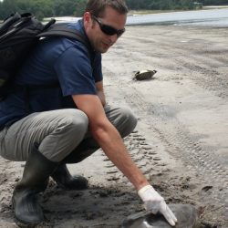
Brian Stacy earned his DVM from the University of Georgia at Athens before completing an Anatomic Pathology Residency at the University of California at Davis and the Zoological Society of San Diego. He has been a Diplomate of the American College of Veterinary Pathologists since 2004 and he earned a PhD from the University of Florida in 2008. Dr. Stacy is a Veterinary Medical Officer for the National Oceanic and Atmospheric Administration (NOAA) Fisheries, Office of Protected Resources. Through a cooperative agreement, Dr. Stacy is based at the University of Florida College of Veterinary Medicine. He works intensively with threatened and endangered marine turtles as part of NOAA’s National Sea Turtle Program and he is involved with a variety of issues related to turtle health, stranding, and mortality. Other species of professional interest for Dr. Stacy include other reptiles, amphibians, and marine mammals. Current and past projects include a variety of infectious and noninfectious disease studies, investigations of animal die-offs, and forensic studies related to human impacts on wildlife.
Richard S. Funk, MA, DVM
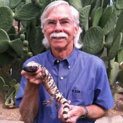
Photo credit: California Turtle and Tortoise Club – Orange County Chapter
Richard S. Funk completed a master’s degree in zoology with a thesis in herpetology. He then earned his DVM at The Ohio State University. Dr. Funk next worked at a busy private practice in Wilmington, North Carolina, treating companion pets and exotics before serving as Clinical Associate Professor at the University of Tennessee College of Veterinary Medicine, where he taught clinical courses and was in charge of the exotics service. Rich then moved to the Tampa, Florida area where he owned an exclusively exotics house call practice. He next moved to the Phoenix, Arizona area and joined a VCA practice treating companion pets and exotics. Dr. Funk later owned his own practice, which offered relief work, house calls, and consults, and he served as an Adjunct Professor of Veterinary Clinical Sciences at Midwestern University College of Veterinary Medicine. Dr. Funk was very active in the Association of Reptile and Amphibian Veterinarians (ARAV), having served as the 2003-2004 President and co-chairman of the Legislation and Welfare Committee. He has also served on the ARAV publications editorial board and he organized and led a number of wet labs at ARAV annual conferences. Dr. Funk had a number of veterinary publications, primarily on reptiles, including chapters in all three volumes of the Mader Reptile Medicine and Surgery texts. His research interests were focused on reptile viruses and on the health and welfare of captive Galapagos tortoises.
The reptile community lost Dr. Funk in August 2022. Please visit the American Veterinary Medical Association website for a brief In Memoriam.
Louisa Asseo DVM DABVP (Canine/Feline Practice)

Louisa Asseo graduated from the University of California at Davis School of Veterinary Medicine in 2002. She has worked in private practices in the Bay and Sacramento areas of California since graduation, focusing on both general practice and emergency medicine. In addition to cats and dogs, her practice focuses on medicine and surgery for reptiles, exotic companion mammals, poultry, and miniature pigs. In 2016, she founded Oasis Veterinary Hospital in the east San Francisco Bay area of California. Louisa has grown the practice to include multiple associates who provide excellent patient care to dogs, cats, reptiles, and exotic mammals. Dr. Asseo received certification as a Diplomate of the American Board of Veterinary Practitioners in canine and feline practice in 2015.
Chris Hanley, DVM, DACZM
![]()
Chris Hanley received his Doctorate of Veterinary Medicine from Tufts University School of Veterinary Medicine in 2000. After completing an avian and exotics internship at the University of Georgia, he completed his zoological medicine residency at the University of Wisconsin School of Veterinary Medicine, Milwaukee County Zoo, and International Crane Foundation. Dr. Hanley became a Diplomate of the American College of Zoological Medicine in 2006. After seven years at the Toledo Zoo, three as an associate and four years as the Director of Animal Health and Nutrition, he moved to the Saint Louis Zoo as a staff veterinarian. In 2019, he became the Assistant Director of Animal Health.
Shannon Martinson, DVM, MVSc, DACVP

Shannon Anne Martinson is an Assistant Professor in Anatomic Pathology in the Department of Pathology and Microbiology at the Atlantic Veterinary College of the University of Prince Edward Island. Dr. Martinson is a Diplomate of the American College of Veterinary Pathology and the author of several publications, including a 2019 article for the Journal of Herpetological Medicine and Surgery. Over the past 10 years she has developed a keen interest and increasing level of expertise in reptile pathology.
Flight Mechanics, Parrot Welfare, & Ethical Concerns
Dr. Todd E. Driggers presented this live webinar event on Flight Mechanics, Parrot Welfare, and Ethical Concerns. The webinar recording begins with a discussion on grooming. Feather trimming birds in captivity has been a common practice performed for many reasons, including fear of loss, safety, and the ability to control and tame. If the gold standard for animal welfare is freedom and feather destructive behavior is a reliable indicator of scientifically studied animal welfare, feather trimming impacts how the animal feels, functions, and prohibits natural responses to positive or aversive stimuli. Perhaps it is time to reflect on the benefits . . .
Pour les vétérinaires. Par les vétérinaires.
Le site Lafervet.com est conçu pour une utilisation par les vétérinaires. Il est ouvert aux vétérinaires diplômés, aux techniciens vétérinaires diplômés, aux animaliers et aux étudiants dans ces domaines.
Créer un compte pour accéder à des articles et des ressources du site.
L'inscription est gratuite.
Para Profesionales Veterinarios. Por Profesionales Veterinarios.
El sitio Lafervet.com es para uso de los profesionales veterinarios. Está abierto a los veterinarios licenciados, técnicos veterinarios licenciados, rehabilitadores licenciados y estudiantes en estos campos.
Cree una cuenta para acceder a los artículos y recursos del sitio.
La registro es gratis.
Already a LafeberVet Member?
Please Login
Snake Anatomy Basics
Snakes are members of the class Reptilia, order Squamata, and suborder Serpentes. There are over 3,500 species of snakes in the world, however, for the most part, the anatomy of the snake is consistent across species.
Snakes have a long narrow body adapted for crawling and their internal anatomy has evolved to fit into a long narrow tube. It is possible to divide this tube into four quadrants (Fig 1). Although the sequence of organs is the same for all species, the relative position and size of the viscera can vary significantly between and within families. The quadrant system . . .
Pour les vétérinaires. Par les vétérinaires.
Le site Lafervet.com est conçu pour une utilisation par les vétérinaires. Il est ouvert aux vétérinaires diplômés, aux techniciens vétérinaires diplômés, aux animaliers et aux étudiants dans ces domaines.
Créer un compte pour accéder à des articles et des ressources du site.
L'inscription est gratuite.
Para Profesionales Veterinarios. Por Profesionales Veterinarios.
El sitio Lafervet.com es para uso de los profesionales veterinarios. Está abierto a los veterinarios licenciados, técnicos veterinarios licenciados, rehabilitadores licenciados y estudiantes en estos campos.
Cree una cuenta para acceder a los artículos y recursos del sitio.
La registro es gratis.
Already a LafeberVet Member?
Please Login
LafeberVet Lit Search: SARS-CoV-2
Introduction
Over 14,000 articles have been posted to PubMed on the 2019 novel coronavirus, known as SARS-CoV-2. Thousands more articles are available as pre-prints. Obviously this explosion of information can be intimidating for the busy veterinarian, but you can use the resources listed in Table 1 to stay current on the latest information. Then turn to our COVID Content Catch-Up review article and this supplemental LafeberVet Literature Search, as well as LafeberVet’s Coronavirus in Animals and Determinants of Viral Infection,to dive a bit deeper.
Many of the references provided below are listed in more than one category. Three strongly recommended citations are bolded. Pre-prints, which should be evaluated with caution, are highlighted in red.
|
Exotic animals
Decaro N, Lorusso A. Novel human coronavirus (SARS-CoV-2): A lesson from animal coronaviruses. Vet Microbiol Vol 244, May 2020. doi: 10.1016/j.vetmic.2020.108693.
Gollakner R, Capua I. Is COVID-19 the first pandemic that evolves into a panzootic? Vet Ital 2020 Apr 24;56(1):7-8. doi: 10.12834/VetIt.2246.12523.1.
Gonultas S, Karabagli M, Bastug Y, et al. COVID-19 and animals: What do we know? Turk J Urol. 2020 May 15. doi: 10.5152/tud.2020.140520. Online ahead of print.
Huang Q, Zhan X, Zeng XT. COVID-19 pandemic: stop panic abandonment of household pets. J Travel Med. 2020 May 18;27(3):taaa046. doi: 10.1093/jtm/taaa046.
Leroy EM, Ar Gouilh M, Brugere-Picoux J. The risk of SARS-CoV-2 transmission to pets and other wild and domestic animals strongly mandates a one-health strategy to control the COVID-19 pandemic. One Health. 2020 Apr 13;100133. doi: 10.1016/j.onehlt.2020.100133.
Loeb J. Covid-19 wake-up call for exotic pet trade. Vet Rec 2020 Apr 18;186(14):432. doi: 10.1136/vr.m1517.
Luan J, Jin X, Lu Y, Zhang L. SARS-CoV-2 spike protein favors ACE2 from Bovidae and Cricetidae. J Med Virol. 2020 Apr 1;10.1002/jmv.25817. doi: 10.1002/jmv.25817. Online ahead of print.
McNamara T, Richt JA, Glickman L. A critical needs assessment for research in companion animals and livestock following the pandemic of COVID-19 in humans. Vector Borne Zoonotic Dis. 2020 May 5. doi: 10.1089/vbz.2020.2650. Online ahead of print.
Shi J, Wen Z, Zhong G, et al. Susceptibility of ferrets, cats, dogs, and different domestic animals to SARS-coronavirus-2. Science. 2020 Apr 8 : eabb7015. doi: org/10.1101/2020.03.30.015347.
Please note: There has been some debate as to the soundness of this study’s protocols and therefore the validity of the conclusions reached by Shi et al have also been questioned.
Tiwari R, Dhama K, Sharun K, et al. COVID-19: animals, veterinary and zoonotic links. Vet Q. 2020 May 12:1-22. doi: 10.1080/01652176.2020.1766725. Online ahead of print.
Zhai X, Sun J, Yan Z, et al. Comparison of SARS-CoV-2 spike protein binding to ACE2 receptors from human, pets, farm animals, and putative intermediate hosts. J Virol. 2020 May 13;JVI.00831-20. doi: 10.1128/JVI.00831-20. Online ahead of print.
Bats
Li C, Yang Y, Ren L. Genetic evolution analysis of 2019 novel coronavirus and coronavirus from other species. Infect Genet Evol. 2020 Mar 10;82:104285. doi: 10.1016/j.meegid.2020.104285. Online ahead of print.
Luan J, Lu Y, Jin X, Zhang L. Spike protein recognition of mammalian ACE2 predicts the host range and an optimized ACE2 for SARS-CoV-2 infection. Biochem Biophys Res Commun. 2020 May 21;526(1):165-169. doi: 10.1016/j.bbrc.2020.03.047. Epub 2020 Mar 19.
Maganga GD, Pinto A, Mobo IM, et al. Genetic diversity and ecology of coronaviruses hosted by cave-dwelling bats in Gabon. Sci Rep. 2020 Apr 30;10(1):7314. doi: 10.1038/s41598-020-64159-1.
Birds
de Wit JJS, Cook JKA. Avian coronaviruses. Avian Pathol. 2020 May 6:1-7. doi: 10.1080/03079457.2020.1761010. Online ahead of print.
Volpato G, Fontefrancescoz MF, Gruppuso P, et al . Baby pangolins on my plate: possible lessons to learn from the COVID-19 pandemic. J Ethnobiol Ethnomed. 2020 Apr 21;16(1):19. doi: 10.1186/s13002-020-00366-4.
Xiu L, Binder RA, Alarja NA, et al. A RT-PCR assay for the detection of coronaviruses from four genera. J Clin Virol. 2020 Apr 30;128:104391. doi: 10.1016/j.jcv.2020.104391. Online ahead of print.
Zhuang Q, Liu S, Zhang X, et al. Surveillance and taxonomic analysis of the coronavirus dominant in pigeons in China. Transbound Emerg Dis. 2020 Mar 12;10.1111/tbed.13541. doi: 10.1111/tbed.13541. Online ahead of print.
Camelids
Wrapp D, De Vlieger D, Corbett KS, et al. Structural basis for potent neutralization of betacoronaviruses by single-domain camelid antibodies. Cell. 2020 Apr 29;S0092-8674(20)30494-3. doi: 10.1016/j.cell.2020.04.031. Online ahead of print.
Civets
Li C, Yang Y, Ren L. Genetic evolution analysis of 2019 novel coronavirus and coronavirus from other species. Infect Genet Evol. 2020 Mar 10;82:104285. doi: 10.1016/j.meegid.2020.104285. Online ahead of print.
Ferrets
Kim YI, Kim SG, Kim SM, et al. Infection and rapid transmission of SARS-CoV-2 in ferrets. Cell Host Microbe 2020 Apr 6. doi: 10.1016/j.chom.2020.03.023 [Epub ahead of print]
Richard, M., Kok, A., de Meulder, D. et al. SARS-CoV-2 is transmitted via contact and via the air between ferrets. Nat Commun 11, 3496 (2020). doi.org/10.1038/s41467-020-17367-2.
Shi J, Wen Z, Zhong G et al. Susceptibility of ferrets, cats, dogs, and other domesticated animals to SARS-coronavirus 2. Science. 2020 Apr 8 : eabb7015. Published online 2020 Apr 8. doi: 10.1126/science.abb7015.
Please note: There has been some debate as to the soundness of this study’s protocols and therefore the validity of the conclusions reached by Shi et al have also been questioned.
Non-human primates
Pre-Print: Deng W, Bao L Gao H, et al. Ocular conjunctival inoculation of SARS-CoV-2 can cause mild COVID-19 in Rhesus macaques. doi: https://doi.org/10.1101/2020.03.13.990036
Gibbons A. Ape researchers mobilize to save primates from coronavirus. Science. 2020 May 8;368(6491):566. doi: 10.1126/science.368.6491.566-a.
Le Bras A. Efficacy of remdesivir in a rhesus macaque model of MERS-CoV infection. Lab Anim (NY). 2020 May;49(5):150. doi: 10.1038/s41684-020-0537-x.
Luan J, Lu Y, Jin X, Zhang L. Spike protein recognition of mammalian ACE2 predicts the host range and an optimized ACE2 for SARS-CoV-2 infection. Biochem Biophys Res Commun. 2020 May 21;526(1):165-169. doi: 10.1016/j.bbrc.2020.03.047. Epub 2020 Mar 19.
Munster VJ, Feldman F, Williamson BN, et al. Respiratory disease in rhesus macaques inoculated with SARS-CoV-2. Nature. 2020 May 12. doi: 10.1038/s41586-020-2324-7. Online ahead of print.
Rockx B, Kuiken T, Herfst S, et al. Comparative pathogenesis of COVID-19, MERS, and SARS in a nonhuman primate model. Science. 2020 Apr 17;eabb7314. doi: 10.1126/science.abb7314. Online ahead of print.
Pre-Print: Williamson BN, Feldmann F, Schwarz B. Clinical benefit of remdesivir in rhesus macaques infected with SARS-CoV-2. bioRXiv. doi: 10.1101/2020.04.15.043166.
Yu P, Qi F, Xu Y, et al. Age-related rhesus macaque models of COVID-19. Animal Model Exp Med. 2020 Mar 30;3(1):93-97. doi: 10.1002/ame2.12108.
Pangolins
Liu P, Jiang JZ, Wan XF, et al. Are pangolins the intermediate host of the 2019 novel coronavirus (SARS-CoV-2)? PLoS Pathog. 2020 May 14;16(5):e1008421. doi: 10.1371/journal.ppat.1008421. eCollection 2020 May.
Volpato G, Fontefrancescoz MF, Gruppuso P, et al . Baby pangolins on my plate: possible lessons to learn from the COVID-19 pandemic. J Ethnobiol Ethnomed. 2020 Apr 21;16(1):19. doi: 10.1186/s13002-020-00366-4.
Cats and dogs
Decaro N, Lorusso A. Novel human coronavirus (SARS-CoV-2): A lesson from animal coronaviruses. Vet Microbiol Vol 244, May 2020. doi: 10.1016/j.vetmic.2020.108693.
Gao T, Pan Xi, Pan C. The fate of house cats during the COVID-19 pandemic. Microbes Infect. 2020 Apr 22. doi: 10.1016/j.micinf.2020.04.006 [Epub ahead of print]
Gollakner R, Capua I. Is COVID-19 the first pandemic that evolves into a panzootic? Vet Ital 2020 Apr 24;56(1):7-8. doi: 10.12834/VetIt.2246.12523.1.
Gonultas S, Karabagli M, Bastug Y, et al. COVID-19 and animals: What do we know? Turk J Urol. 2020 May 15. doi: 10.5152/tud.2020.140520. Online ahead of print.
Halfmann PJ, Hatta M, Chiba S, et al. Transmission of SARS-CoV-2 in domestic cats. N Engl J Med. 2020 May 13. doi: 10.1056/NEJMc2013400. Online ahead of print.
He HJ, Zhang W, Liang J, et al. Etiology and genetic evolution of canine coronavirus circulating in five provinces of China, during 2018-2019. Microb Pathog. 2020 Apr 18;145:104209. doi: 10.1016/j.micpath.2020.104209. Online ahead of print.
Huang Q, Zhan X, Zeng XT. COVID-19 pandemic: stop panic abandonment of household pets. J Travel Med. 2020 May 18;27(3):taaa046. doi: 10.1093/jtm/taaa046.
Leroy EM, Ar Gouilh M, Brugere-Picoux J. The risk of SARS-CoV-2 transmission to pets and other wild and domestic animals strongly mandates a one-health strategy to control the COVID-19 pandemic. One Health. 2020 Apr 13;100133. doi: 10.1016/j.onehlt.2020.100133.
Li X. Can cats become infected with Covid-19? Vet Rec. 2020 Apr 18;186(14):457-458. doi: 10.1136/vr.m1455.
Li X. Cats under the shadow of the SARS-CoV-2 pandemic. Transbound Emerg Dis. 2020 Apr 28. doi: 10.1111/tbed.13599. Online ahead of print.
Luan J, Lu Y, Jin X, Zhang L. Spike protein recognition of mammalian ACE2 predicts the host range and an optimized ACE2 for SARS-CoV-2 infection. Biochem Biophys Res Commun. 2020 May 21;526(1):165-169. doi: 10.1016/j.bbrc.2020.03.047. Epub 2020 Mar 19.
Mallapaty S. Coronavirus can infect cats – dogs, not so much. Nature 2020. PMID: 32238897. No abstract available.
McNamara T, Richt JA, Glickman L. A critical needs assessment for research in companion animals and livestock following the pandemic of COVID-19 in humans. Vector Borne Zoonotic Dis. 2020 May 5. doi: 10.1089/vbz.2020.2650. Online ahead of print.
Parry NMA. COVID-19 and pets: When pandemic meets panic. Forensic Science International: Reports Volume 2, Dec 2020. doi: 10.1016/j.fsir.2020.100090.
Shi J, Wen Z, Zhong G, et al. Susceptibility of ferrets, cats, dogs, and other domesticated animals to SARS-coronavirus 2. Science. 2020 Apr 8 : eabb7015. Published online 2020 Apr 8. doi: 10.1126/science.abb7015.
Sit THC, Brackman CJ, Ip SM, et al. Infection of dogs with SARS-CoV-2. Nature. 2020 May 14. doi: 10.1038/s41586-020-2334-5. Online ahead of print.
Pre-Print: Temmam S, Barbarino A, Maso D, et al. Absence of SARS-CoV-2 infection in cats and dogs in close contact with a cluster of COVID-19 patients in a veterinary campus. bioRxiv. doi: 10.1101/2020.04.07.029090.
Wang H, Wang F, Wang H, Zhao Q. Potential infectious risk from the pets carrying SARS-CoV-2. Travel Med Infect Dis. 2020 May 5;101737. doi: 10.1016/j.tmaid.2020.101737.
Zhai X, Sun J, Yan Z, et al. Comparison of SARS-CoV-2 spike protein binding to ACE2 receptors from human, pets, farm animals, and putative intermediate hosts. J Virol. 2020 May 13;JVI.00831-20. doi: 10.1128/JVI.00831-20. Online ahead of print.
Mechanism
Underlying cause(s) of COVID-19 infections
Bao L, Deng W, Huang B, et al. The pathogenicity of SARS-CoV-2 in hACE2 transgenic mice. Nature. 2020 May 7. doi: 10.1038/s41586-020-2312-y. Online ahead of print.
Barton MC, Bennett KV, Cook JR, et al. Hypothesized behavioral host manipulation by SARS-CoV2/COVID-19 infection. Med Hypotheses. 2020 Apr 22;141:109750. doi: 10.1016/j.mehy.2020.109750. Online ahead of print.
Decaro N, Lorusso A. Novel human coronavirus (SARS-CoV-2): A lesson from animal coronaviruses. Vet Microbiol Vol 244, May 2020. doi: 10.1016/j.vetmic.2020.108693.
Deng J, Jin Y, Liu Y, et al. Serological survey of SARS-CoV-2 for experimental, domestic, companion and wild animals excludes intermediate hosts of 35 different species of animals. Transbound Emerg Dis. 2020 Apr 17. doi: 10.1111/tbed.13577. Online ahead of print.
Franklin AB, Bevins SN. Spillover of SARS-CoV-2 into novel wild hosts in North America: A conceptual model for perpetuation of the pathogen. Sci Total Environ. 2020 May 12;733:139358. doi: 10.1016/j.scitotenv.2020.139358. Online ahead of print.
Gao Y, Yan L, Huang Y, et al. Structure of the RNA-dependent RNA polymerase from COVID-19 virus. Science 368(6492):779-782, 2020. doi: 10.1126/science.abb7498.
He HJ, Zhang W, Liang J, et al. Etiology and genetic evolution of canine coronavirus circulating in five provinces of China, during 2018-2019. Microb Pathog. 2020 Apr 18;145:104209. doi: 10.1016/j.micpath.2020.104209. Online ahead of print.
Li C, Yang Y, Ren L. Genetic evolution analysis of 2019 novel coronavirus and coronavirus from other species. Infect Genet Evol. 2020 Mar 10;82:104285. doi: 10.1016/j.meegid.2020.104285. Online ahead of print.
Liu P, Jiang JZ, Wan XF, et al. Are pangolins the intermediate host of the 2019 novel coronavirus (SARS-CoV-2)? PLoS Pathog. 2020 May 14;16(5):e1008421. doi: 10.1371/journal.ppat.1008421. eCollection 2020 May.
Luan J, Jin X, Lu Y, Zhang L. SARS-CoV-2 spike protein favors ACE2 from Bovidae and Cricetidae. J Med Virol. 2020 Apr 1;10.1002/jmv.25817. doi: 10.1002/jmv.25817. Online ahead of print.
Luan J, Lu Y, Jin X, Zhang L. Spike protein recognition of mammalian ACE2 predicts the host range and an optimized ACE2 for SARS-CoV-2 infection. Biochem Biophys Res Commun. 2020 May 21;526(1):165-169. doi: 10.1016/j.bbrc.2020.03.047. Epub 2020 Mar 19.
Maganga GD, Pinto A, Mobo IM, et al. Genetic diversity and ecology of coronaviruses hosted by cave-dwelling bats in Gabon. Sci Rep. 2020 Apr 30;10(1):7314. doi: 10.1038/s41598-020-64159-1.
McNamara T, Richt JA, Glickman L. A critical needs assessment for research in companion animals and livestock following the pandemic of COVID-19 in humans. Vector Borne Zoonotic Dis. 2020 May 5. doi: 10.1089/vbz.2020.2650. Online ahead of print.
Munster VJ, Feldman F, Williamson BN, et al. Respiratory disease in rhesus macaques inoculated with SARS-CoV-2. Nature. 2020 May 12. doi: 10.1038/s41586-020-2324-7. Online ahead of print.
Rockx B, Kuiken T, Herfst S, et al. Comparative pathogenesis of COVID-19, MERS, and SARS in a nonhuman primate model. Science. 2020 Apr 17;eabb7314. doi: 10.1126/science.abb7314. Online ahead of print.
Su H, Yang M, Wan C, et al. Renal histopathological analysis of 26 postmortem findings of patients with COVID-19 in China. Kidney Int. 2020 Apr 9;S0085-2538(20)30369-0. doi: 10.1016/j.kint.2020.04.003. Online ahead of print.
Wang X, Xu W, Hu G, et al. SARS-CoV-2 infects T lymphocytes through its spike protein-mediated membrane fusion. Cell Mol Immunol. 2020 Apr 7;1-3. doi: 10.1038/s41423-020-0424-9. Online ahead of print.
Wölfel R, Corman VM, Guggemos W, et al. Virological assessment of hospitalized patients with COVID-2019. Nature. 2020 Apr 1. doi: 10.1038/s41586-020-2196-x. Online ahead of print.
Yu P, Qi F, Xu Y, et al. Age-related rhesus macaque models of COVID-19. Animal Model Exp Med. 2020 Mar 30;3(1):93-97. doi: 10.1002/ame2.12108.
Zhai X, Sun J, Yan Z, et al. Comparison of SARS-CoV-2 spike protein binding to ACE2 receptors from human, pets, farm animals, and putative intermediate hosts. J Virol. 2020 May 13;JVI.00831-20. doi: 10.1128/JVI.00831-20. Online ahead of print.
Zhuang Q, Liu S, Zhang X, et al. Surveillance and taxonomic analysis of the coronavirus dominant in pigeons in China. Transbound Emerg Dis. 2020 Mar 12;10.1111/tbed.13541. doi: 10.1111/tbed.13541. Online ahead of print.
Transmission
Decaro N, Lorusso A. Novel human coronavirus (SARS-CoV-2): A lesson from animal coronaviruses. Vet Microbiol Vol 244, May 2020. doi: 10.1016/j.vetmic.2020.108693.
Pre-Print: Deng W, Bao L Gao H, et al. Ocular conjunctival inoculation of SARS-CoV-2 can cause mild COVID-19 in Rhesus macaques. doi: 10.1101/2020.03.13.990036.
Halfmann PJ, Hatta M, Chiba S, et al. Transmission of SARS-CoV-2 in domestic cats. N Engl J Med. 2020 May 13. doi: 10.1056/NEJMc2013400. Online ahead of print.
He X, Lau EHY, Wu P, et al. Temporal dynamics in viral shedding and transmissibility of COVID-19. Nat Med. 2020 May;26(5):672-675. doi: 10.1038/s41591-020-0869-5. Epub 2020 Apr 15.
Huang Q, Zhan X, Zeng XT. COVID-19 pandemic: stop panic abandonment of household pets. J Travel Med. 2020 May 18;27(3):taaa046. doi: 10.1093/jtm/taaa046.
Kim YI, Kim SG, Kim SM, et al. Infection and rapid transmission of SARS-CoV-2 in ferrets. Cell Host Microbe 2020 Apr 6. doi: 10.1016/j.chom.2020.03.023 [Epub ahead of print]
Kissler SM, Tedijanto C, Goldstein E, et al. Projecting the transmission dynamics of SARS-CoV-2 through the postpandemic period. Science. 2020 Apr 14;eabb5793. doi: 10.1126/science.abb5793. Online ahead of print.
Leroy EM, Ar Gouilh M, Brugere-Picoux J. The risk of SARS-CoV-2 transmission to pets and other wild and domestic animals strongly mandates a one-health strategy to control the COVID-19 pandemic. One Health. 2020 Apr 13;100133. doi: 10.1016/j.onehlt.2020.100133.
Li X. Can cats become infected with Covid-19? Vet Rec. 2020 Apr 18;186(14):457-458. doi: 10.1136/vr.m1455.
Li X. Cats under the shadow of the SARS-CoV-2 pandemic. Transbound Emerg Dis. 2020 Apr 28. doi: 10.1111/tbed.13599. Online ahead of print.
Liu P, Jiang JZ, Wan XF, et al. Are pangolins the intermediate host of the 2019 novel coronavirus (SARS-CoV-2)? PLoS Pathog. 2020 May 14;16(5):e1008421. doi: 10.1371/journal.ppat.1008421. eCollection 2020 May.
Luan J, Lu Y, Jin X, Zhang L. Spike protein recognition of mammalian ACE2 predicts the host range and an optimized ACE2 for SARS-CoV-2 infection. Biochem Biophys Res Commun. 2020 May 21;526(1):165-169. doi: 10.1016/j.bbrc.2020.03.047. Epub 2020 Mar 19.
McNamara T, Richt JA, Glickman L. A critical needs assessment for research in companion animals and livestock following the pandemic of COVID-19 in humans. Vector Borne Zoonotic Dis. 2020 May 5. doi: 10.1089/vbz.2020.2650. Online ahead of print.
Richard, M., Kok, A., de Meulder, D. et al. SARS-CoV-2 is transmitted via contact and via the air between ferrets. Nat Commun 11, 3496 (2020). doi.org/10.1038/s41467-020-17367-2.
Scorzolini L, Corpolongo A, Castilletti C. Comment of the potential risks of sexual and vertical transmission of Covid-19 infection. Clin Infect Dis. 2020 Apr 16;ciaa445. doi: 10.1093/cid/ciaa445. Online ahead of print.
Shi J, Wen Z, Zhong G et al. Susceptibility of ferrets, cats, dogs, and other domesticated animals to SARS-coronavirus 2. Science. 2020 Apr 8 : eabb7015. Published online 2020 Apr 8. doi: 10.1126/science.abb7015.
Please note: There has been some debate as to the soundness of this study’s protocols and therefore the validity of the conclusions reached by Shi et al have also been questioned.
Sit THC, Brackman CJ, Ip SM, et al. Infection of dogs with SARS-CoV-2. Nature. 2020 May 14. doi: 10.1038/s41586-020-2334-5. Online ahead of print.
Sun J, Zhu A, Li H, et al. Isolation of infectious SARS-CoV-2 from urine of a COVID-19 patient. Emerg Microbes Infect. 2020 Dec;9(1):991-993. doi: 10.1080/22221751.2020.1760144.
Tiwari R, Dhama K, Sharun K, et al. COVID-19: animals, veterinary and zoonotic links. Vet Q. 2020 May 12:1-22. doi: 10.1080/01652176.2020.1766725. Online ahead of print.
Wang H, Wang F, Wang H, Zhao Q. Potential infectious risk from the pets carrying SARS-CoV-2. Travel Med Infect Dis. 2020 May 5;101737. doi: 10.1016/j.tmaid.2020.101737.
Yu P, Qi F, Xu Y, et al. Age-related rhesus macaque models of COVID-19. Animal Model Exp Med. 2020 Mar 30;3(1):93-97. doi: 10.1002/ame2.12108.
Zhai X, Sun J, Yan Z, et al. Comparison of SARS-CoV-2 spike protein binding to ACE2 receptors from human, pets, farm animals, and putative intermediate hosts. J Virol. 2020 May 13;JVI.00831-20. doi: 10.1128/JVI.00831-20. Online ahead of print.
Diagnosis
Pre-Print: Adams ER, Ainsworth M, Anand R, et al. Antibody testing for COVID-19: A report from the National COVID Scientific Advisory Panel. medRxiv doi: 10.1101/2020.04.15.20066407.
Chang L, Zhao L, Gong H, Wang Lunan, Wang L. Severe acute respiratory syndrome coronavirus 2 RNA detected in blood donations. Emerg Infect Dis. 2020 Apr 3;26(7). doi: 10.3201/eid2607.200839. Online ahead of print.
Chen C, Guiju G, Yanli Xu, et al. SARS-CoV-2–positive sputum and feces after conversion of pharyngeal samples in patients with COVID-19. Annals of Internal Medicine March 30, 2020. doi: 10.7326/M20-0991.
Chen X, Zhao B, Qu Y, et al. Detectable serum SARS-CoV-2 viral load (RNAaemia) is closely correlated with drastically elevated interleukin 6 (IL-6) level in critically ill COVID-19 patients. Clin Infect Dis. 2020 Apr 17;ciaa449. doi: 10.1093/cid/ciaa449. Online ahead of print.
Helms J, Kremer S, Merdji H, et al. Neurologic features in severe SARS CoV-2 infection. N Engl J Med. 2020 Apr 15:NEJMc2008597. doi: 10.1056/NEJMc2008597. Online ahead of print.
Huang Y, Chen S, Yang Z, et al. SARS-CoV-2 viral load in clinical samples of critically ill patients. Am J Respir Crit Care Med. 2020 Apr 15. doi: 10.1164/rccm.202003-0572LE. Online ahead of print.
Pre-Print: Lassaunière R, Frische A, Haboe ZB, et al. Evaluation of nine commercial SARS-CoV-2 immunoassays. medRxiv. doi: 10.1101/2020.04.09.20056325.
Lin C, Xiang J, Yan M, et al. Comparison of throat swabs and sputum specimens for viral nucleic acid detection in 52 cases of novel coronavirus (SARS-Cov-2)-infected pneumonia (COVID-19). Clin Chem Lab Med. 2020 Apr 16;/j/cclm.ahead-of-print/cclm-2020-0187/cclm-2020-0187.xml. doi: 10.1515/cclm-2020-0187. Online ahead of print.
Mao L, Huijuan J, Wang M, et al. Neurologic manifestations of hospitalized patients with coronavirus disease 2019 in Wuhan, China. JAMA Neurol. 2020 Apr 10:e201127. doi: 10.1001/jamaneurol.2020.1127. Online ahead of print.
McNamara T, Richt JA, Glickman L. A critical needs assessment for research in companion animals and livestock following the pandemic of COVID-19 in humans. Vector Borne Zoonotic Dis. 2020 May 5. doi: 10.1089/vbz.2020.2650. Online ahead of print.
Pal M, Berhanu G, Desalegn C, et al. Severe acute respiratory syndrome coronavirus-2 (SARS-CoV-2): an update. Cureus. 2020 Mar 26;12(3):e7423. doi: 10.7759/cureus.7423.
Pasomsub E, Watcharananan SP, Boonyawat K, et al. Saliva sample as a non-invasive specimen for the diagnosis of coronavirus disease-2019 (COVID-19): a cross-sectional study. Clin Microbiol Infect. 2020 May 15;S1198-743X(20)30278-0. doi: 10.1016/j.cmi.2020.05.001. Online ahead of print.
Peng L, Liu J, Xu W, et al. SARS-CoV-2 can be detected in urine, blood, anal swabs, and oropharyngeal swabs specimens. J Med Virol. 2020 Apr 24. doi: 10.1002/jmv.25936. Online ahead of print.
Su H, Yang M, Wan C, et al. Renal histopathological analysis of 26 postmortem findings of patients with COVID-19 in China. Kidney Int. 2020 Apr 9;S0085-2538(20)30369-0. doi: 10.1016/j.kint.2020.04.003. Online ahead of print.
Sun J, Xiao J, Sun R, et al. Prolonged persistence of SARS-CoV-2 RNA in body fluids. Emerg Infect Dis. 2020 May 8;26(8). doi: 10.3201/eid2608.201097. Online ahead of print.
To KKW, Tsang OTY, Leung WS, et al. Temporal profiles of viral load in posterior oropharyngeal saliva samples and serum antibody responses during infection by SARS-CoV-2: an observational cohort study. Lancet Infect Dis. 2020 May;20(5):565-574. doi: 10.1016/S1473-3099(20)30196-1.
Wang W, Xu Y, Gao R, et al. Detection of SARS-CoV-2 in different types of clinical specimens. JAMA. 2020;323(18):1843-1844. doi: 10.1001/jama.2020.3786.
Wölfel R, Corman VM, Guggemos W, et al. Virological assessment of hospitalized patients with COVID-2019. Nature. 2020 Apr 1. doi: 10.1038/s41586-020-2196-x. Online ahead of print.
Pre-Print: Wylie AL, Fournier J, Casanovas-Massana A, et al. Saliva is more sensitive for SARS-CoV-2 detection in COVID-19 patients than nasopharyngeal swabs. medRxiv. doi: 10.1101/2020.04.16.20067835.
Xiao AT, Tong YX, Zhang S. Profile of RT-PCR for SARS-CoV-2: a preliminary study from 56 COVID-19 patients. Clin Infect Dis. 2020 Apr 19;ciaa460. doi: 10.1093/cid/ciaa460. Online ahead of print.
Xiu L, Binder RA, Alarja NA, et al. A RT-PCR assay for the detection of coronaviruses from four genera. J Clin Virol. 2020 Apr 30;128:104391. doi: 10.1016/j.jcv.2020.104391. Online ahead of print.
Zhou B, She J, Wang Y, Ma X. The duration of viral shedding of discharged patients with severe COVID-19. Clin Infect Dis. 2020 Apr 17:ciaa451. doi: 10.1093/cid/ciaa451. Online ahead of print.
Zhuang Q, Liu S, Zhang X, et al. Surveillance and taxonomic analysis of the coronavirus dominant in pigeons in China. Transbound Emerg Dis. 2020 Mar 12;10.1111/tbed.13541. doi: 10.1111/tbed.13541. Online ahead of print.
Treatment
Pre-Print: Chen C, Xhang Y, Huang J, et al. Favipiravir versus arbidol for COVID-19: A randomized clinical trial. medRxiv doi: 10.1101/2020.03.17.20037432.
Chen X, Zhao B, Qu Y, et al. Detectable serum SARS-CoV-2 viral load (RNAaemia) is closely correlated with drastically elevated interleukin 6 (IL-6) level in critically ill COVID-19 patients. Clin Infect Dis. 2020 Apr 17;ciaa449. doi: 10.1093/cid/ciaa449. Online ahead of print.
Gao Y, Yan L, Huang Y, et al. Structure of the RNA-dependent RNA polymerase from COVID-19 virus.Science 368(6492):779-782, 2020. doi: 10.1126/science.abb7498.
Grein J, Ohmagari N, Shin D, et al. Compassionate use of remdesivir for patients with severe Covid-19. N Engl J Med. 2020 Apr 10;NEJMoa2007016. doi: 10.1056/NEJMoa2007016. Online ahead of print.
Klok FA, Kruip MJHA, van der Meer NJM, et al. Incidence of thrombotic complications in critically ill ICU patients with COVID-19. Thromb Res. 2020 Apr 10:S0049-3848(20)30120-1. doi: 10.1016/j.thromres.2020.04.013. Online ahead of print.
Le Bras A. Efficacy of remdesivir in a rhesus macaque model of MERS-CoV infection. Lab Anim (NY). 2020 May;49(5):150. doi: 10.1038/s41684-020-0537-x.
Pre-Print: Magagnoli J, Narendran S, Pereira F, et al. Outcomes of hydroxychloroquine usage in United States veterans hospitalized with Covid-19. medRxiv. doi: 10.1101/2020.04.16.20065920.
Pre-Print: Mahevas M, Tran VTT, Roumier M, et al. No evidence of clinical efficacy of hydroxychloroquine in patients hospitalized for COVID-19 infection with oxygen requirement: results of a study using routinely collected data to emulate a target trial. medRxiv. doi: 10.1101/2020.04.10.20060699.
McNamara T, Richt JA, Glickman L. A critical needs assessment for research in companion animals and livestock following the pandemic of COVID-19 in humans. Vector Borne Zoonotic Dis. 2020 May 5. doi: 10.1089/vbz.2020.2650. Online ahead of print.
Mehra MR, Desai SS, Ruschitzka F, Patel AN. Hydroxychloroquine or chloroquine with or without a macrolide for treatment of COVID-19: a multinational registry analysis. The Lancet. May 22, 2020. doi: doi: 10.1016/S0140-6736(20)31180-6.
Moore JB, June CH. Cytokine release syndrome in severe COVID-19. Science. 2020. PMID: 32303591. doi: 10.1126/science.abb8925.
Pal M, Berhanu G, Desalegn C, et al. Severe acute respiratory syndrome coronavirus-2 (SARS-CoV-2): an update. Cureus. 2020 Mar 26;12(3):e7423. doi: 10.7759/cureus.7423.
Rockx B, Kuiken T, Herfst S, et al. Comparative pathogenesis of COVID-19, MERS, and SARS in a nonhuman primate model. Science. 2020 Apr 17;eabb7314. doi: 10.1126/science.abb7314. Online ahead of print.
Sheahan TP, Sims AC, Zhou S, et al. An orally bioavailable broad-spectrum antiviral inhibits SARS-CoV-2 in human airway epithelial cell cultures and multiple coronaviruses in mice. Sci Transl Med. 2020 Apr 29;12(541):eabb5883. doi: 10.1126/scitranslmed.abb5883. Epub 2020 Apr 6.
Pre-Print: Silva Borba MG, de Almedia F, Sampaio VS, et al. Chloroquine diphosphate in two different dosages as adjunctive therapy of hospitalized patients with severe respiratory syndrome in the context of coronavirus (SARS-CoV-2) infection: Preliminary safety results of a randomized, double-blinded, phase IIb clinical trial (CloroCovid-19 Study). medRxiv. doi: 10.1101/2020.04.07.20056424.
Tang W, Cao Z, Han M, et al. Hydroxychloroquine in patients mainly with mild to moderate COVID-19: an open-label, randomized, controlled trial. BMJ. 2020 May 14;369:m1849. doi: 10.1136/bmj.m1849.
Wang Y, Zhang D, Du G, et al. Remdesivir in adults with severe COVID-19: a randomised, double-blind, placebo-controlled, multicentre trial. Lancet 2020; 395: 1569–78 Published Online April 29, 2020. doi: 10.1016/ S0140-6736(20)31022-9.
Pre-Print: Williamson BN, Feldmann F, Schwarz B. Clinical benefit of remdesivir in rhesus macaques infected with SARS-CoV-2. bioRXiv. doi: 10.1101/2020.04.15.043166.
Prevention & management
Gao T, Pan Xi, Pan C. The fate of house cats during the COVID-19 pandemic. Microbes Infect. 2020 Apr 22. doi: 10.1016/j.micinf.2020.04.006 [Epub ahead of print]
Gollakner R, Capua I. Is COVID-19 the first pandemic that evolves into a panzootic? Vet Ital 2020 Apr 24;56(1):7-8. doi: 10.12834/VetIt.2246.12523.1.
Gonultas S, Karabagli M, Bastug Y, et al. COVID-19 and animals: What do we know? Turk J Urol. 2020 May 15. doi: 10.5152/tud.2020.140520. Online ahead of print.
He X, Lau EHY, Wu P, et al. Temporal dynamics in viral shedding and transmissibility of COVID-19. Nat Med. 2020 May;26(5):672-675. doi: 10.1038/s41591-020-0869-5. Epub 2020 Apr 15.
Huang Q, Zhan X, Zeng XT. COVID-19 pandemic: stop panic abandonment of household pets. J Travel Med. 2020 May 18;27(3):taaa046. doi: 10.1093/jtm/taaa046.
Leroy EM, Ar Gouilh M, Brugere-Picoux J. The risk of SARS-CoV-2 transmission to pets and other wild and domestic animals strongly mandates a one-health strategy to control the COVID-19 pandemic. One Health. 2020 Apr 13;100133. doi: 10.1016/j.onehlt.2020.100133.
Lorusso A, Calistri P, Mercante MT, et al. A “One-Health” approach for diagnosis and molecular characterization of SARS-CoV-2 in Italy. One Health.. 2020 Apr 19;100135. doi: 10.1016/j.onehlt.2020.100135. Online ahead of print.
Pal M, Berhanu G, Desalegn C, et al. Severe acute respiratory syndrome coronavirus-2 (SARS-CoV-2): an update. Cureus. 2020 Mar 26;12(3):e7423. doi: 10.7759/cureus.7423.
Shi J, Wen Z, Zhong G et al. Susceptibility of ferrets, cats, dogs, and other domesticated animals to SARS-coronavirus 2. Science. 2020 Apr 8 : eabb7015. Published online 2020 Apr 8. doi: 10.1126/science.abb7015.
Please note: There has been some debate as to the soundness of this study’s protocols and therefore the validity of the conclusions reached by Shi et al have also been questioned.
Sun J, Zhu A, Li H, et al. Isolation of infectious SARS-CoV-2 from urine of a COVID-19 patient. Emerg Microbes Infect. 2020 Dec;9(1):991-993. doi: 10.1080/22221751.2020.1760144.
Tiwari R, Dhama K, Sharun K, et al. COVID-19: animals, veterinary and zoonotic links. Vet Q. 2020 May 12:1-22. doi: 10.1080/01652176.2020.1766725. Online ahead of print.
Volpato G, Fontefrancescoz MF, Gruppuso P, et al . Baby pangolins on my plate: possible lessons to learn from the COVID-19 pandemic. J Ethnobiol Ethnomed. 2020 Apr 21;16(1):19. doi: 10.1186/s13002-020-00366-4.
Case reports
Alzamora MC, Paredes T, Caceres D, et al. Severe COVID-19 during pregnancy and possible vertical transmission. Am J Perinatol. 2020 Apr 18. doi: 10.1055/s-0040-1710050. Online ahead of print.
Colavita F, Lapa D, Carletti F. SARS-CoV-2 isolation from ocular secretions of a patient with COVID-19 in Italy with prolonged viral RNA detection. Ann Intern Med 2020 Apr 17;M20-1176. doi: 10.7326/M20-1176. Online ahead of print.
Magro C, Mulvey J, Berlin D, et al. Complement associated microvascular injury and thrombosis in the pathogenesis of severe COVID-19 infection: a report of five cases. Transl Res. 2020 Apr 15;S1931-5244(20)30070-0. doi: 10.1016/j.trsl.2020.04.007. Online ahead of print.
Sala S, Peretto G, Gramegna M, et al. Acute myocarditis presenting as a reverse Tako-Tsubo syndrome in a patient with SARS-CoV-2 respiratory infection. Eur Heart J . 2020 May 14;41(19):1861-1862. doi: 10.1093/eurheartj/ehaa286.
Su H, Yang M, Wan C, et al. Renal histopathological analysis of 26 postmortem findings of patients with COVID-19 in China. Kidney Int. 2020 Apr 9;S0085-2538(20)30369-0. doi: 10.1016/j.kint.2020.04.003. Online ahead of print.
Tavazzi G, Pellegrini C, Maurelli M, et al. Myocardial localization of coronavirus in COVID‐19 cardiogenic shock. Eur J Heart Fail. 2020 Apr 10. doi: 10.1002/ejhf.1828. Online ahead of print.
Varga Z, Flammer AJ, Steiger P, et al. Endothelial cell infection and endotheliitis in COVID-19. Lancet. 2020 May 2;395(10234):1417-1418. doi: 10.1016/S0140-6736(20)30937-5. Epub 2020 Apr 21.
Epidemic forecasting
Kissler SM, Tedijanto C, Goldstein E, et al. Projecting the transmission dynamics of SARS-CoV-2 through the postpandemic period. Science. 2020 Apr 14;eabb5793. doi: 10.1126/science.abb5793. Online ahead of print.
References
Coronavirus in Animals: Implications for Veterinary Staff
Coronaviruses are a large group of viruses named for the crown-like spike proteins on their surface (Fig 1). These viruses belong to the enveloped RNA virus family Coronaviridae. Coronaviruses consist of single-stranded, positive-sense genomes, and are classified into four genera based on differences in their protein sequences: alphacoronavirus, betacoronavirus, gammacoronavirus, or deltacoronavirus.1
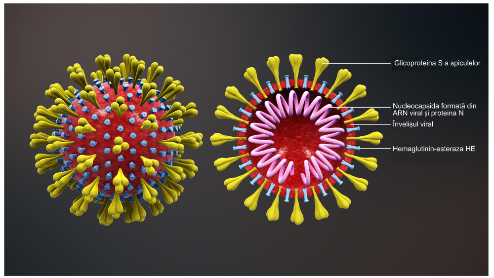
Figure 1. Coronavirus diagram. Photo credit: Danvasilis via Wikimedia Commons (Click to enlarge)
Coronaviruses include many pathogens of mammals and birds that individually cause a remarkable variety of diseases, including pneumonia, reproductive disease, enteritis, polyserositis, sialodacryoadenitis, hepatitis, encephalomyelitis, nephritis, and various other disorders. Coronavirus and coronavirus-like infections have been described in swine, cattle, horses, camels, cats, dogs, rodents, birds, bats, rabbits, ferrets, mink, and various wildlife species, although many coronavirus infections are subclinical. In humans, coronaviruses are included in the spectrum of viruses that cause the common cold and more severe respiratory disease — specifically Severe Acute Respiratory Syndrome (SARS), caused by SARS-CoV-1, and Middle East respiratory syndrome (MERS), which are both zoonoses.
Severe Acute Respiratory Syndrome (SARS-CoV) likely originated in exotic animals from a Guangdong marketplace 2, specifically Himalayan palm civets (Paguma larvata) and raccoon dogs (Nyctereutes procyonoides).3,4 The disease then emerged to infect people and was first reported in Asia in 2002. Symptoms included fever, chills, and body aches, which sometimes progressed to pneumonia and acute respiratory distress, with a mortality rate of about 10%. SARS shocked the world by its high virulence and efficient transmissibility among humans 5, causing the first large-scale pandemic of the 21st century. The illness spread to over two dozen countries in North America, South America, Europe, and Asia; however, no human SARS cases have been reported anywhere in the world since 2004.2
Middle East Respiratory Syndrome or MERS-CoV originated in camels and emerged to infect people. This syndrome was first reported in 2012 in Saudi Arabia and spread to over 25 additional countries. Symptoms usually include fever, cough, and shortness of breath, which often progress to pneumonia. Approximately 30-40% of patients reported with MERS have died. MERS cases continue to occur, primarily in the Arabian Peninsula; however, as of 2019, there have been only two confirmed cases of MERS in the US, both in 2014.6
In 2019, a novel coronavirus (COVID-19 or SARS-CoV-2) emerged in Wuhan, China. Since then, cases have been detected in most countries worldwide and on March 11, 2020, the World Health Organization characterized the outbreak as a pandemic.7 The COVID-19 outbreak appears to have originated from a Wuhan seafood market where wild animals, including marmots, birds, rabbits, bats and snakes, are traded illegally. The first people infected with the disease, a group primarily made up of seafood market stallholders, contracted infection through animal contact.8
It is improbable that SARS-CoV-2 emerged through laboratory manipulation of a related SARS-CoV-like coronavirus. If genetic manipulation had been performed, one of the several reverse-genetic systems available for coronaviruses would probably have been used. However, the genetic data irrefutably show that COVID-19 is not derived from any previously used virus backbone. Instead, two scenarios that can plausibly explain the origin of COVID-19: (1) natural selection in an animal host before zoonotic transfer, and (2) natural selection in humans following zoonotic transfer.9,10
The host range is defined as the range of species susceptible to a virus. One of the critical determinants of coronavirus host range is the interaction between the coronavirus spike protein and the host cell receptor. Angiotensin-converting enzyme 2 (ACE2) has been identified as a receptor for the attachment and uptake of SARS-CoV-1 and COVID-19 in host cells.11,12 The distribution of ACE2 in human tissues corresponds mainly to the cell types in which SARS-CoV replication has been observed. Besides type II pneumocytes and bronchial epithelial cells, ACE2 expression has been found in type I pneumocytes, endothelial cells, and smooth muscle cells of blood vessels, but not alveolar macrophages.13
Looking for additional information on coronaviruses and the structure of COVID-19? See “Determinants of Viral Infection: An Addendum to Coronaviruses in Animals” shared by Dr. Giacomo Rossi.
My colleagues (SO, GR, KJD) and I are deeply concerned by reports of animal shelters being overwhelmed with abandoned and surrendered ferrets (Mustela putorius furo). We believe this is due to misdirected fear of infection by their owners, and we are concerned that a similar situation may arise in cats with reporting of non-peer-reviewed preprints about cats and COVID-19*. People in the Middle Ages killed many cats because they feared they were associated with transmission of the Black Death (Yersinia pestis plague). So let’s examine what we know about ferrets and COVID-19.
The reason that ferrets were among the first animals to be considered as COVID-19 hosts is because ferrets are the animal model for studying influenza and developing annual flu vaccines. Many laboratories around the world have colonies of ferrets for experimental research. These virus laboratories have skills in infecting animal models (including mice, hamsters, and monkeys), investigating clinical and pathological changes in infected animals, and measuring transmission of virus after experimental infection (duration of transmission and quantity of virus transmitted). Two reviews in the journal Veterinary Pathology describe SARS and MERS infection in animal models, including ferrets.14,15
Ferrets are susceptible to SARS-CoV-1 infection16 and can also transmit the virus at low levels by direct contact.12,15,17 In different studies, ferrets have shown diverse clinical signs, but in general the pathology seen was milder than that seen in humans and without significant mortality.16,18 Ferrets can develop a fever in response to infection, which is characteristic of SARS-CoV-1-infected human patients.19 However, there are conflicting reports regarding the histopathological lesions and severity of clinical signs in the ferret model that require further investigation.
For MERS-CoV, the host cell receptor is dipeptidyl peptidase IV (DPPIV) 20 , a ubiquitously expressed cell surface protein that functions in immune homeostasis. Interestingly, whereas MERS-CoV can utilize bat DPP4 (bDPP4), camel DPP4 (cDPP4), and human DPP4 (hDPP4) molecules for entry, it cannot infect cells using the DPP4 molecules from traditional small mammal models, including mice, ferrets, guinea pigs, and hamsters. The inability of MERS-CoV to infect these species in vivo and in vitro is primarily due to spike-receptor incompatibilities and not due to other species-specific host cell factors. Therefore ferrets are not susceptible to experimental MERS-CoV infection because their homologous DPP4 molecules do not function as receptors for MERS-CoV entry.21 Even after administering a high dose of MERS-CoV, no viral replication can be detected.22
As pathogenicity and host susceptibility may differ for different coronaviruses, it is currently unknown whether ferrets are susceptible to COVID-19 pathology and transmission. Wan et al 2020 23 looked at the spike protein of the COVID-19 virus, and the ACE2 receptor that SARS uses as its binding site to invade cells. Their analysis “confidently predicts” that the COVID-19 virus also uses ACE2 as its receptor. The authors predict that a single genetic mutation at one location in the virus’ genome could significantly enhance its ability to bind, and that surveillance for this mutation should be performed. The study also predicts that the COVID-19 virus can bind to ACE2 in ferrets (and pigs, cats and some non-human primates) with similar efficiency as it does in people.
Conflicting reports, seen only as non-peer reviewed preprints, show ferrets can be infected with very large amounts of virus (multiple log 10 times than seen in natural human infections) and with laboratory attenuated strains of COVID-19 that may not occur in nature. Some laboratory-infected ferrets show a fever, mild histopathological lesions, and low virus transmission. It is important not to make generalizations about the susceptibility of pet ferrets to natural COVID-19 infection, as research on the potential use of ferrets as an animal model for COVID-19 is being performed around the world, and all the findings are not yet submitted for publication.
Looking for additional information on coronavirus in ferrets or the role of other species in the receptivity of COVID-19? See “Determinants of Viral Infection: An Addendum to Coronaviruses in Animals” shared by Dr. Giacomo Rossi.
Also read the article authored by Dr. Donnelly for the Association of Exotic Mammal Veterinarians, made available on April 9, 2020: “From SARS to MERS, Thrusting Coronavirus into the Spotlight“.
Also see “COVID-19 And Pet Birds” by Dr. Stephanie Lamb on our sister site, Lafeber Pet Birds.
*Footnote: The preprint article by Zhang et al “SARS-CoV-2 neutralizing serum antibodies in cats: a serological investigation” submitted on April 3, 2020, states “A cohort of serum samples were collected from cats in Wuhan, including 102 sampled after COVID-19 outbreak, and 39 prior to the outbreak. Fifteen of 102 (14.7%) cat sera collected after the outbreak were positive for the receptor-binding domain (RBD) of SARS-CoV-2 by indirect enzyme-linked immunosorbent assay (ELISA)” has significant problems. First, while 15% of cats were found positive in Wuhan, only 0.5% of the human population (55,000 people) were found positive (Wuhan has a population of 11 million). Second, the results are based on an indirect ELISA. But there is not yet a validated human serological assay available for SARS-CoV-2, let alone an assay for cats. The commercially available human serology tests are not yet specific or sensitive enough to be used by public health authorities in the West because they are giving either high percentages of false positives or false negatives. Until further validation is conducted and published for domesticated animals, the cat serology results are meaningless.
Public health concerns
COVID-19 is primarily transmitted by direct person-to-person contact. There is no evidence of pets, domestic species, or wildlife in the United States playing any role in disease transmission. Coronaviruses are constantly mutating – they accumulate point mutations because of polymerase errors (infidelity) during transcription (genetic drift), and genetic recombination occurs at high frequency between the genomes of different but related coronaviruses during co-infection situations. This is why caution must be used when interpreting experimental animal infection with COVID-19, as multiple laboratory strains of COVID-19 exist due to natural coronavirus mutation through multiple cell cultures and/or collection from fluids and tissues of experimentally infected animals. Out of an abundance of caution, the U.S. Centers for Disease Control and Prevention recommends individuals positive for COVID-19 limit contact with pets and other animals.24
“Specifically, while a person infected with COVID-19 is symptomatic, they should maintain separation from household animals as they would with other household members, and avoid direct contact with pets, including petting, snuggling, being kissed or licked, sleeping in the same location, and sharing food…If possible, another household member should be designated to care for pets in the home and should follow standard handwashing practices before and after interacting with the household animal. If a person with COVID-19 must care for pets, they should ensure they wash their hands before and after caring for pets.”—Centers for Disease Control, Interim Guidance for Public Health Professionals Managing People with COVID-19 in Home Care and Isolation Who Have Pets or Other Animals 24
Any investigation into the contacts and exposures of infected or quarantined people should also include animals.
What should you do?
Pet owners should consult their veterinarian if a ferret develops respiratory illness . Telemedicine may be appropriate to provide veterinary consultation if the owner is positive for COVID-19.24 The US Food and Drug Administration recently announced that it intends to “temporarily suspend enforcement of portions of the federal veterinarian-client-patient relationship requirements” in order to allow veterinarians to better utilize telemedicine to address animal health needs during the pandemic.25 In turn, veterinarians should contact their State Veterinarian and Public Health Director if they encounter a companion animal with “a new, concerning illness” that lives in the same home as a person with COVID-19.24 Proper hygiene must be maintained during all human-animal contact.
Also refer pet owners to “If You Have Animals” by the US Centers for Disease Control and Prevention.
Stay informed
COVID-19 is an emerging infectious disease and our understanding is rapidly evolving. Get the latest public health information from:
- American Veterinary Medical Association (AVMA)
- US Centers for Disease Control and Prevention (CDC)
- National Institutes of Health (NIH)
- World Health Organization (WHO)
- World Organisation for Animal Health (OIE)
- Worms & Germs Blog
Acknowledgments
Thank you to Drs. Yvonne van Zeeland and J.M.A. van den Brand for helpful input.
References
Determinants of Viral Infection: An Addendum to Coronaviruses in Animals
One of the critical determinants of viral host range is the interaction between the coronavirus spike (S) protein (Fig 1) and the host cell receptor. Angiotensin-converting enzyme 2 (ACE2), a receptor for the attachment and uptake of the coronavirus that caused Severe Acute Respiratory Syndrome (SARS-CoV-1), is widely expressed with conserved primary structures throughout the animal kingdom, from fish, amphibians, and reptiles, to birds and mammals. Structural analysis also suggests that ACE2 from these animals can potentially bind to the receptor binding domain (RBD) of COVID-19’s spike protein (Fig 2). By itself, the structural analysis suggests these animals would all be possible natural hosts for the virus.
Fortunately, in order for a coronavirus to bind to a host cell releasing their genomic material inside it and starting the infection of the host, two conditions must be met. First, there must be homology between the receptor site of the host cell’s receptor, ACE2, and the RBD on the spike protein of the viral envelope. Additionally, a specific endogenous protease must be present on the host cell that is capable of “activating” the fusion between the RBD and ACE2. This second mechanism, which highlights the need for specific proteases for viral activation, greatly reduces the spectrum of “permissive” hosts to a specific virus, even if there are strong receptor analogies. Based on which and how much protease is present in a tissue, the infection can vary in severity, and the virus may have a greater or lesser virulence.
Understanding this concept is essential, as it explains why, in different animal species, a virus can have different organ tropism, but above all it can induce milder or more serious clinical signs.

Figure 1. Coronaviruses possess crown-like spike (S) proteins on their surface. Click image to enlarge.

Figure 2. In order for a host cell to be receptive to COVID-19, there must be homology between the receptor binding domain (located on the spike of the viral envelope) and the host cell’s angiotensin-converting enzyme 2 receptor or the receptor binding site. Photo credit: ScientificAnimations.com/C&EN. Click image to enlarge.
The peculiar structure of the spike “S” in COVID-19
“The spike is the business part as far as viral entry is concerned…It is in charge not only of attachment at the host cell surface, but also of fusing the viral and host cell membranes to allow the infection to start…”– David Veesler 1
A specific endogenous protease cleaves the S protein of COVID-19 into two subunits: the S1 and S2 subunits (Fig 3). A small RBD on the S1 subunit (amino acids 318–510) can then combine with the host cell receptor, ACE2. The entire receptor-binding loop, known as the receptor-binding motif (RBM) (amino acids 424–494), is located on the RBD and is responsible for complete contact with ACE2.

Figure 3. A small receptor binding domain on the S1 subunit of the spike (S) protein binds with the host cell receptor, angiotensin-converting enzyme 2. The receptor binding domain displays a concave surface during this interaction with the receptor. Photo credit: Bio Connect.com.
Glycosylation can also affect the interaction of the RBD with ACE2. N-glycosylation represents the attachment of a polysaccharide or glycan to a nitrogen atom or the amide nitrogen of an asparagine (Asn) residue of a protein. This type of binding is important for both the structure and function of virus spike and host cell receptor binding site (RBS) proteins. Among the 23 glycosylation sites on spike glycoprotein, two are in the RBD. In contrast with other coronaviruses, in COVID-19 an adjunctive glycosylation has been detected on asparagine Asn330 2, but not in N357. In addition, these N-linked glycosylation sites are fundamental not only for the ability of coronavirus to infect a novel species but also as a site for the attack of neutralizing antibodies. This is demonstrated in the case of the highly specific S230 antibody for COVID-19 (Fig 4).3 This type of binding is important both for the structure and for the function of virus spike and host-cell RBS protein N-glycosylation is acquired in the endoplasmic reticulum and Golgi apparatus during the intracellular phases of virus replication (Fig 5).

Figure 4. Schematic of complex glycans in the interplay between virus and host. Photo credit: SemanticScholar.org. Click image to enlarge.

Figure 5. N-glycosylation is acquired in the endoplasmic reticulum and Golgi apparatus during the intracellular phases of virus replication. Photo credit: ScienceDirect.com. Click image to enlarge.
The presence and role of the host cell specific endogenous protease
Many human respiratory viruses rely on interactions with host cell proteases for their activation, and this activation is necessary for their successful replication within host cells. Type II transmembrane protease, serine S1 member 2 (TMPRSS2 or epitheliasin) competently activates coronaviruses, influenza A, and other viruses, including members of Paramyxoviridae (Fig 6) Epitheliasin or TMPRSS2 is present in human airway epithelium as well as other organs and in the corresponding structures of different animal species.4
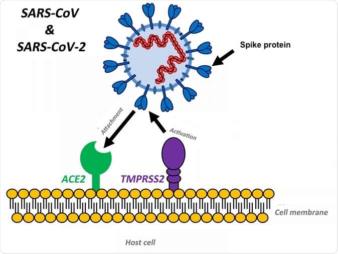
Figure 6. In addition to the host cell receptor angiotensin-converting enzyme 2 (ACE2), the host tissue must express a specific endogenous protease (TMPRSS2) that activates fusion of the virus receptor binding domain with the host cell membrane receptor binding site. Photo credit: SoundHealthandLastingWealth.com. Click image to enlarge.
Moreover, as polymorphisms in the TMPRSS2 gene were found to be associated with influenza severity in human patients (Cheng et al 2015), it appears that different influenza viruses might also take advantage of TMPRSS2 for spread in the human host. Non-human hosts, such as ferrets, swine, bats and probably others, could show a different receptivity to the COVID-19 virus, and different organ pathologies.
In order for a host to be receptive to COVID-19, there must be a homology between the spike’s RBD and the ACE2 receptor. In addition, the host tissue must express the specific serine protease TMPRSS2 to activate the fusion of the virus with the host cell.
Coronavirus infections are initiated by spike (S) protein activation by TMPRSS2 attachment to cell surface receptors by RBD, and fusion of the viral lipid envelope and endosomal membranes upon endocytosis to release the virus genome into the cytosol (Fig 7).
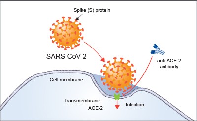
Figure 7. Angiotensin-converting enzyme 2 (ACE-2) is the host cell receptor responsible for mediating infection by SARS-CoV-2, the novel coronavirus responsible for coronavirus disease 2019 (COVID-19). Treatment with anti-ACE-2 antibodies disrupts the interaction between virus and receptor. Photo credit: RNDSystems.com
COVID-19 or SARS-CoV-2 uses the receptor ACE2 for entry and the serine protease TMPRSS2 for S protein priming. Protein priming is a generalized mechanism to initiate RNA synthesis of the virus within the host cell.
Some coronaviruses, like the SARS coronavirus of 2003 and the novel coronavirus COVID-19, are activated by TMPRSS2 and can thus be inhibited by TMPRSS2 inhibitors.5 A TMPRSS2 inhibitor approved for clinical use blocked entry and might constitute a treatment option (Fig 8).5

Figure 8. The novel coronavirus COVID-19 is activated by TMPRSS2 and can be inhibited by TMPRRSS2 inhibitors. Photo credit: ScienceDirect.com. Click image to enlarge.
What is the role of animals in the receptivity of COVID-19?
As described above, a high degree of homology between the viral spike protein and the host ACE2 receptor is not enough to allow the virus to enter the host cell. The serine protease TMPRSS2 must be simultaneously expressed on the cell to activate the fusion of viral cell membranes with the host cell wall.
FERRETS
To our knowledge, to date there are no published studies showing the expression and distribution of TMPRSS2 in ferret tissues. This data would allow us to identify the target organs in the ferret, as well as the real potential for pathogenicity of COVID-19 for this species.
Instead, we have data regarding the presence of TMPRSS2 in the tissues of the dog, cat, and pig, which indicates that the ACE2 receptors of these species are unable to bind COVID-19.
RODENTS
As with SARS, mice and rats are likely resistant to infection.
BIRDS
Birds are not believed to be susceptible to infection with COVID-19.
DOGS
Canine respiratory coronavirus (CRCoV) was first identified in 2003 in samples obtained from the respiratory tracts of dogs with canine infectious respiratory disease (CIRD), also known as kennel cough).6 Szczepanski et al demonstrated that infection sustained by CRCoV is characterized by a preliminary endocytic internalization, prior to membrane fusion, a process that requires caveolin-1 and dynamin but not TMPRSS2, which is missing in canine cells and tissues.6 Furthermore, in this species, fusion of viral and cellular membranes occurs before the endosome progresses to the late phase, indicating that the dog is not a permissive host for COVID-19.
CATS
Feline coronavirus (FCoV) (Alphacoronavirus 1 genus Alphacoronaivirus) 7, is divided into serotypes I and II based on the amino acid sequence of the spike (S) protein.8,9 Type I FCoV is the dominant serotype (80–90%) in Europe and Asia, and can also be divided into two biotypes, feline infectious peritonitis virus (FIP virus, FIPV; virulent FCoV) and feline enteric coronavirus (FECV; avirulent FCoV), according to its pathogenesis in animals.10
Feline coronavirus cell surface attachment and entry is not mediated by TMPRSS2, but through feline aminopeptidase N (fAPN), which acts as the RBS for viral infections. Feline coronaviuruses enter the cell through endocytosis after attachment to the feline aminopeptidase-N (fAPN).11 It is speculated that low endosomal pH triggers the conformation change of the spike protein of FCoV that releases the virus from the endosome to the cytosol. These studies are important in helping to exclude the domestic cat and others felids as permissive hosts to COVID-19 12, but this type of characterization regarding the receptor and its protease has never been performed in other wild felids. Therefore it cannot be excluded that the recent episodes of infection occurring in some tigers of the Wildlife Conservation Society’s Bronx Zoo, may be linked to a different receptor structure in these big felids, which were also sensitive to SARS and influenza viruses.13
PIGS
Porcine epidemic diarrhea virus (PEDV) and porcine deltacoronavirus (PDCoV) are important infectious disease concerns in pigs. These are alpha and delta coronaviruses, respectively.
Porcine TMPRSS2 is expressed through out the entire respiratory tract.14 In addition, TMPRSS2 protein expression has been identified in the digestive tract, some lymphoid organs, kidneys, and reproductive system. Using immunohistochemistry and reverse transcription PCR assays to evaluate TMPRSS2 mRNA expression, the author showed some interesting cell type localization in this species’ tissues.
Pigs are important natural hosts of influenza A viruses, and due to their susceptibility to swine, avian, and human viruses, they may serve as intermediate hosts supporting adaptation and genetic reassortment. Cleavage of the influenza virus surface (HA) by host cell proteases is essential for viral infectivity. Most influenza viruses, including human and swine viruses, are activated at a monobasic glycoprotein hemagglutinin (HA) cleavage site, in association with TMPRSS2 cleavage site. Peitsch et al showed that swine proteases homologous to TMPRSS2 and HAT, designated swTMPRSS2 and swAT, respectively, were expressed in several parts of the porcine respiratory tract, but also in different organs.15
Despite the expression of the TMPRSS2 protease in the swine respiratory tract, small changes or replacements of amino acids cause the spike protein of COVID-19 to not bind well to the cellular receptor of the pig. Therefore even though TMPRSS2 is well distributed in the pig, this species is not a good host for COVID-19 for this reason.7
Also view “Susceptibility of ferrets, cats, dogs, and different domestic animals to SARS-coronavirus-2”, a preliminary report that has not been peer-reviewed, which reports that SARS-CoV-2 replicates poorly in dogs, pigs, chickens, and ducks, but efficiently in ferrets and cats.*
References
Giacomo Rossi, DVM, PhD, MS, DECZM (WHP), EBVS
 Giacomo Rossi earned his doctorate from The University of Pisa (Italy) in 1996, and his Master of Sciences in Rome in 1997. Researcher in Avian Pathology and Pathology of exotic animals at University of Pisa starting to 1998, he was Associate Professor at University of Camerino (Italy) until 2008. Dr. Rossi is a Full Professor of General Pathology, Physiopathology and Immunopathology at the University of Camerino (Italy). He serves as the Director of the Animal Pathology Laboratory. From 2006-2013, he was Director of the Scientific Committee of AIVPA (Italian Association of Vets for Small Animals), and then AIVPA President from 2014-2017. Professor Rossi is a member of the Editorial Board of some international journals, and in the last twenty years he has participated in many international scientific meetings and conferences focusing on different aspects of animal pathology and immunopathology, with special emphasis on avian species. Giacomo is the author and editor of several books related to avian and exotic animal medicine and he has published over 300 scientific and veterinary papers. He is also a known national and international speaker on topics related to the pathology and medicine of pet birds, zoo animals and wildlife. The principal topics of his research are on avian ganglioneuritis (also known as proventricular dilatation disease), megabacteriosis, and flavivirus infections, like West Nile disease, Japanese encephalitis virus, and Usutu virus.
Thomas M. Donnelly, DVM, MS, DACLAM, DABVP (Exotic Companion Mammal Practice), DECZM
 Thomas Donnelly received degrees in veterinary medicine and pathology from the University of Sydney and then moved to New York to study comparative medicine and pathology. His work links the understanding of animal diseases and their complexities with the development of novel therapies for humans and animals. Tom also enjoys seeing exotic pets and is a Veterinary Information Network consultant for Mammals Small and Exotic. He was a member of the Avian & Exotic Pet Services at the Animal Medical Center and later at Tufts Veterinary School. He currently works at the French National Veterinary School in Alfort, Maisons-Alfort, France where he is Research Professor and Co-service head of the exotic animal service. Tom is the author of over 100 scientific and clinical publications, including chapters in “Ferrets, Rabbits and Rodents: Clinical Medicine & Surgery” and co-editor of “Veterinary Clinical Advisor: Birds and Exotic Pets.” Tom is allergic to many animals. Although his veterinary medicine career and allergies collide, he is fortunate in finding the challenges and diversity of working with veterinarians, exotic pets and their often equally unusual owners constantly gratifying.
In Memoriam: Dr. Pete Sakas
Everyone at Lafeber Company was greatly saddened to learn of the passing of Dr. Pete Sakas.
Dr. Peter Sakas joined Niles Animal Hospital in 1980 while still a veterinary medical student, working under Dr. T.J. Lafeber during his college breaks. Dr. Lafeber was so impressed with Dr. Sakas that he was hired immediately after he graduated in 1983. When Dr. Lafeber left clinical practice to build Lafeber Company in 1985, he sold his practice, Niles Animal Hospital, to Dr. Sakas.

Niles Animal Hospital and Bird Medical Center was established by Dr. T.J. Lafeber in 1949. Dr. Sakas became a staff veterinarian in 1983 and purchased the practice in 1985.
Dr. Sakas died on March 30, 2020 from COVID-19-related illness.
After an employee tested positive for COVID-19 (SARS-CoV-2), Niles Animal Hospital closed for a self-imposed quarantine period on March 26. When Dr. Sakas fell ill and was admitted to the hospital, doctors placed him on dialysis because of his past medical history of kidney loss due to cancer. Initially he appeared to improve, but then he suddenly developed respiratory difficulty. “By Saturday or Sunday, things went downhill pretty rapidly,” his brother told the Chicago Tribune. “All of a sudden, he was having trouble breathing and his kidney started developing blood clots”. Dr. Sakas was placed on a ventilator in a medically-induced coma before he passed away. He was 67 years old.
Dr. Sakas was deeply admired and he will be greatly missed, not only for his medical excellence, but also for his kindness and compassion to all animals, wild and domestic. His family has asked that donations go to the University of Illinois College of Veterinary Medicine’s Companion Animal Memorial Fund.
Do you have a favorite memory or story of Pete Sakas? The family has asked that you share your stories on his personal Facebook page or the Niles Animal Hospital Facebook page.
Todd Driggers, DVM
 Todd Eric Driggers is a 1994 graduate of Purdue University School of Veterinary Medicine. After graduation, he interned at the Avian and Exotic Animal Clinic in Indianapolis and he eventually moved to Arizona, starting the exotic practice at Mesa Veterinary Hospital. In 1996, he started Arizona’s first exclusive exotic animal practice, serving clients and small zoos. Currently, he has both a mobile practice and a stationary three-doctor practice, The Avian and Exotic Animal Clinic of Arizona. Dr. Driggers was a 2016 finalist for the T.J. Lafeber Avian Practitioner of the Year Award. He has presented at Annual Conferences of the Association of Avian Veterinarians and the Association of Exotic Mammal Veterinarians and he has written several chapters in various peer-reviewed journals and books. Dr. Driggers vacations in places to view and learn about the animals that he treats at his office, including Ecuador, Galapagos, Costa Rica, Dominican Republic, Madagascar, Australia, and of course Arizona. Dr. Driggers is also an avid herper. His pets include chameleons, Gila monsters, shingleback skinks, a ball python, leopard tortoise, radiated tortoise, and desert tortoise, as well as wallabies, horses, bunnies, a dog, and three indoor cats.
Avian Anesthesia post test
The Avian Anesthesia webinar course was reviewed and approved by the American Association of Veterinary State Boards (AAVSB) Registry of Approved Continuing Education (RACE) program for 1 hour of continuing education.
You have three chances to take this post-test and pass. With a passing grade of 75% or . . .
Pour les vétérinaires. Par les vétérinaires.
Le site Lafervet.com est conçu pour une utilisation par les vétérinaires. Il est ouvert aux vétérinaires diplômés, aux techniciens vétérinaires diplômés, aux animaliers et aux étudiants dans ces domaines.
Créer un compte pour accéder à des articles et des ressources du site.
L'inscription est gratuite.
Para Profesionales Veterinarios. Por Profesionales Veterinarios.
El sitio Lafervet.com es para uso de los profesionales veterinarios. Está abierto a los veterinarios licenciados, técnicos veterinarios licenciados, rehabilitadores licenciados y estudiantes en estos campos.
Cree una cuenta para acceder a los artículos y recursos del sitio.
La registro es gratis.
Already a LafeberVet Member?
Please Login
Avian Anesthesia Webinar
Dr. Lorenzo Crosta presented this live webinar on the clinical perspectives of avian anesthesia. View the RACE-approved webinar recording, then take a brief quiz to earn 1 hour of continuing education credit. After briefly reviewing clinically relevant avian anatomy and physiology, Dr. Crosta touches upon injectable anesthesia, then discusses in detail preanesthesia and inhalation anesthesia in clinical practice. The discussion then moves onto monitoring the avian patient, from vital parameters to capnography, doppler, electrocardiography, and pulse oximetry. Dr. Crosta also discusses analgesia, intra-operative fluid therapy, as well as specific concerns related to avian anesthesia, such as positioning the . . .
Pour les vétérinaires. Par les vétérinaires.
Le site Lafervet.com est conçu pour une utilisation par les vétérinaires. Il est ouvert aux vétérinaires diplômés, aux techniciens vétérinaires diplômés, aux animaliers et aux étudiants dans ces domaines.
Créer un compte pour accéder à des articles et des ressources du site.
L'inscription est gratuite.
Para Profesionales Veterinarios. Por Profesionales Veterinarios.
El sitio Lafervet.com es para uso de los profesionales veterinarios. Está abierto a los veterinarios licenciados, técnicos veterinarios licenciados, rehabilitadores licenciados y estudiantes en estos campos.
Cree una cuenta para acceder a los artículos y recursos del sitio.
La registro es gratis.
Already a LafeberVet Member?
Please Login
Lorenzo Crosta med vet, PhD, GP Cert (Exotic Animal), DECZM, EBVS
 Lorenzo Crosta med vet, PhD, GP Cert (Exotic Animal), DECZM, EBVS® European Veterinary Specialist in Zoo Health Management. In 2019, he was named the T.J. Lafeber Avian Practitioner of the Year. Dr. Crosta earned his Doctorate of Veterinary Medicine from the University of Milan in 1989. From 1990-1999, he worked in private practice in Milan, before serving as the Veterinary Director of Loro Parque in Tenerife, Spain from 2000 to 2005. Dr. Crosta also served as the Consulting Veterinarian to the Brazilian Government for the Spix’s and Lear’s Macaws Recovery Program from 2000-2014. After Loro Parque, Lorenzo returned to his homeland where he earned a Ph.D in Veterinary Clinical Sciences from the University of Turin in 2009. From 2018 to 2021, he was an Associate Professor of Avian and Zoological Medicine at the University of Sydney School of Veterinary Science, Australia. He served as the Director of the Avian, Reptile, and Exotic Pet Hospital. Lorenzo has paired his love of teaching and his enjoyment of public speaking with his ability to speak four languages. He has lectured on avian medicine in many countries, including the United States, Brazil, Australia, Hungary, Portugal, Israel, and Arab nations.
Ready for the Holidays?

Unfortunately, emergency medicine and critical care don’t stop for the holiday season, so we are just sending a little reminder to make sure that your cupboard contains enough EmerAid for any crisis over the holidays.

Lafeber Company will be CLOSED from Tuesday, December 24 through Wednesday, January 1, 2020.
Unlike past years, a skeleton crew will be processing all orders placed through LafeberVet’s* Online Store (USA) during the holiday break.*
*Only online orders placed from existing accounts with shopping cart access will be processed. Orders by new registrants will not be processed over the holiday break.
The Lafeber Company office will not answer phones during the holiday break or process orders left by voicemail or email.
Wishing you a safe and joyous holiday season!
Critical Care Techniques for Avian Wildlife Emergencies Post Test
. . .
Pour les vétérinaires. Par les vétérinaires.
Le site Lafervet.com est conçu pour une utilisation par les vétérinaires. Il est ouvert aux vétérinaires diplômés, aux techniciens vétérinaires diplômés, aux animaliers et aux étudiants dans ces domaines.
Créer un compte pour accéder à des articles et des ressources du site.
L'inscription est gratuite.
Para Profesionales Veterinarios. Por Profesionales Veterinarios.
El sitio Lafervet.com es para uso de los profesionales veterinarios. Está abierto a los veterinarios licenciados, técnicos veterinarios licenciados, rehabilitadores licenciados y estudiantes en estos campos.
Cree una cuenta para acceder a los artículos y recursos del sitio.
La registro es gratis.
Already a LafeberVet Member?
Please Login
Critical Care Techniques for Avian Wildlife Emergencies
Dr. Heather Barron presented this webinar on avian critical care. View a recording of the live, interactive event, then take the brief post-test to earn 1 hour of continuing education credit. The goal of wildlife medicine is always eventual release and therefore triage of avian wildlife may vary based on case load, regulations, and the presenting situation. Dr. Barron examines the guidelines used to set triage policy and the reasons a bird may not be releasable or have a good quality of life in captivity. She then discusses practical measures intended to alleviate suffering and improve the odds of . . .
Pour les vétérinaires. Par les vétérinaires.
Le site Lafervet.com est conçu pour une utilisation par les vétérinaires. Il est ouvert aux vétérinaires diplômés, aux techniciens vétérinaires diplômés, aux animaliers et aux étudiants dans ces domaines.
Créer un compte pour accéder à des articles et des ressources du site.
L'inscription est gratuite.
Para Profesionales Veterinarios. Por Profesionales Veterinarios.
El sitio Lafervet.com es para uso de los profesionales veterinarios. Está abierto a los veterinarios licenciados, técnicos veterinarios licenciados, rehabilitadores licenciados y estudiantes en estos campos.
Cree una cuenta para acceder a los artículos y recursos del sitio.
La registro es gratis.
Already a LafeberVet Member?
Please Login
Clinical Avian Nutrition for Veterinary Health Professionals Post-Test
. . .
Pour les vétérinaires. Par les vétérinaires.
Le site Lafervet.com est conçu pour une utilisation par les vétérinaires. Il est ouvert aux vétérinaires diplômés, aux techniciens vétérinaires diplômés, aux animaliers et aux étudiants dans ces domaines.
Créer un compte pour accéder à des articles et des ressources du site.
L'inscription est gratuite.
Para Profesionales Veterinarios. Por Profesionales Veterinarios.
El sitio Lafervet.com es para uso de los profesionales veterinarios. Está abierto a los veterinarios licenciados, técnicos veterinarios licenciados, rehabilitadores licenciados y estudiantes en estos campos.
Cree una cuenta para acceder a los artículos y recursos del sitio.
La registro es gratis.
Already a LafeberVet Member?
Please Login
Post Test: Avian Nutrition Basics
View the on-demand recording of this non-interactive webinar, then take the brief quiz. With a passing grade of 70% or higher, you will receive a continuing education certificate for 1 hour of continuing education credit in jurisdictions that recognize American Association of Veterinary State Boards Registry of Approved Continuing Education approval . . .
Pour les vétérinaires. Par les vétérinaires.
Le site Lafervet.com est conçu pour une utilisation par les vétérinaires. Il est ouvert aux vétérinaires diplômés, aux techniciens vétérinaires diplômés, aux animaliers et aux étudiants dans ces domaines.
Créer un compte pour accéder à des articles et des ressources du site.
L'inscription est gratuite.
Para Profesionales Veterinarios. Por Profesionales Veterinarios.
El sitio Lafervet.com es para uso de los profesionales veterinarios. Está abierto a los veterinarios licenciados, técnicos veterinarios licenciados, rehabilitadores licenciados y estudiantes en estos campos.
Cree una cuenta para acceder a los artículos y recursos del sitio.
La registro es gratis.
Already a LafeberVet Member?
Please Login
Clinical Avian Nutrition for Veterinary Health Professionals
Dr. Susan Orosz presented this live, interactive webinar event on the clinical perspectives of avian nutrition. How can veterinary health professionals best address the nutritional needs of the companion bird in the exam room? View the RACE-approved, non-interactive webinar recording, the pass the brief post-test to earn 1 hour of continuing education credit . . .
Pour les vétérinaires. Par les vétérinaires.
Le site Lafervet.com est conçu pour une utilisation par les vétérinaires. Il est ouvert aux vétérinaires diplômés, aux techniciens vétérinaires diplômés, aux animaliers et aux étudiants dans ces domaines.
Créer un compte pour accéder à des articles et des ressources du site.
L'inscription est gratuite.
Para Profesionales Veterinarios. Por Profesionales Veterinarios.
El sitio Lafervet.com es para uso de los profesionales veterinarios. Está abierto a los veterinarios licenciados, técnicos veterinarios licenciados, rehabilitadores licenciados y estudiantes en estos campos.
Cree una cuenta para acceder a los artículos y recursos del sitio.
La registro es gratis.
Already a LafeberVet Member?
Please Login
Avian Nutrition Basics
This 1-hour, R.A.C.E.-approved webinar recording is designed to impart a basic understanding of avian nutrition for the veterinary health professional as well as students in these fields. Viewing of this recorded is strongly recommended before viewing the recording of the live webinar event Clinical Avian Nutrition for Veterinary Health Professionals by Susan Orosz, PhD, DVM, DABVP (Avian Practice), DECZM . . .
Pour les vétérinaires. Par les vétérinaires.
Le site Lafervet.com est conçu pour une utilisation par les vétérinaires. Il est ouvert aux vétérinaires diplômés, aux techniciens vétérinaires diplômés, aux animaliers et aux étudiants dans ces domaines.
Créer un compte pour accéder à des articles et des ressources du site.
L'inscription est gratuite.
Para Profesionales Veterinarios. Por Profesionales Veterinarios.
El sitio Lafervet.com es para uso de los profesionales veterinarios. Está abierto a los veterinarios licenciados, técnicos veterinarios licenciados, rehabilitadores licenciados y estudiantes en estos campos.
Cree una cuenta para acceder a los artículos y recursos del sitio.
La registro es gratis.
Already a LafeberVet Member?
Please Login
2019 Avian Practitioner of the Year
Dr. Lorenzo Crosta named Avian Practitioner of the Year

Eighteen exceptional avian veterinarians from all around the world were nominated for the 2019 T.J. Lafeber Avian Practitioner of the Year. The independent Selection Committee narrowed this list to five finalists and the Award recipient, Dr. Lorenzo Crosta, was announced during the Plenary Session at the 2019 ExoticsCon.

Lorenzo Crosta, med vet, PhD, GP Cert (Exotic Animal), DECZM, EBVS European Veterinary Specialist in Zoo Health Management is an Associate Professor of Avian and Zoological Medicine at the University of Sydney School of Veterinary Science, Australia.

Download and share the press release.

Award recipients
Visit Lafeber.com to learn more about Dr. Crosta and for a list of previous Award recipients.

Did you know…?
The T.J. Lafeber Avian Practitioner of the Year is nominated by their peers: YOU. The Awardee is NOT, and has never been, selected by Lafeber Company.
This autonomous committee, consisting of Association of Avian Veterinarians members, is led by Dr. Kenneth Welle, Professor and Chief of the Zoological Medicine Service at the University of Illinois School of Veterinary Medicine. A Lafeber Company Veterinary Consultant, who does NOT vote on the Award recipient, manages committee paperwork and scheduling only.
Jessica Elbert, DVM

Jessica Elbert received her Doctorate of Veterinary Medicine from Iowa State University in 2018. She is currently an Anatomic Pathology Resident and PhD student at the University of Georgia College of Veterinary Medicine with special interest in zoo and exotic animal species and paramyxoviruses.
Jessica Comolli , DVM
Jessica Comolli received her Doctorate of Veterinary Medicine from the University of Georgia College of Veterinary Medicine. She completed a zoological medicine internship at Iowa State University and is currently a zoological medicine resident at the University of Georgia, Zoo Atlanta, and the Georgia Aquarium.
A Guide to Avian Necropsy
The postmortem examination is a valuable part of the diagnostic work-up. Shared by a veterinary pathologist with a special interest in birds, this guide to avian necropsy provides comprehensive instructions for the avian postmortem exam. This article offers step-by-step guidance on avian necropsy with a variety of photographs and video clips that illustrate useful clinical techniques and normal avian anatomy. Feel confident in your knowledge of avian anatomy? You can also “Test Yourself” by identifying the structures shown in four separate images . . .
Pour les vétérinaires. Par les vétérinaires.
Le site Lafervet.com est conçu pour une utilisation par les vétérinaires. Il est ouvert aux vétérinaires diplômés, aux techniciens vétérinaires diplômés, aux animaliers et aux étudiants dans ces domaines.
Créer un compte pour accéder à des articles et des ressources du site.
L'inscription est gratuite.
Para Profesionales Veterinarios. Por Profesionales Veterinarios.
El sitio Lafervet.com es para uso de los profesionales veterinarios. Está abierto a los veterinarios licenciados, técnicos veterinarios licenciados, rehabilitadores licenciados y estudiantes en estos campos.
Cree una cuenta para acceder a los artículos y recursos del sitio.
La registro es gratis.
Already a LafeberVet Member?
Please Login
Nobuko Wakamatsu-Utsuki, DVM, PhD, DACVP, DACPV
 Nobuko Wakamatsu-Utsuki earned her Bachelor of Veterinary Science from Kitasato University, School of Veterinary Medicine in Towada, Japan and her PhD from the University of Georgia. She is currently a Clinical Associate Professor in the Department of Comparative Pathobiology and Indiana Animal Disease Diagnostic Laboratory at Purdue University College of Veterinary Medicine. Dr. Wakamatsu-Utsuki previously served as an Associate Professor in the Department of Pathobiological Sciences and the Louisiana Animal Disease Diagnostic Laboratory at Louisiana State University School of Veterinary Medicine. Dr. Wakamatsu-Utsuki has a special interest in avian pathology and poultry medicine. Her research interests focus on avian diseases, particularly the pathogenesis of infectious diseases and she has published extensively on these topics.
Radial Vein Blood Collection in the Miniature Pig
Blood collection in miniature pigs can be a challenge. Peripheral veins are not readily accessible and some vessels, such as the auricular vein, are inadequate for obtaining sufficient volumes. The radial vein is located along the medial aspect of the forelimb. This vessel is relatively straight and generally superficial . . .
Pour les vétérinaires. Par les vétérinaires.
Le site Lafervet.com est conçu pour une utilisation par les vétérinaires. Il est ouvert aux vétérinaires diplômés, aux techniciens vétérinaires diplômés, aux animaliers et aux étudiants dans ces domaines.
Créer un compte pour accéder à des articles et des ressources du site.
L'inscription est gratuite.
Para Profesionales Veterinarios. Por Profesionales Veterinarios.
El sitio Lafervet.com es para uso de los profesionales veterinarios. Está abierto a los veterinarios licenciados, técnicos veterinarios licenciados, rehabilitadores licenciados y estudiantes en estos campos.
Cree una cuenta para acceder a los artículos y recursos del sitio.
La registro es gratis.
Already a LafeberVet Member?
Please Login
Valarie Tynes, DVM, DACVB, DACAW

Valarie Tynes owns Premier Veterinary Behavior Consulting in Sweetwater, Texas. Dr. Tynes is a native of Fort Worth, Texas and she earned her Doctorate of Veterinary Medicine in 1987 from Texas A&M University College of Veterinary Medicine. After several years in private practice, Valarie completed a residency in clinical animal behavior at the University of California at Davis in 2003. Her special interests include miniature pigs and other exotic pets as well as animal behavior. Dr. Tynes is a Diplomate of the American College of Veterinary Behaviorists and a frequent speaker at professional meetings around the world. She has authored numerous articles and chapters on a variety of behavior-related topics and she is the editor of Behavior of Exotic Pets. Dr. Tynes is also the author of Miniature Pig Pet Care, part of A Quick Reference Guide to Unique Pet Species, on the Veterinary Information Network.
Basic Information Sheet: Miniature Pig
Miniature Pig (Sus scrofa domesticus )
Natural history
Pigs were domesticated approximately 9,000 years ago from wild boars (Sus scrofa). In the 1960s, farmers developed a smaller dwarf pig from wild Vietnamese breeds that was more easily housed. Vietnamese potbellied pigs (also known as Asian or Chinese potbellied pigs or just potbellies) were first introduced to the United States in 1985. Since that time, the popularity of miniature pigs has waxed and waned.
The term “miniature” is used to describe a variety of smaller pig breeds as well as crossbreeds. There are at least 14 recognized breeds of miniature pigs, including the Vietnamese potbellied pig, the Juliana pig, the KuneKune, and others; many pets are mixed breed, of unknown origin. Terms such as “teacup”, “micro mini” and “nano pig” are marketing ploys rather than true breeds.
Taxonomy
Class: Mammalia
Order: Artiodactyla
Sub Order or clade: Suina
Family: Suidae
Physical description
Miniature pigs are stout, barrel-shaped animals with relatively large heads and short limbs.
Purebred Vietnamese potbellied pigs are usually all black with upright ears and a straight tail and grow to be 36-50 cm (14-20 in) tall at the shoulders. The adult size of other miniature pigs is much more variable.
Note: Adult size is not reached until 3-4 years of age
Sexual dimorphism
Both males and females have tusks (upper and lower canine teeth), however, the tusk root of the female closes at maturity and ceases to grow. In contrast, the male tusk retains an open root allowing continued growth, which is more rapid in intact boars than castrated males.
In addition, intact boars exhibit a few distinctly male characteristics, including: excessive facial hair, excessive hair on the distal limbs and tail, densely fibrous shoulder plates, a strong, offensive odor, and specific behavioral displays (see Behavior below) .
Diet
Wild pigs are opportunistic omnivores, eating roots, berries, and whatever else they can expose with their snout and hooves.
The pet pig diet should consist of a specially formulated miniature pig feed, such as Ralston Purina’s Mazuri®, Ross Mill Farm’s Champion™ Pig Food, Heartland, or Manna Pro. Mini pigs can also be offered an all-purpose livestock or equine feed. Commercial swine feed is not recommended for miniature pet pigs.
Feeds are often classified as a starter ration (for piglets in the process of being weaned), grower (fed until puberty), breeder, or maintenance. Maintenance rations are started at about 8 weeks of age.
Pigs are extremely food motivated and always appear hungry. Never feed a pig ad libitum; rather, carefully ration food, dividing the daily volume into at least two small meals.
| Young piglet | ½ cup per 15-20 lb/day | ½ cup per 7-9 kg/day |
| Adult | 1 cup per 50-80 lb/day | 1 cup per 23-36 kg/day |
Also offer fiber, in the form of grass hay or allow the pig to graze, to help control hunger.
Treats are unnecessary but, if offered, vegetables are the best option. The pig should be asked to perform a task (even as simple as coming when called) before feeding a treat. Pigs notice WHEN they receive treats, not HOW MUCH they receive – an important tip for owners dieting their overfed pets.
Secure food and water bowls to the ground so the pig cannot climb in the dishes or tip them over. Provide a bowl with a weighted bottom and/or wide base; alternatively, secure the bowl within the center of a tire (select image below to enlarge). Be sure bowls are low enough for the pig to easily access. Place bowls on an easily cleaned surface, like a shower stall or a large, shallow tray. Fresh water should be available at all times (see below).

Husbandry
| Indoor
|
Strict indoor life (e.g. apartment dwelling) is never a good option for pet pigs. Pigs restricted to the indoors will root on objects like carpet and walls. In addition, hoof overgrowth tends to be more extreme in indoor-only pigs (select image left to enlarge) and boredom is an issue as well. Confine unsupervised, indoor pigs to a “pig-safe room” and provide a designated rooting area and/or rooting box (see enrichment below) to minimize destruction. |
| Outdoor pen | All pet pigs should spend several hours each day outdoors exploring and rooting the ground. The pen should be at least 2.5 x 4.5 m (8 x 15 feet) per pig, although more space promotes more exploration and exercise. Shade should be provided to prevent sunburn.
Fencing must be strong and secure. Suggested materials include wood, chain link, hog panels, and T posts. At least 30 cm (1 foot) of mesh fence should extend beneath the ground to prevent digging beneath the fence. Note: Fencing must not only keep pigs in but keep predators such as dogs out. Rough surfaces, like a concrete patio or pavers in the feeding area or at the entrance to a shelter, will help to keep the hooves worn (see hoof care below). |
| Pig house | The pen should include a clean, dry, draft-free, insulated “pig house” for sleeping and nesting, measuring approximately 2 square meters (6 sq ft). Flooring should provide good traction (e.g. rubber stall mat or outdoor carpet). During cold weather, a commercial pig heating blanket or a heat lamp safely placed out of reach may be used with caution.
Provide proper nesting material such as straw/hay, blankets (fleece recommended as these are more resistant to ripping), sleeping bags, and/or aspen shavings (avoid cedar). Bedding must be deep enough to allow rooting or for the animal to completely cover itself. Replace wet or dirty bedding daily. Replace ripped blankets to prevent accidental foreign body ingestion. |
| Environmental temperature | Adult potbellied pigs are most comfortable at environmental temperatures between 15–24°C (60–75°F).
Pigs are incapable of sweating (see Integument below) and are susceptible to overheating at temperatures exceeding 30°C (85°F). Pigs will begin to pant as they get warm, but this is often insufficient for cooling. Pigs require fans and water for soaking when it is hot; mud puddles are a great option as these allow for both cooling and sun protection via a coating of mud. Shade is also important because pigs have sensitive skin that can sunburn. |
| Relative humidity | 50% |
| Drinking water | Many pigs prefer to move back and forth between their water and food bowls at feeding time. Some pigs have difficulty swallowing dry pellets and may require water added directly to feed to prevent choking.
Water intake sometimes decreases during the winter or in overweight pigs. Restricted access to water can lead to health problems such as cystitis and urolithiasis. Offering warm or room temperature water may encourage drinking, especially during colder weather. Adding a few tablespoons of juice (cranberry, orange) may also encourage drinking. |
| Water for soaking
|
Water for soaking should be provided in a separate container from drinking water. A child’s wading pool is suitable for this purpose, but the sides may need to be cut down to accommodate the pig’s short legs (select images left to enlarge). It may also be helpful to provide flooring with traction in the pool (e.g. outdoor carpet, rubber mat, bathtub appliqués). There are also heavy duty, rectangular, non-slip, vinyl pools with low, soft, padded sides made specifically for miniature pigs. |
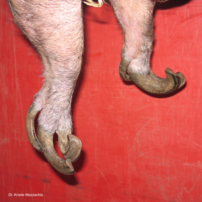


Activity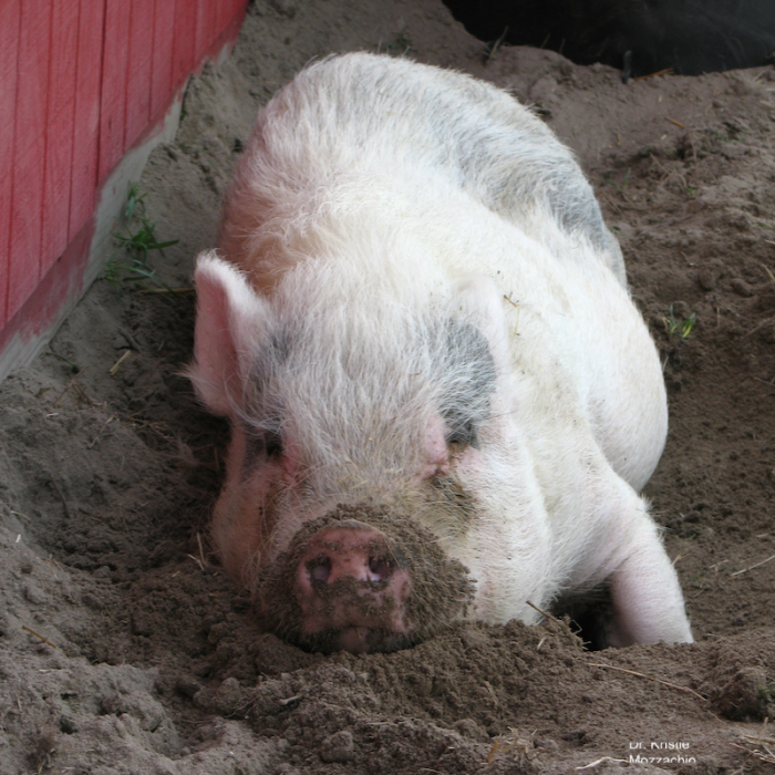 |
Rooting is common and is normal pig behavior (select image left to enlarge). Pigs are also good runners and jumpers and skilled swimmers, although they generally prefer less-athletic activities once adulthood is reached.
Pigs are also normally quite vocal, with a large vocabulary of sounds. |
| Social | Pigs are highly social and intelligent animals that do best in a herd of conspecifics (at least 2 pigs). Aggression can stem from territorial disputes over sleeping quarters or food or may simply be an effort to gain status in the herd. In single-pig households, the human family is the pig’s “herd” and aggressive behaviors are common if not recognized early and appropriately modified. These behaviors tend to emerge around 2 years of age when a pig develops its social standing within the herd. |
| Enrichment
|
Free-ranging pigs forage for food by grazing and rooting for approximately 7 hours each day (V. Tynes, email message to author, June 12, 2019). Offer pet pigs the opportunity to graze on grass or grass hay, which provides an excellent way to hide food, slow feeding, and promote foraging. For pigs that spend some time indoors, a rooting box is also recommended. Feeder puzzles can also provide a challenge to pet pigs (select image left to enlarge). Pigs don’t “play” with toys the way a dog or cat might; enrichment revolves around exploration of new and interesting items or environments and, of course, food. |
| Reproductive | Sexual behavior begins very early in pigs—before weaning—and can include: unpredictable behavior, aggression, rooting, as well as mounting and destructive behaviors. Intact pigs can be difficult to house train.
Females can display cyclic moodiness, mounting, and urinary marking every 21 days or so. Intact males display behaviors, such as kneading the ground (similar to a cat), mounting, and intense foaming at the mouth (unrelated to anticipation of food), as well as frequent vocalizations or “huffing” noises. |

Normal physiologic data
| Life expectancy | 14-21 years (average 15-18) |
| Body weight | Miniature pigs typically weigh between 32-68 kg (70-150 lb). Mini pigs reach half their adult weight by about 1 year of age and will continue to grow until 3-4 years of age when growth plates completely fuse. |
| Body temperature | 37.6-39°C (99.7-102.2°F) |
| Heart rate | 70-80 bpm, often higher during restraint Up to 200 bpm in piglets |
| Respiratory rate | 12-18/minute Up to 40/minute in piglets |
| Reproductive data | See Anatomy/physiology below |
| Weaning age | 6 weeks or more |
Anatomy/ physiology
| Dentition | I3/3 C1/1 PM 4/4 M 3/3 = 44
Piglets are born with eight teeth called “needle teeth”. In commercial pigs, these teeth are sometimes clipped to prevent damage to littermates or to the sow’s teats. This practice is not typically necessary in miniature pigs. Deciduous teeth erupt until about 3 months of age; permanent teeth are present by about 20 months. Tooth loss of premolars and molars is common in older pigs. |
| Gastrointestinal |
|
Integument |
Although pigs possess some sweat glands, they are not functional and do not respond to thermoregulatory cues. Therefore, pigs are susceptible to overheating at temperatures exceeding 30°C (85°F). Overheated pigs begin to pant as they warm, but this is often insufficient for cooling, and this is one reason pigs wallow in mud.
Carpal glands are scent marking glands found on the medial aspect of the forelegs (select image left to enlarge). White to brown, pasty debris can sometimes be expressed from the glands and inspissation of secretion is common. |
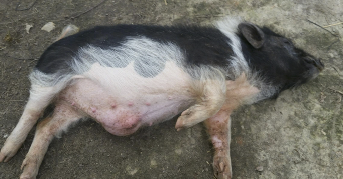 |
The preputial diverticulum fills with foul-smelling smegma in intact males. This sac can become quite large and prominent and should not be confused with an umbilical hernia (select image left to enlarge). |
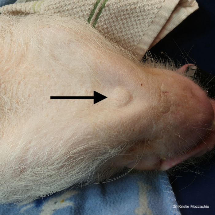 |
The mental gland found on the ventral aspect of the chin has both scent-marking and tactile functions (select image left to enlarge). |
| Musculoskeletal | Adaptations of the skull for rooting behavior include:
Pigs have one pair of principle digits (the two main toes that they walk on) and one pair of accessory digits (declaws) per leg. |
Reproduction |
Penis: Curved with a sigmoid shape and a corkscrew tip (select image left to enlarge), which correlates with the interdigitating structure of the long cervix.
Puberty: As early as 2-4 months of age. Pigs exhibit sexual behavior before they are weaned and are fertile by 3 months of age. Estrous cycle: 17-25 day intervals (average 21) Estrus: 1-3 days
Gestation: 112-116 days
Litter size: average 6-8, range 2-15
Birthweight: 340-794 g (0.75-1.75 lb)
|
| Respiratory | See Therapeutics below for a description of laryngeal anatomy. |
| Special Senses
|
Pigs possess a good sense of hearing. The ear canal is extremely narrow, filled with debris, and otoscopic examination is generally not feasible. Miniature pigs often produce large amounts of dark brown, flaky to waxy, aural secretions (see eyes-ears-nose below). Although this resembles the secretions produced in dogs or cats with ear mite infestation, this secretion is normal for a miniature pig.
Pigs have generally poor vision. Pigs possess a keen sense of smell. The nostrils are located at the end of the snout and are capable of closing when the pig is rooting. |
Restraint
| Manual
|
Restraint is likely the most difficult aspect of dealing with miniature pig patients. Pigs are a prey species: sows do not pick up or restrain their babies, but predators do…
Pigs are susceptible to exertional myopathy, and although uncommon in pet pigs, this condition is extremely painful and difficult to treat. Minimize restraint whenever possible, so there is less squealing and less stress for owners and veterinary health staff. Oral trazodone (10 mg/kg PO) and gabapentin (20 mg/kg PO) may be helpful when administered together the night before and again 2 hours prior to the veterinary visit. It also helps if the pig is harness trained (pig-specific harnesses are commercially available, e.g. Comfort Fit Safety Harness) and/or crate-trained. Crates can be partially dismantled, so the pig can be examined (or sedated) while confined in the bottom half of the pet carrier. Always expect squealing when restraining a pig. Hearing protection is recommended. It is helpful to learn to differentiate between different pig vocalizations (anger/annoyance versus fear versus pain). It is much safer to perform exams on the floor instead of a table as many pigs will try to leap off. Solid, non-slip flooring is also a must when working with pigs, as slippery surfaces guarantee a panicked patient even before the procedure begins. Rubber-backed bath mats work well for this purpose. DO NOT use a hog snare OR pick up mini pigs by the legs as shoulder, lower back, or leg injuries can occur. Also, subclinical degenerative joint disease may be present and can become clinical after inappropriate restraint. Restrain a small to medium-sized pig by placing one hand beneath the rump and the other hand in front of the forelimbs and under the neck (rather than under the chest) (select top image left to enlarge). Larger pigs can be handled in narrow chutes or cornered using crowd boards or sorting panels (select middle image left to enlarge), which are commercially available through various suppliers, including Ross Mill Farm. |
| Chemical | Deep sedation or light general anesthesia is often required for most diagnostic or therapeutic procedures.
See Therapeutics below. |


Preventive medicine
| Physical examination: | Recommend 6-12 hours of fasting prior to physical exam in case sedation is necessary. Also instruct owners to bring their pig’s daily food ration or favorite treats.
Visual examination should include observation of gait, conformation, body condition score, skin health, hoof condition, respiratory rate and character. |
| Vaccination | Vaccination protocols vary, and there is no widely accepted standard for miniature pigs. Recommendations are based on diseases common in the area, potential exposure, age, breeding status, zoonotic potential. Minimum recommendations for miniature pigs may include:
Begin vaccinations at 8-12 weeks of age; administer two doses 14 days apart, then annually. |
| Fecal parasite testing | Fecal flotation should be performed during yearly routine physical exam to monitor for gastrointestinal parasites. In larger “open” herds such as a pig sanctuary, fecal monitoring should be performed more frequently, a minimum of twice yearly. |
| Dental
(tusk trim)
|
Routine dental care should be part of suid preventive health care, and routine cleaning should ideally be performed as in a dog or cat (select top image left to enlarge).
Tusks can become long and extremely sharp, and tusk trims may be necessary to prevent injury to humans and other animals or damage to household furniture or flooring. Tusks can also become caught on caging or they may even grow long enough to penetrate the flesh of the face (select middle image left to enlarge). Tusks should be trimmed well above the gum line using obstetric saw (Gigli) wire and edges can be smoothed with a file or Dremel tool. Intact males may require a tusk trim every 6-12 months and neutered males every 1-5 years. Females typically do not require tusk trims. Dental abscesses are a potential complication (select bottom image left to enlarge), therefore tusk trims should be kept to a minimum and performed only when needed. |
| Hoof care
|
A normal healthy pig will require a hoof trim about once a year, typically beginning at 1-2 years of age. The frequency of trimmings may increase with indoor-only environment, hoof deformities, cracks, or the development of arthritis.
The frequency of hoof trims depends on genetics, age, diet, activity level, and the environmental substrate provided. Rough surfaces, like concrete or patio pavers in the feeding area or at the entrance to a shelter, will help to wear the hooves. Some owners are able to train their pigs to allow hoof trim, and this is strongly encouraged. Small nippers are recommended for owners and are available from online suppliers such as RossMillFarm.com |
| Surgical sterilization
|
Spaying and neutering is recommended to improve pet quality, reduce odor in males, and lessen some destructive or undesirable behaviors. Surgical sterilization also prevents uterine infections and tumors in female pigs as well as testicular tumors in male pigs.
Castration
Ovariohysterectomy
|


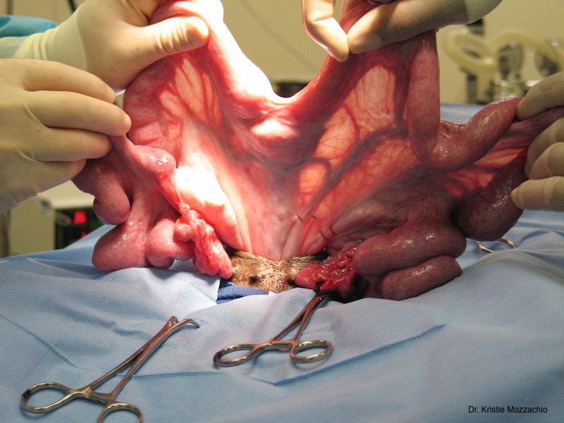
Important medical conditions
| Obesity | Pigs easily gain weight, and obesity is a common problem in pet pigs. The condition is often associated with free choice feeding (or overfeeding of inappropriate items such as dog food or table scraps) and lack of exercise.
Entropion and arthritis are common problems in obese pigs, and obesity contributes to cardiopulmonary disease as well. Vision and hearing are often impaired due to physical interference of fat deposits over the eyes and around the ears. Obesity can also be correlated with very low water consumption, which can lead to cystitis and urolithiasis. The risk of obesity in pet pigs can be minimized with:
|
| Musculoskeletal
|
Erysipelas can cause lameness due to polyarthritis.
Osteoarthritis is extremely common in elbow joints, carpus, tarsus, phalanges, and spine, particularly in obese pigs. Clinical signs can include difficulty rising, reduced activity, and lameness. Some animals walk on their “knees” (carpus), while others sink onto their haunches, displaying a hunched posture that can resemble constipation (select image left to enlarge). Management relies upon regular hoof trims, non-slip surfaces, bedding to minimize exposure to cold or slippery surfaces, glucosamine supplements, anti-inflammatories, and pain medications. Alternative therapies such as acupuncture or laser may be beneficial. Degenerative joint disease is an important reason for euthanasia in older, otherwise healthy pigs. |
| Eyes-Ears-Nose
|
Miniature pigs normally produce large amounts of dark brown, flaky to waxy, ear secretions (select image left to enlarge) that can be removed with a gauze square or cotton ball, if desired, but ear cleaning is not necessary unless there is a problem like otitis.
Otitis media/interna is secondary to ascending infection (caused by Streptococcus sp., Pasteurella multocida, rarely Mycoplasma) along the Eustachian tubes. DO NOT place fluids of any kind into a pig’s ear. Some pigs develop a head tilt (temporary or permanent) after ear cleaning solutions are used. Entropion can be genetic and involve either upper or lower lids; surgical correction may be necessary. It is also a common problem in obese pigs; primary treatment in these cases involves weight loss first and foremost. Atrophic rhinitis is a condition in young, growing pigs caused by Pasteurella multocida and Bordetella bronchiseptica. Clinical signs initially include epistaxis, sneezing, nasal discharge; affected pigs may later develop snout deviation as a result of nasal turbinate destruction. Many pigs develop a red-brown ocular discharge (select image left to enlarge). Although this is frequently a normal finding, rule out mechanical irritation caused by entropion or foreign material. Allergies may also play a role. Gently remove discharge with a warm, wet cloth or baby wipe as this secretion can build up to a thick crust and cause irritation to the underlying skin. |
| Integumentary
|
Diamond skin disease (see Erysipelas below)
“Dippity pig” or erythema multiforme: The cause of this condition is unknown, but the disorder may run in families. It tends to affect animals <2 years of age, and is precipitated by a stressful event (e.g. transport, new human or animal addition to household, fight with another pig, thunderstorm). Disease presents as acute onset of severe lumbar pain–causing the pig to repeatedly “dip” into a sitting position while making distress vocalizations–and extremely painful, serosanguineous, burn-like lesions that may develop along the spine over the course of several hours (akin to shingles in people). Although this illness has been described as erythema multiforme, it is not entirely consistent with the disease seen in other species. Various medications have been tried in the treatment of “dippity pig”, including antibiotics, steroids, and pain relievers, but the stress of veterinary exam and medicating may not be worthwhile. “Dippity pig” usually resolves on its own within 24-48 hours, and the pig returns to normal. This condition is best managed by a low-stress environment (i.e. alone in a warm, quiet, dim room with a pile of blankets or isolated in a stall with a pile of hay). This condition can recur. Pig louse (Hematopinus suis) |
| Gastrointestinal |
Constipation Endoparasites Gastrointestinal foreign body Gastric ulceration Tusk abscess |
| Urogenital
|
Urolithiasis
Struvite, calcium carbonate, and calcium apatite uroliths can develop in males. Surgical treatment consists of bladder decompression via cystotomy and clearance of the urethra by retro- and normograde flushing or by tube cystostomy (16-20 Fr Foley catheter placed into the bladder apex). Uterine tumor Older intact females often develop cystic endometrial hyperplasia and uterine tumors (select image left to enlarge). Approximately 75% of intact females >10 years of age develop uterine tumors, most commonly leiomyomas. These tumors may grow large enough to cause illness and death and surgical removal of these masses is technically difficult, expensive, and stressful for the patient. Therefore early spay is highly recommended for non-breeding females. |
| Toxic exposure | Pigs can frequently get into protected areas (like the area behind the refrigerator) due to their voracious appetite and their predilection for rooting paired with their strength.
Gastric lavage is the safest management technique if ingestion of a toxin is recent (<4-6 hours) and lavage is not contraindicated (e.g. caustic substance). Activated charcoal can also be hidden in baby food. DO NOT administer hydrogen peroxide or mineral oil via syringe or drench due to the danger of aspiration. |
| Erysipelas | This systemic disease, caused by the soil bacteria Erysipelothrix rhusiopathae, is associated with anorexia, lethargy, high fever (>40°C or 104°F), malaise, lameness due to polyarthritis, discolored skin (diamond skin disease), and potentially death. Slightly raised, diamond-shaped skin lesions do not occur commonly but are pathognomonic for this disease. Acute, subacute, and chronic forms of disease are described. This disease also has zoonotic potential.
Immediate treatment is very effective. Historically, penicillin is the drug of choice although a long-acting beta-lactam antibiotic such as Excede® (ceftiofur sodium) reduces the need for frequent injections. A non-steroidal anti-inflammatory agent, such as flunixin or meloxicam, is also indicated to reduce fever. Tetracycline, chlortetracycline, lincomycin, and tylosin are also effective. |


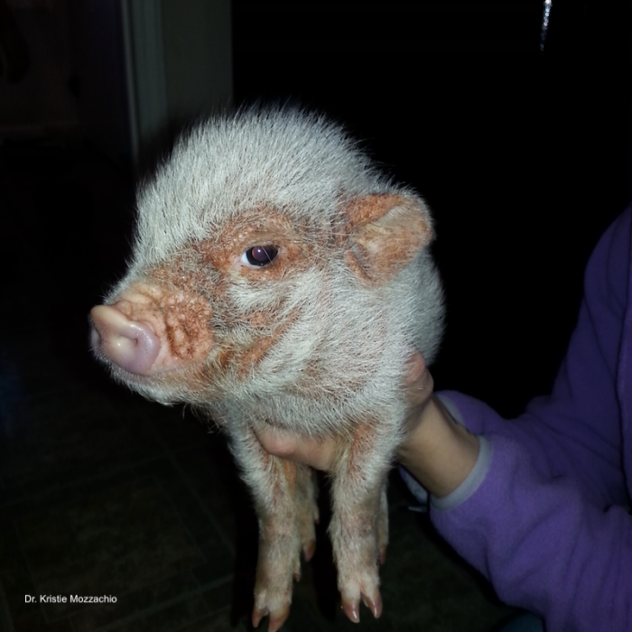

Zoonotic potential
Erysipelas
Sarcoptic mange
Swine influenza virus infections have been documented in people. Human influenza viruses have also been occasionally isolated from pigs.
Diagnostics
| Venipuncture | An ideal location for blood collection in miniature pigs is the radial vein located on the medial aspect of the forelimb.
Jugular venipuncture is performed by placing the pig on its back (see pig flip). Pull the forelimbs caudally and collect blood from the right jugular furrow, a “trough” to the right of the manubrium of the sternum, with the needle angled toward the opposite (left) shoulder. Small blood volumes can be collected from the tail vein in larger pigs (ventral midline of tail base, similar to collection in the bovine). Other venipuncture sites include saphenous, femoral, cephalic veins as well as the cranial vena cava. Auricular veins are difficult to access in a conscious animal and allow for only limited volume collection due to the small, collapsible vessels. Auricular veins are also one of the only readily accessible sites for intravenous catheterization so should be conserved, especially in sick animals. |
| Anesthesia
|
Intubation is challenging because the pig has a narrow “gape” (mouth doesn’t open wide) and a long, narrow oropharyngeal cavity (select image left to enlarge). Also, the relatively small larynx is loosely attached and kinks ventrally, which creates a sharp angle that prevents easy insertion of an endotracheal tube. The pharyngeal diverticulum and blind sacs formed by the large ventral and two lateral ventricles also make intubation more difficult. Laryngospasm can further complicate intubation. That said, intubation is readily performed with proper instruction and equipment!
A long laryngoscope blade and stylet is needed to facilitate intubation. Intubation is best performed with the pig in sternal recumbency. |
Catheterization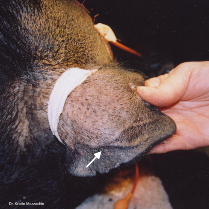 |
For low-volume infusions, intravenous catheters are most readily placed in the auricular vein (select image left to enlarge). Catheters can also be placed in cephalic and saphenous veins. |

References and further reading
Body Condition Scoring the Miniature Pig
Miniature pigs reach half their adult weight (32-68 kg) by about 1 year of age and will continue to grow until 3-4 years of age. Pigs easily gain weight and obesity is a very common problem in pet pigs, especially when animals are fed free-choice and not exercised. The risk of obesity in pet pigs can be minimized with client education on body condition scoring as well as regular weighing . . .
Pour les vétérinaires. Par les vétérinaires.
Le site Lafervet.com est conçu pour une utilisation par les vétérinaires. Il est ouvert aux vétérinaires diplômés, aux techniciens vétérinaires diplômés, aux animaliers et aux étudiants dans ces domaines.
Créer un compte pour accéder à des articles et des ressources du site.
L'inscription est gratuite.
Para Profesionales Veterinarios. Por Profesionales Veterinarios.
El sitio Lafervet.com es para uso de los profesionales veterinarios. Está abierto a los veterinarios licenciados, técnicos veterinarios licenciados, rehabilitadores licenciados y estudiantes en estos campos.
Cree una cuenta para acceder a los artículos y recursos del sitio.
La registro es gratis.
Already a LafeberVet Member?
Please Login
Reading Bird Body Language
Most parrots are highly vocal, using calls and a variety of sounds to establish and protect territory, attract mates, warn of danger, and maintain social connections. Parrots also communicate visually, using flashes of their colorful plumage as well as eye contact and body language. Careful observation of parrot body language can provide clues when a companion bird is receptive to play and handling or should be allowed to calm down.
Download the PDF version of the Reading Bird Body Language client education handout, or modify the DOCX version or DOC version for your veterinary hospital. Then view the YouTube video “Sleepy Red-Bellied Parrot” for an illustration of tongue wiggling and beak grinding, two behaviors mentioned in the handout.
Post-Test: Minimizing Stress to Avian Patients
View the recording of this live webinar event, then take the brief quiz. With a passing grade of 70% or higher, you will receive a continuing education certificate for 1 hour of continuing education credit in jurisdictions that recognize American Association of Veterinary State Boards Registry of Approved Continuing Education approval . . .
Pour les vétérinaires. Par les vétérinaires.
Le site Lafervet.com est conçu pour une utilisation par les vétérinaires. Il est ouvert aux vétérinaires diplômés, aux techniciens vétérinaires diplômés, aux animaliers et aux étudiants dans ces domaines.
Créer un compte pour accéder à des articles et des ressources du site.
L'inscription est gratuite.
Para Profesionales Veterinarios. Por Profesionales Veterinarios.
El sitio Lafervet.com es para uso de los profesionales veterinarios. Está abierto a los veterinarios licenciados, técnicos veterinarios licenciados, rehabilitadores licenciados y estudiantes en estos campos.
Cree una cuenta para acceder a los artículos y recursos del sitio.
La registro es gratis.
Already a LafeberVet Member?
Please Login
Minimizing Stress to Avian Patients During the Veterinary Visit
The recording of this live, interactive webinar was presented by Dr. Alicia McLaughlin, a certified Fear Free™ veterinarian who is spearheading the development of an avian-focused Fear Free™ course. This presentation explores the reasons stress should be minimized during avian veterinary visits and the challenges that must be overcome. Dr. McLaughlin also shares practical tips for clinical implementation as they relate to clinic design, staff training, client education, as well as tips for working with avian patients in an exam room or hospital setting. Alicia has also provided two client education handouts for download that she uses in her . . .
Pour les vétérinaires. Par les vétérinaires.
Le site Lafervet.com est conçu pour une utilisation par les vétérinaires. Il est ouvert aux vétérinaires diplômés, aux techniciens vétérinaires diplômés, aux animaliers et aux étudiants dans ces domaines.
Créer un compte pour accéder à des articles et des ressources du site.
L'inscription est gratuite.
Para Profesionales Veterinarios. Por Profesionales Veterinarios.
El sitio Lafervet.com es para uso de los profesionales veterinarios. Está abierto a los veterinarios licenciados, técnicos veterinarios licenciados, rehabilitadores licenciados y estudiantes en estos campos.
Cree una cuenta para acceder a los artículos y recursos del sitio.
La registro es gratis.
Already a LafeberVet Member?
Please Login
2019 AEMV Student Case Report Contest
Lafeber Company was proud to sponsor the Association of Exotic Mammal Veterinarians Student Case Report Contest. Veterinary students from all over the world were encouraged to write a 2-page case report about an exotic companion mammal seen at their college of veterinary medicine or during a clinical experience. Submissions closed March 22 and judges from the Research Committee evaluated the 14 case reports received. Judges were blinded to the students, mentors, co-authors, and institutions at which the cases were seen. See the brief summaries of each winning case report. Each student has been encouraged to submit their paper . . .
Pour les vétérinaires. Par les vétérinaires.
Le site Lafervet.com est conçu pour une utilisation par les vétérinaires. Il est ouvert aux vétérinaires diplômés, aux techniciens vétérinaires diplômés, aux animaliers et aux étudiants dans ces domaines.
Créer un compte pour accéder à des articles et des ressources du site.
L'inscription est gratuite.
Para Profesionales Veterinarios. Por Profesionales Veterinarios.
El sitio Lafervet.com es para uso de los profesionales veterinarios. Está abierto a los veterinarios licenciados, técnicos veterinarios licenciados, rehabilitadores licenciados y estudiantes en estos campos.
Cree una cuenta para acceder a los artículos y recursos del sitio.
La registro es gratis.
Already a LafeberVet Member?
Please Login
Alicia McLaughlin, DVM
 Alicia McLaughlin received her Doctorate of Veterinary Medicine from Oklahoma State University. She completed a veterinary internship in exotic animal medicine at The Center for Bird & Exotic Animal Medicine in Bothell, Washington and she is now an associate veterinarian at this practice. Dr. McLaughlin has been involved in veterinary research and leadership throughout her career, and has authored or co-authored articles in both professional journals and national conference proceedings. Dr. McLaughlin is passionate about providing low-stress veterinary visits for all of her patients. She is a certified Fear FreeTM veterinarian, and is spearheading the development of an avian-focused Fear Free™ course. In her spare time, Dr. McLaughlin likes to relax with her veterinarian husband and young daughter in the great outdoors and provide environmental enrichment for her pets. She currently shares her home with two giant schnauzers, two white-bellied caiques, one blue-throated macaw, four leopard geckos, one gargoyle gecko, one New Caledonian giant gecko, two ball pythons, one rainbow boa, and multiple freshwater fish.
.
Post Test: Basic Wildlife Rehabilitation Triage
The Basic Wildlife Rehabilitation Triage webinar was reviewed and approved by the American Association of Veterinary State Boards (AAVSB) Registry of Approved Continuing Education (R.A.C.E.) program for 1 hour of continuing education, in jurisdictions which recognize AAVSB R.A.C.E. approval.
You have three chances to take this post-test and pass. If you pass, you will see your results and a link to your continuing education certificate. Good luck!
Post-test: Routine Veterinary Care of the Miniature Pig
View the recording of this live webinar event, then take the brief quiz. With a passing grade of 70% or higher, you will receive a continuing education certificate for 1 hour of continuing education credit in jurisdictions that recognize American Association of Veterinary State Boards Registry of Approved Continuing Education approval . . .
Pour les vétérinaires. Par les vétérinaires.
Le site Lafervet.com est conçu pour une utilisation par les vétérinaires. Il est ouvert aux vétérinaires diplômés, aux techniciens vétérinaires diplômés, aux animaliers et aux étudiants dans ces domaines.
Créer un compte pour accéder à des articles et des ressources du site.
L'inscription est gratuite.
Para Profesionales Veterinarios. Por Profesionales Veterinarios.
El sitio Lafervet.com es para uso de los profesionales veterinarios. Está abierto a los veterinarios licenciados, técnicos veterinarios licenciados, rehabilitadores licenciados y estudiantes en estos campos.
Cree una cuenta para acceder a los artículos y recursos del sitio.
La registro es gratis.
Already a LafeberVet Member?
Please Login
Miniature Pig Care
This client education handout reviews basic care of the miniature pig. Topics covered include diet, housing, training and proper handling, as well as common health problems and preventive care measures, like vaccination and surgical sterilization.
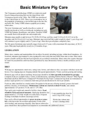
Download the PDF version of the Basic Mini Pig Care client education handout, or modify the DOCX version or DOC version for your veterinary hospital.
Considering bringing a mini pig into a home? Refer to Are You the Right Pet For Me? Miniature Pigs.
Are You the Right Pet For Me? Miniature Pigs
Pot-belled or miniature pigs are interesting, complex animals that are sometimes kept as pets in urban areas. Unfortunately, there are many misleading claims associated with miniature pet pigs that can eventually lead to these animals being surrendered to shelters or rescues by owners that “leapt before they looked”. Although it is always important to educate oneself before bringing an exotic pet into the home, this is never more true than with the pot-bellied pig. Use this client education material to review the challenges of pet pig care, then refer to LafeberVet’s basic care handout for advice on managing a pet pig. 
Download the PDF version of the Are You the Right Pet For Me? client education handout, or modify the DOCX version for your veterinary hospital.
Basic Wildlife Rehabilitation Triage
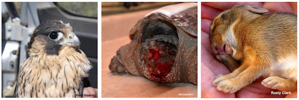
Lecture outline
 |
|
Abstract
Sick and injured wild animals often present to veterinary clinics and wildlife rehabilitators. It can be difficult to know which wild animals will survive the rehabilitation process to release and which won’t. This presentation will use data from one of the largest wildlife rehabilitation centers in the country to help predict which patients will make it to release and which won’t. This will help those in involved in wildlife rehabilitation know when to euthanize on admission and when to attempt rehabilitation. This case-based, interactive presentation aims to bring beginners up to speed, but also offers helpful tips, tricks and insights for the experienced rehabilitator and veterinarian.
About the presenter
Renée Schott is the Medical Director and a Senior Veterinarian at the Wildlife Rehabilitation Center of Minnesota (WRCMN), one of the largest rehabilitation centers in the country. She has been involved in wildlife rehabilitation for over 15 years and has worked at WRCMN for over 8 years. Additionally, Renée is involved in teaching courses at the University of Minnesota-College of Veterinary Medicine and she is a Course Instructor for the International Wildlife Rehabilitation Council. Renee also does relief work at The Raptor Center in St. Paul and she is a former member of the National Wildlife Rehabilitators Association Board of Directors.
Webinar recording
Post-test
With a passing grade of 70% or higher, you will receive a continuing education certificate for 1 hour of continuing education credit in jurisdictions that recognize AAVSB RACE approval.
Test your knowledge
Expert Q&A
Dr. Schott was able to answer most questions submitted during the live event, however remaining questions were answered by email and are posted below.
About spinal trauma (with deep pain present) in wild patients, how many days should we wait for improvement to make a decision if to carry on treatment or to euthanize?I usually give patients 5-7 days–if i see noticeable improvement, I will give 5-7 more days. If at any time there is no improvement in that 5-7 days, it is time to euthanize because they have plateaued.
What causes those stripped like primary flight feathers on CAGO? I have two like this currently, also frozen into ponds and appear to be elders…The stripped feathers in the CAGO I showed were from a droopy wing that the goose would chronically step on (wing droop was due to an old unhealed fracture).
What treatments would you use for torticollis? Would treatment change if the resolution of symptoms did not progress past a certain point early on, even after 6 months?Torticollis indicates a problem in the brain. usually in wildlife this is due to trauma, but we cannot rule out other things (infectious things like West Nile virus or Baylisascaris) but the treatment is the same–time and pain medications, low stress environment. I give them 5-7 days and if i don’t see neurological improvement (i.e. slightly less torticollis) then I euthanize. If they show slight improvement, they get 5-7 more days. If at any time there is no improvement in that 5-7 days, it is time to euthanize because they have plateaued.
RACE approval
This program is approved by the American Association of Veterinary State Boards (AAVSB) Registry of Continuing Education (R.A.C.E.) to offer a total of 1.00 CE credits to any one veterinarian and/or 1.00 veterinary technician CE credit. This RACE approval is for Category Two: Non-Scientific Clinical using the delivery method of Interactive-Distance/Non-Interactive Distance. This approval is valid in jurisdictions which recognize AAVSB RACE.
Renée Schott, DVM, CWR
 Renée Schott is the Medical Director and a Senior Veterinarian at the Wildlife Rehabilitation Center of Minnesota (WRCMN), one of the largest rehabilitation centers in the country. Renée has been involved in wildlife rehabilitation for over 15 years and she has worked at WRCMN for over 10 years. Additionally, Renée is involved in teaching courses at the University of Minnesota College of Veterinary Medicine and she is a Course Instructor for the International Wildlife Rehabilitation Council. Renée is also a former member of the National Wildlife Rehabilitators Association Board of Directors.
Routine Veterinary Care of the Miniature Pig
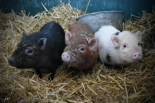
Lecture outline
Lecture topics include:
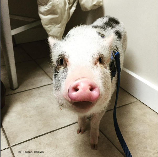 |
|
- Routine veterinary procedures
-
- Physical examination
- Hoof trim
- Tusk trim (males)
- Vaccination
- Deworming
- Nutrition
Abstract
Mini pigs remain a surprisingly common pet, with a resurgence in popularity every few years. Owners typically have a “small animal mindset”, and as such, seek like-minded veterinarians, but lack of training and paucity of resources leads many veterinarians to shy away from these unique pets.
Pigs are prey animals and behave accordingly, making handling and restraint one of the most difficult aspects of patient care. Crowd or sorting boards are the most important piece of specialized handling equipment, although smaller pigs can be held, larger pigs can be flipped, and if necessary, safe sedation protocols exist to make these stubborn and vocal patients more manageable. Once the hurdle of restraint has been surmounted, veterinary care is relatively straightforward and often extrapolated from other pet species.
Physical exam of the pet pig is largely visual, with observation of body and hoof condition, attitude, ambulation and mentation. Knowledge of normal anatomy such as carpal glands and preputial diverticulum is helpful as these may cause concern for the uneducated owner. Nutritional advice is often needed as well since some commercially prepared mini pig feeds may provide inaccurate label instructions that lead to obesity. On the contrary, unscrupulous breeders may suggest feeding protocols that lead to malnutrition and emaciation in an effort to keep the pet at the small size promised. Owners must not only be taught to “feed to body condition” but must understand what constitutes good body condition in a mini pig.
Hoof trim, tusk trim, deworming and vaccination are the most common procedures performed by the veterinarian. Hoof trim, with proper shaping and balancing, is needed every year at a minimum; tusk trim may be occasionally needed for males but, as risk of tooth infection is high, only certain conditions warrant trimming. There is no universally accepted vaccination protocol, so the pet pig practitioner must develop one suited to the pig’s environment and potential exposure, all while adhering to label instructions geared to much larger swine. Even deworming protocols vary, although evaluation is similar to other species and drug dosages specific to swine are available.
Overall, mini pigs can be a challenging species to vet, but with some guidance on their uniquities, pet pig health can be easily maintained.
About the presenter
Kristie Mozzachio operates a mobile “pet pig exclusive” practice serving the states of North Carolina, Virginia, Maryland, Pennsylvania, and New Jersey. She provides consults on pet pigs as well as research mini pigs, both nationally and internationally. She also serves as a veterinary advisor for Ross Mill Farm & Piggy Camp and Refuge GroinGroin in France. Dr. Mozzachio has also lectured on mini pigs at numerous meetings, and most recently she has co-authored mini pig chapters in the Exotic Animal Formulary and Diseases of Swine. [MORE]
Dr. Mozzachio can be contacted directly at [email protected].
Webinar recording
Download the presentation
Download Dr. Mozzachio’s presentation: PDF (14 MB). Contact us if you would like a copy of the PowerPoint file with annotated notes.
Post-test
With a passing grade of 70% or higher, you will receive a certificate for 1 hour of continuing education credit in jurisdictions that recognize AAVSB R.A.C.E. approval.
Expert Q+A
Although Dr. Mozzachio was able to answer most questions submitted during the live event, remaining questions were answered by email and are posted below.
How often do you see struvite stones that require surgery?
Infrequently, about 20 cases in the last 10+ years (although I suspect that there are more that have gone undiagnosed/untreated). Unfortunately, surgery is difficult due to the anatomy of the penis, and there is always the possibility of recurrence. I feel that routine urinalysis + survey radiographs would be very useful in males over about 8 years of age. A veterinarian at the University of Tennessee has postulated that age-related bladder atony predisposes to the condition since older males are typically affected.
Should pigs have their teeth hand scaled and polished routinely and do you recommend owners learn to brush their teeth with dog toothpaste products?
Absolutely! Care of the teeth should be similar to that recommended for dogs and cats. However, very few veterinary clinics are willing to perform dental procedures on mini pigs, in part because intubation can be a challenge. It’s also extremely difficult to access all teeth because pigs have such a narrow gape – much more difficult than a dog or cat dental. Owners should be encouraged to brush the teeth with dog toothpaste products or baking soda paste (IF possible – depends on the pig) . Some owners report success with dental chews designed for dogs. I’m more cautious about using dental chews since they must be the right size and consistency to prevent choking and allow for prolonged chewing. Many pigs eat like little sharks and simply “gulp” any food offered. So… it depends on the pig and the dental product both. At a minimum, all sedated pigs should have a brief oral exam and removal of any large tartar chunks. Pigs typically form tartar so severe that the build-up is the only thing holding a rotten tooth in place!
Do you recommend regular external and internal deworming or [do you] always check for clinical signs/examine feces and skin?
At the first wellness visit, I typically examine for skin parasites (visual at least + skin scrape of any suspicious areas), perform a fecal float, then give a round of dewormer regardless (i.e. dewormer dose every 10-12 days for a total of 3 doses). After that, I do fecal flotation every 6-12 months to monitor and only deworm as necessary. However, in a sanctuary/rescue with continuous influx of new pigs, I typically run fecal floats every 6 months to monitor egg count, and also deworm every 6 months. I switch dewormers every few years or if fecal float shows increasing egg numbers that might indicate resistance.
Is there any indication that male pigs will be more likely to obstruct from urethral stones if they are castrated too young?
I haven’t seen any evidence that castration at a young age predisposes to urinary obstruction. I believe that age-related bladder atony, with incomplete emptying of urine, is the greatest predisposing factor.
How often do miniature pigs shed?
Once or twice a year, usually during warmer weather, although they may shed when scared (i.e. thunderstorm) at any time of year.
What joint supplementation do you recommend?
Any human, canine, or equine supplement is fine in minipigs. Dasuquin® (Nutramax Laboratories) and Cosequin® (Nutramax) as well as injectable Adequan® (Luitpold Pharmaceuticals) have had good results.
RACE approval
This program 776-36251 is approved by the American Association of Veterinary State Boards (AAVSB) Registry of Continuing Education (R.A.C.E.) to offer a total of 1.00 CE credits to any one veterinarian and/or 1.00 veterinary technician CE credit. This RACE approval is for Category Two: Non-Scientific Clinical using the delivery method of Interactive-Distance/Non-Interactive Distance. This approval is valid in jurisdictions which recognize AAVSB RACE; however, participants are responsible for ascertaining each board’s CE requirements. RACE does not “accredit” or “endorse” or “certify” any program or person, nor does RACE approval validate the content of the program.
Kristie Mozzachio, DVM, DACVP, CVA
Kristie Mozzachio received her Doctorate of Veterinary Medicine from North Carolina State University (NCSU) in 2001. Kristie completed an anatomic pathology residency at NCSU in 2004, and she became certified in veterinary acupuncture at the Chi Institute in 2015. Dr. Mozzachio currently works as a freelance toxicologic pathologist and she operates a mobile pet pig exclusive practice serving the states of North Carolina, Virginia, Maryland, Pennsylvania, and New Jersey. She provides consults on pet pigs as well as research mini pigs, both nationally and internationally. She also serves as a veterinary advisor for Ross Mill Farm & Piggy Camp and Refuge GroinGroin in France. Dr. Mozzachio has also lectured on mini pigs at numerous meetings, and most recently she has co-authored mini pig chapters in the Exotic Animal Formulary and Diseases of Swine. She currently owns 2 mini pigs, the oldest 21 years of age.
Post Test: Reproductive Disease in Reptiles
The Reproductive Disease in Reptiles webinar recording was reviewed and approved by the American Association of Veterinary State Boards (AAVSB) Registry of Approved Continuing Education (RACE) program for 1 hour of continuing education credit for veterinarians and veterinary technicians in jurisdictions that recognize AAVSB RACE approval. You have three chances to take this post-test and pass. With a passing grade, you will see your results and a link to your continuing education certificate . . .
Pour les vétérinaires. Par les vétérinaires.
Le site Lafervet.com est conçu pour une utilisation par les vétérinaires. Il est ouvert aux vétérinaires diplômés, aux techniciens vétérinaires diplômés, aux animaliers et aux étudiants dans ces domaines.
Créer un compte pour accéder à des articles et des ressources du site.
L'inscription est gratuite.
Para Profesionales Veterinarios. Por Profesionales Veterinarios.
El sitio Lafervet.com es para uso de los profesionales veterinarios. Está abierto a los veterinarios licenciados, técnicos veterinarios licenciados, rehabilitadores licenciados y estudiantes en estos campos.
Cree una cuenta para acceder a los artículos y recursos del sitio.
La registro es gratis.
Already a LafeberVet Member?
Please Login
Alejandro Morales, DVM, MS
![]()
Alejandro Morales earned his veterinary degree from San Carlos University in Guatemala City, with a master’s degree in One Health from Ross University. Dr. Morales has worked as the assistant director and veterinarian for the ARCAS Wildlife Rescue and Rehabilitation Center in Petén, Guatemala since 2007. Alejandro’s passion for working with wildlife started at a young age when he worked as a guide in the Natural History Museum in Guatemala City, participating with the Educational department of La Aurora Zoo and leading the volunteer programs in the Environmental Library for Youngsters, La Jungla Zoo. Working in the realm of wildlife rehabilitation has been a good mixture of his callings in life: conservation, education, veterinary medicine, and wildlife. Alejandro serves in the International Primatological Societies’ Captive Care Committee. He has presented on wildlife medicine and rehabilitation in multiple events. Alejandro believes that conservation without education is void, and that veterinary medicine plays a key role in the welfare and preservation of the species that share the world with us.
Tim Reichard, DVM
Dr. Tim Reichard is the owner of Dr. Tim’s Wildlife and Exotics Care in Toledo, Ohio. Dr. Reichard obtained his Doctorate of Veterinary Medicine at Washington State University in 1980. He then completed an internship and residency in zoological medicine at San Diego Zoo before serving as a Toledo Zoo veterinarian for over 25 years. Dr. Reichard has been recognized for his expertise in preventative medicine programs in a variety of zoo species, and he has also been involved in a number of conservation projects with native snake and amphibian species. Tim also served as an instructor for the veterinary technician program at Stautzenberger College from 2005 to 2014, and he has authored or coauthored more than 35 professional and popular publications.
Reproductive Disease in Reptiles
Reptile reproduction can be a confusing topic due to the variety of normal reproductive strategies found throughout different reptile species. Nevertheless it important to understand normal reproductive processes to correctly approach some commonly seen problems. This webinar recording reviews normal reproductive strategies of reptiles, including important reproductive anatomy and physiology. Three clinically important conditions, pre-ovulatory or follicular stasis, post-ovulatory dystocia, and cloacal prolapse, are also explored in detail . . .
Pour les vétérinaires. Par les vétérinaires.
Le site Lafervet.com est conçu pour une utilisation par les vétérinaires. Il est ouvert aux vétérinaires diplômés, aux techniciens vétérinaires diplômés, aux animaliers et aux étudiants dans ces domaines.
Créer un compte pour accéder à des articles et des ressources du site.
L'inscription est gratuite.
Para Profesionales Veterinarios. Por Profesionales Veterinarios.
El sitio Lafervet.com es para uso de los profesionales veterinarios. Está abierto a los veterinarios licenciados, técnicos veterinarios licenciados, rehabilitadores licenciados y estudiantes en estos campos.
Cree una cuenta para acceder a los artículos y recursos del sitio.
La registro es gratis.
Already a LafeberVet Member?
Please Login
Central Venous Catheter Placement in Chelonians
Long-term vascular access is difficult to obtain and maintain in chelonians. Fortunately, central venous catheters provide flexibility and length to avoid catheter dislodgement. Central lines are an effective tool that allow serial blood measurements and can be used for anesthesia administration, intravenous drug delivery, blood product transfusions, and continuous fluid therapy or continuous rate infusions.This photo tutorial article describes this simple technique step-by-step . . .
Pour les vétérinaires. Par les vétérinaires.
Le site Lafervet.com est conçu pour une utilisation par les vétérinaires. Il est ouvert aux vétérinaires diplômés, aux techniciens vétérinaires diplômés, aux animaliers et aux étudiants dans ces domaines.
Créer un compte pour accéder à des articles et des ressources du site.
L'inscription est gratuite.
Para Profesionales Veterinarios. Por Profesionales Veterinarios.
El sitio Lafervet.com es para uso de los profesionales veterinarios. Está abierto a los veterinarios licenciados, técnicos veterinarios licenciados, rehabilitadores licenciados y estudiantes en estos campos.
Cree una cuenta para acceder a los artículos y recursos del sitio.
La registro es gratis.
Already a LafeberVet Member?
Please Login
Olivia Petritz, DVM, DACZM
 Olivia Petritz earned her doctorate at Purdue University College of Veterinary Medicine in 2008. She then completed a small animal medicine and surgery residency at a specialty hospital in San Diego, California followed by an exotic animal internship at Gulf Coast Avian and Exotics in Houston, Texas. Dr. Petritz then returned to California to complete a 3-year residency in companion exotic animals and zoo medicine at the University of California, Davis. She achieved board certification in November 2013. Olivia then started an exotic animal medicine service at a large specialty hospital in Los Angeles. She worked in California for 3 years before becoming an Assistant Clinical Professor at North Carolina State University, where she works in the Exotic Animal Medicine Service.
Emergency and Critical Care of Rabbits
The free, live, RACE-approved continuing education webinar was presented by Charly Pignon, DVM, DECZM (Small Mammal) on November 7, 2018. Lecture topics include emergency triage, cardiopulmonary resuscitation, analgesia, fluid therapy, and critical care nutrition. RACE approval has expired for the webinar recording, but STAY TUNED for renewal . . .
Pour les vétérinaires. Par les vétérinaires.
Le site Lafervet.com est conçu pour une utilisation par les vétérinaires. Il est ouvert aux vétérinaires diplômés, aux techniciens vétérinaires diplômés, aux animaliers et aux étudiants dans ces domaines.
Créer un compte pour accéder à des articles et des ressources du site.
L'inscription est gratuite.
Para Profesionales Veterinarios. Por Profesionales Veterinarios.
El sitio Lafervet.com es para uso de los profesionales veterinarios. Está abierto a los veterinarios licenciados, técnicos veterinarios licenciados, rehabilitadores licenciados y estudiantes en estos campos.
Cree una cuenta para acceder a los artículos y recursos del sitio.
La registro es gratis.
Already a LafeberVet Member?
Please Login
Emergency and Critical Care in Rabbits Post Test
. . .
Pour les vétérinaires. Par les vétérinaires.
Le site Lafervet.com est conçu pour une utilisation par les vétérinaires. Il est ouvert aux vétérinaires diplômés, aux techniciens vétérinaires diplômés, aux animaliers et aux étudiants dans ces domaines.
Créer un compte pour accéder à des articles et des ressources du site.
L'inscription est gratuite.
Para Profesionales Veterinarios. Por Profesionales Veterinarios.
El sitio Lafervet.com es para uso de los profesionales veterinarios. Está abierto a los veterinarios licenciados, técnicos veterinarios licenciados, rehabilitadores licenciados y estudiantes en estos campos.
Cree una cuenta para acceder a los artículos y recursos del sitio.
La registro es gratis.
Already a LafeberVet Member?
Please Login
Pignon Webinar Abstract

The presenter, Charly Pignon, is getting ready to welcome you to this live interactive continuing education event
The webinar will begin at 1 pm EST (New York)
What time is that in my time zone?
Visit the webinar page for more information or read the abstract below
Abstract
Intensive care in rabbits requires a thorough knowledge of rabbit anatomy and physiology along with an understanding of basic clinical procedures. Although there is extensive data available on the laboratory rabbit, studies focusing on companion rabbits in an intensive care setting are needed to optimize prognosis and improve quality of care.
When a rabbit is presented on an emergency basis, the first step is triage. As in other species, first observe the rabbit in his carrier and pay particular attention to the animal’s breathing, position, and the level of awareness. If the animal is in respiratory distress or not breathing at all, if the animal is not responding to manipulation, then the rabbit requires immediate stabilization or a resuscitation protocol should be initiated. In other cases, as rabbits hide clinical signs, a precise history should be obtained and a thorough physical examination should be performed.
In cardiopulmonary arrest, mechanical cardiac activity halts leading to hemodynamic collapse. As this is an absolute emergency, the clinic staff must to be prepared. As has been described in carnivores, the “ABCDEFG” sequence can be used, but is adapted to the anatomical and physiological particularities of rabbits.
As rabbits are very sensitive to pain and its consequences can be deadly, one of the first treatments to give to rabbit are painkillers. The analgesic potency of non-steroidal anti-inflammatory drugs is often insufficient and some of their side effects can be dangerous for in the debilitated patient. Opioids, such as buprenorphine, morphine, or fentanyl, can be safely used in most cases. More and more data are also available on co-analgesics like alpha-2 agonists, dissociative agents, and local anesthetics, which could be very interesting to use in critical care.
In many critical cases, clinical signs are often secondary to dehydration or alterations of the electrolytes equilibrium. Fluid therapy should be performed in three steps: deficit correction (hypertonic bolus in order to reach 90 mmHg of arterial blood pressure), rehydration (using isotonic crystalloids to reach normal hydration status), and maintenance (to cover the loss of fluid, electrolytes, and energy in a normally hydrated animal) with electrolytes supplementation as needed.
Anke Stöhr, med. vet., ZB Reptilien
![]() Anke Stöhr was a 2017-2020 Zoological Medicine Resident and masters student at the Louisiana State University School of Veterinary Medicine with a focus in herpetological medicine. Dr. Stöhr studied veterinary medicine at the University of Leipzig, Germany (2002-2008). After graduation, she worked in various small animal veterinary hospitals with a focus on reptile medicine. From 2010-2014, she worked on her doctoral thesis “Diagnosis and characterization of ranaviruses in reptiles and amphibians in Germany” at the University of Hohenheim, Germany. In 2012, Dr. Stöhr became a certified specialist for reptile medicine (ZB Reptilien) in Germany. She has published various articles in international peer-reviewed journals, co-authored a book chapter in “Ranaviruses: Lethal Pathogens of Ectothermic Vertebrates” and presented her work at international conferences. She has been a member of the Association of Reptilian and Amphibian Veterinarians (ARAV) since 2011 and has been elected as an ARAV member-at-large in 2017. Her current research interest includes thyroid disease in chelonians and blood transfusion medicine in reptiles.
Anke Stöhr was a 2017-2020 Zoological Medicine Resident and masters student at the Louisiana State University School of Veterinary Medicine with a focus in herpetological medicine. Dr. Stöhr studied veterinary medicine at the University of Leipzig, Germany (2002-2008). After graduation, she worked in various small animal veterinary hospitals with a focus on reptile medicine. From 2010-2014, she worked on her doctoral thesis “Diagnosis and characterization of ranaviruses in reptiles and amphibians in Germany” at the University of Hohenheim, Germany. In 2012, Dr. Stöhr became a certified specialist for reptile medicine (ZB Reptilien) in Germany. She has published various articles in international peer-reviewed journals, co-authored a book chapter in “Ranaviruses: Lethal Pathogens of Ectothermic Vertebrates” and presented her work at international conferences. She has been a member of the Association of Reptilian and Amphibian Veterinarians (ARAV) since 2011 and has been elected as an ARAV member-at-large in 2017. Her current research interest includes thyroid disease in chelonians and blood transfusion medicine in reptiles.
Mariana Pardo, MV, BVSc, DACVECC

Mariana Pardo is a board-certified veterinary emergency and critical care specialist at Veterinary Medical Center of Long Island. Dr. Pardo earned her Doctorate of Veterinary Medicine in 2009 from the Universidad Mayor in Santiago, Chile. She finished two emergency and critical care internships at the University of Georgia (UGA) and then the University of Florida. She recently completed her emergency and critical residency at Cornell University. She has a strong interest in exotic critical care medicine, and during her time at UGA, Dr. Pardo worked on a manuscript with Dr. Stephen Divers: “Jugular central venous catheter placement through a modified Seldinger technique for long-term venous access in chelonians”.
2018 Avian Practitioner of the Year
Forty-five exceptional avian veterinarians from all around the world were nominated for the 2018 T.J. Lafeber Avian Practitioner of the Year. The independent Selection Committee narrowed this list to six finalists and the Award recipient, Dr. Robert Doneley, was announced during the Plenary Session at the 2018 ExoticsCon . . .
Pour les vétérinaires. Par les vétérinaires.
Le site Lafervet.com est conçu pour une utilisation par les vétérinaires. Il est ouvert aux vétérinaires diplômés, aux techniciens vétérinaires diplômés, aux animaliers et aux étudiants dans ces domaines.
Créer un compte pour accéder à des articles et des ressources du site.
L'inscription est gratuite.
Para Profesionales Veterinarios. Por Profesionales Veterinarios.
El sitio Lafervet.com es para uso de los profesionales veterinarios. Está abierto a los veterinarios licenciados, técnicos veterinarios licenciados, rehabilitadores licenciados y estudiantes en estos campos.
Cree una cuenta para acceder a los artículos y recursos del sitio.
La registro es gratis.
Already a LafeberVet Member?
Please Login
Lucile Chassang, DVM, IPSAV

Dr. Lucile Chassang graduated from Alfort National Veterinary School (France) in 2013. After 1 year of small animal practice, she performed a 1-year zoological medicine internship in the Faculty of Veterinary Medicine of the University of Montreal, in Saint Hyacinthe, Québec, Canada. In 2015, she joined Dr. Minh Huynh’s team at Fregis Veterinary Hospital in Arcueil, France. She recently completed a European College of Zoological Medicine residency program in small mammal medicine. This 4-year alternate program was undertaken at both Fregis Veterinary Hospital with Dr. Minh Huynh and Alfort National Veterinary School with Dr. Tom Donnelly and Dr. Charly Pignon.
Edward Ramsay, DVM, DACZM
Edward Ramsay is a Professor Emeritus at the University of Tennessee College of Veterinary Medicine. Dr. Ramsay completed a residency at the University of California at Davis in zoological medicine and is board-certified in zoological medicine. He served as the 2007-2008 President of the American Association of Zoo Veterinarians, and he is the author of numerous publications related to zoo medicine. In addition to content review, Dr. Ramsay has donated a variety of images for LafeberVet.
Kirk Suedmeyer, DVM, DACZM

Dr. Kirk Suedmeyer is the Director of Animal Health and Research at the Kansas City Zoo in Kansas City, Missouri, where he has worked since 1992. Dr. Suedmeyer is a Diplomate of the American College of Zoological Medicine and Past-President of the American Association of Zoo Veterinarians (2012-2013) and the Association of Reptile and Amphibian Veterinarian (1993-1995). Kirk is a graduate of the University of Missouri-Columbia College of Veterinary Medicine (UMC-CVM) and he currently serves as an Adjunct Assistant Professor at UMC-CVM. He also serves as an associate editor of the Journal of Zoo and Wildlife Medicine, and has published over 100 articles on exotic animal medicine and surgery. In his spare time, Kirk is a master falconer and wildlife artist.
Kathy Duncan
 Kathy Duncan has been an active wildlife rehabilitator with Pacific Wildlife Care (PWC) since 1999. Ms. Duncan previously served on the PWC Board of Directors for 7 years and she has been Education Director since 2006. Kathy is currently a staff rehabilitation technician at PWC’s wildlife clinic, where she has worked since its opening in 2007. Kathy specializes in the care of opossums, squirrels, and small mammals. She also provides home care to orphaned and injured opossums from neonates through release. Pacific Wildlife Care takes in close to 200 opossums a year. With their full-time veterinarian, Kathy helps to establish diet and housing guidelines for opossums, both at the clinic and in the home care program. Kathy also conducts annual training classes in opossum care for PWC volunteers. She also regularly conducts educational outreach programs using PWC’s educational opossums and raptors since 2002. Kathy is a member of the California Council of Wildlife Rehabilitators and the Opossum Society of the United States, and she is a past-member of the National Opossum Society.
Natalie Antinoff, DVM, DABVP (Avian Practice)

Dr. Natalie Antinoff is the owner of Antinoff Veterinary Services, which provides veterinary relief and consulting for various practices, primarily specialty facilities. Dr. Antinoff also regularly provides scheduled per diem care for non-traditional pets at Texas Avian & Exotic Animal Hospital in Grapevine, Texas and Mountain West Veterinary Specialists in Layton, Utah. Dr. Antinoff has also been a consultant for the Veterinary Information Network since 1997. She was also the Director of the bird and exotic pet referral practice, Gulf Coast Avian and Exotics, a member of Gulf Coast Veterinary Specialists in Houston, Texas. Dr. Antinoff is board certified in avian practice through the American Board of Veterinary Practitioners. She completed an internship in small animal medicine and surgery, and a residency in avian and exotic pet medicine and surgery at The Animal Medical Center in New York.
Miguel Saggese, DVM, MSc, PhD

Dr. Miguel Saggese: A native of Argentina, Dr. Miguel Saggese joined Western University of Health Sciences in 2007 as an assistant professor in microbiology and avian diseases. He obtained his veterinary degree in 1995 at the University of Buenos Aires, Argentina. In 2004, Dr. Saggese completed a 3-year residency and a masters program at The Raptor Center, University of Minnesota. During these years, he studied the effect of West Nile virus (WNV) on birds of prey and the efficacy of WNV vaccines. Between 2004 and 2007, his research focused on avian tuberculosis while completing his Ph.D. in microbiology at The Schubot Center, Texas A&M University. Dr. Saggese defines himself as an ornithologist as well as veterinarian and he combines both disciplines to better understand the effect of diseases in avian populations. He is an associate editor of the Journal of Zoo and Wildlife Medicine and is actively involved in several organizations, including the Raptor Research Foundation, the Association of Avian Veterinarians, and the Eagle Conservation Alliance.
Michael Kent, DVM, DACVIM, DACVR

Dr. Michael S. Kent is an assistant professor in the department of surgery and radiology at the University of California at Davis (UCD) School of Veterinary Medicine. Dr. Kent received his DVM at UCD and completed an internship in small animal medicine at the University of Pennsylvania. He then went on to complete residencies in both Medical and Radiation Oncology at UCD as well as an NIH sponsored 2-year clinical research training program. He is now a diplomate of both the American College of Veterinary Radiology and the American College of Veterinary Internal Medicine. The Morris Animal Foundation and the National Canine Cancer Foundation have funded Dr. Kent’s research investigations into canine neoplasia, including studies on innovative therapy for lymphoma.
Michael E. Comella
 Michael E. Comella is a reptile rehabilitator and director of Wichita Falls Reptile Rescue. Mike is also a professional development instructor with Vernon College in Wichita Falls, TX, where he administers a Texas Department of State Health Services accredited animal control course on reptile handling; with special emphasis on venomous snakes and large constrictors. He has devoted 25 years to field and husbandry studies of herptiles, including 20 years in safe handling of native venomous snakes. In 2003, Michael was presented with his first case of an endangered reptile, displaced by construction activities. He soon became known locally as an ad hoc rescuer and was presented with many turtles injured by vehicle traffic. These events sparked a paradigm shift from observer and enthusiast, to a decade spent in pursuit of an intense passion for conservation, reptile medicine, and development of rehabilitation protocols for reptiles and amphibians. Michael also administers a social media page related to rehabilitation of reptile and amphibian wildlife.
Michael Dutton, DVM, MS, DABVP (Canine & Feline Practice), DABVP (Avian Practice), DABVP (Exotic Companion Mammal Practice), CVPP
 Michael Dutton is a Diplomate of the American Board of Veterinary Practitioners (ABVP) in three specialties– Avian Practice, Exotic Companion Mammal Practice, and Canine/Feline Practice. Dr. Dutton is a graduate of Michigan State University and he has completed a post-doctoral Master’s Degree in Pain Management. He is also a Certified Pain Practitioner by the International Veterinary Academy of Pain Management (IVAPM). He currently serves on both ABVP and American Animal Hospital Association (AAHA) committees. Dr. Dutton owns the Exotic and Bird Clinic of New Hampshire and the Weare Animal Hospital. He also co-founded the Association of Exotic Mammal Veterinarians, which now has over 900 members. Dr. Dutton lectures and publishes primarily on exotic pet medicine/surgery and small animal pain management.
Maureen Murray, DVM, DABVP (Avian Practice)
Dr. Maureen Murray is a Clinical Assistant Professor at Tufts Cummings School of Veterinary Medicine. She is a clinician at the Tufts University Wildlife Clinic, which treats numerous species of native New England wildlife. She is board certified in avian medicine. Along with clinical work and teaching, Dr. Murray pursues research in the area of wildlife health, including the effects of rodenticides on birds of prey.
Marla Lichtenberger, DVM, DACVECC
 Dr. Marla Lichtenberger* was a Diplomate of the American College of Veterinary Emergency and Critical Care. Dr. Lichtenberger completed an internship at the Animal Medical Center in New York City and a residency at the Animal Emergency Center in Milwaukee, Wisconsin. Dr. Lichtenberger continued on as the Senior Emergency and Critical Care Specialist at the Animal Emergency Center for 6 years. During this time, Dr. Lichtenberger earned board certification in Veterinary Emergency and Critical Care. Marla was a frequent international speaker on emergency and critical care for small animals and exotics, as well as a published researcher. Dr. Lichtenberger also served on the board of an exotic companion mammal committee that prepared the first American Board of Veterinary Practitioners exam for small mammal medicine in 2009. Dr. Lichtenberger was formerly the owner of the Milwaukee Animal Emergency Center for Animals, a critical care facility offering 24-hour care to not only dogs and cats but also reptiles, small mammals, birds, and other exotic animals.
*Dr. Lichtenberger passed away on April 4, 2019. Please visit AAV.org for a detailed In Memoriam.