ExoticsCon 2015

Lafeber Company was proud to serve as THE platinum sponsor of ExoticsCon 2015

Lafeber Company is proud to serve as THE platinum sponsor of ExoticsCon 2015


What did you think?

Be sure to complete the ExoticsCon Evaluation Form to make next year’s conference even better! Complete the survey by Friday, September 25 and you will automatically be entered into a drawing. Three winners will be randomly selected and awarded a $100 Visa gift card.

Join us in Portland

Join us in Portland, Oregon August 26-September 1, 2016 for the next Joint Conference of the Association of Avian Veterinarians, Association of Exotic Mammal Veterinarians, and Association of Reptilian and Amphibian Veterinarians. Click here for a fun video by Katie Lennox on all Portland, Oregon has to offer.

Understanding avian cognition

African grey with head in bowl

Learning opportunities at ExoticsCon were available for ALL experience levels including TWO master classes by Irene Pepperberg, research associate at Harvard University in the field of avian cognition.

What has Griffin the grey parrot been up to? Get information on the latest study from Dr. Pepperberg’s lab in “Wait For It…A Grey Parrot Demonstrates Self-Control“.


Santiago Díaz DVM

Santiago Diaz
Dr. Santiago Díaz is the owner of the Exotic Animal Hospital of Orlando in Orlando, Florida. Santiago previously worked at Broward Avian & Exotic Animal Hospital for 5 years as an associate veterinarian. Dr. Díaz completed his veterinary degree at the University of Florida in 2011. During his short career, Dr. Díaz has lectured at the Western Veterinary Conference in Las Vegas and at veterinary technician schools in the South Florida region. Santiago is currently translating LafeberVet’s large collection of medical documents from English to Spanish. These documents will benefit veterinarians in the Latin American countries and Spain, providing them with the knowledge to care for special species.

Palawan Turtle Crisis

Did you hear about the Palawan turtle crisis? In June 2015, Philippine authorities confiscated over 4,000 turtles, many of them critically endangered Philippine forest turtles (Siebenrockiella leytensis), intended for the illegal pet trade. Enjoy LafeberVet’s brief, fun slideshow that explores our Emeraid donation for the Palawan turtle crisis as well as care of many, many, many turtles. Although medical supplies are not currently required, financial contributions are still needed for this important conservation effort . . .


To continue you need to be a LafeberVet.com member. (Français), (Español)

Pour continuer, vous devez être un membre LafeberVet.com

Para continuar, debe ser miembro de LafeberVet.com

Already a LafeberVet Member?

Please Login

Rabbit GI Case Challenge Discussion

Gastric dilatation or “bloat” and gastrointestinal obstruction is an acute and life-threatening condition of pet rabbits commonly caused by an obstruction with pellets of compressed hair. The discussion portion of this Case Challenge reviews onset, clinical signs, and diagnostic test results of obstructive and non-obstructive gastrointestinal disease. This condition is considered a surgical emergency and key points of urgent care strive to stabilize the patient through analgesia, decompression when indicated, and supportive care. Surgery is discussed as well as recommendations for patients that cannot go to surgery due to clinical or financial constraints. Follow-up care as well . . .


To continue you need to be a LafeberVet.com member. (Français), (Español)

Pour continuer, vous devez être un membre LafeberVet.com

Para continuar, debe ser miembro de LafeberVet.com

Already a LafeberVet Member?

Please Login

Bloat and Gastrointestinal Blockage in Rabbits Client Handout

Gastrointestinal obstruction and a stomach distended with gas and fluid or “bloat” is a serious health problem of pet rabbits. Use this client educational handout to answer owner questions:  What causes bloat and obstruction? Why is bloat a serious condition? What does bloat look like in the rabbit? This handout also explains the basics of a diagnostic workup, treatment, follow-up care, and prevention for this critical condition.

Download LafeberVet’s Bloat and Gastrintestinal Blockage Handout.


bloat obstruction handout

Case Challenge: A 5-Year-Old Rabbit With Anorexia and Lethargy

A 5-year old female spayed lop rabbit presents with a history of acute anorexia . . .


To continue you need to be a LafeberVet.com member. (Français), (Español)

Pour continuer, vous devez être un membre LafeberVet.com

Para continuar, debe ser miembro de LafeberVet.com

Already a LafeberVet Member?

Please Login

Are You Listed? Find An Avian Veterinarian

Bird Collage Panel cropped width 800

Find An Avian Veterinarian

Are you an avian veterinarian in the USA or Canada?

Are you listed in the Find An Avian Veterinarian locator on our sister site, Lafeber Pet Birds? Pet bird owners all across North America use this online service.


Find an Avian Vet screenshot

Contact Us

…to add your information to this popular Lafeber Pet Birds feature

OR

….if your practice information needs to be updated.

Contact Us


Veterinary Locator Services

More extensive veterinary locators are also available through the American Board of Veterinary Practitioners and the Association of Avian Veterinarians. LafeberVet routinely refers pet owners to these locator services through our Veterinary Locator Services post.

Basic Information Sheet: European Hedgehog

Western European Hedgehog (Erinaceus europaeus)

European-hedgehog-Radoslaw-Ziomber

Natural history


The European hedgehog (Erinaceus europaeus) is commonly associated with humans in rural, suburban, and urban habitats. Its native range spans from Ireland, Great Britain, southern Scandinavia, and Western Europe to the Czech Republic, where its range overlaps with that of the Northern white-breasted hedgehog (Erinaceus roumanicus). In addition to its native range, E. europaeus was introduced in 1974 to the Scottish island of Uist and in the late 19th Century to New Zealand, where it is considered an invasive pest species.

European hedgehogs are nocturnal animals, and their dominant senses are hearing and olfaction. Hedgehogs prefer habitats that provide cover like hedgerows, shrub borders in suburban gardens, and leaf litter. Hedgehogs tend to avoid thick forests, although they will use forest edge habitat. Adult hedgehogs are solitary, but they may share overlapping home ranges. Each home range is large, and hedgehogs can travel approximately 3-4 km (1.9-2 miles) each night. Each hedgehog builds several nests within its home range, concealing itself in a nearby nest at the end of a night’s foraging.

Sharp spines that cover the dorsum and sides as well as the ability to roll up serve as a highly effective defense mechanism against predators. European hedgehogs have few natural predators, with the badger likely serving as their most important. Other known predators include dogs, foxes, snakes and large owls.


Conservation status


The International Union for Conservation of Nature lists Erinaceus europaeus as a species of “Least Concern” because animals are common and abundant throughout its wide range (Amori 2008).


Taxonomy


Class: Mammalia

Order: Erinaceomorpha: gymnures and hedgehogs

Family: Erinaceidae

Subfamily: Erinaceinae: spiny hedgehogs

Genus: Erinaceus: woodland hedgehogs

Genus: Erinaceus europaeus


Physical description


The dorsal surface and sides of the European hedgehog are covered in pale spines that have a wide brown band and a white tip. The spines are relatively long, measuring 1.9-2.5 cm (0.75-1 in) in length. The face, throat, chest, abdomen, and legs are covered with coarse, grey-brown or yellow-brown fur.

Head and body length of the adult hedgehog averages 20-30 cm (7.8-11.8 in). Males are generally larger than females, however the presence of a prepuce on the mid-ventral abdomen is the best means of gender identification.


Diet


The European hedgehog is primarily insectivorous. Insects frequently eaten by free-ranging animals include beetles, caterpillars, earwigs, flies, and centipedes, however European hedgehogs can be more omnivorous and somewhat opportunistic in their feeding behavior. Therefore the diet also includes snails and slugs, earthworms, woodlice, mollusks, and sometimes small vertebrates like frogs, toads, snakes, birds and their eggs, as well as small mammals in the form of carrion.

The bulk of most captive adult diets consist of a protein source such as meat-based dry or canned cat or dog/puppy food (free of gravy), and/or specialist hedgehog food. Some rehabilitators add a commercially available diet for insect-eating songbirds. Some rehabilitators also mix the protein source with a small amount of crushed, unsweetened cereal (oat, bran, moistened muesli, or whole grain wheat). The diet may also be supplemented with a multivitamin and/or even a pancreatic enzyme supplement to aid digestion and promote a more rapid build-up of body reserves, particularly in underweight juveniles. Offer food once or twice daily to most adults (British Hedgehog Preservation Society 2013, Pfäffle 2010, Robinson 2002, British Hedgehog Preservation Society year unknown). A once daily ration, offered in the evening to these nocturnal animals, may reduce the risk of obesity in healthy animals (Bexton 2003).

Treats can include fresh fruit such as banana, raisins and sultanas, dry cat or hedgehog kibble, unsweetened crushed “digestive biscuits” (hard, cereal-based treats), or small amounts of cooked chicken or raw liver (British Hedgehog Preservation Society 2013, British Hedgehog Preservation Society year unknown).

NEVER offer cow’s milk as hedgehogs cannot digest lactose (Bexton 2003).


Husbandry


Water Provide fresh water daily. Heavy crocks can be offered to prevent tipping. Ensure the water level is shallow enough to prevent accidental drowning and cleaned frequently, as hedgehogs tend to foul water bowls with shavings or other cage bedding materials.
Cage design Important housing criteria include:

  • Space:  Provide the largest enclosure reasonably possible that affords the hedgehog opportunities to hide, forage, and build nests measuring at least 30-60 cm (1-2 ft.) in diameter. Unless hibernating, long-term patients should ideally not be retained in small pens as a percentage of hospitalized hogs exhibit obsessive caged behaviors (Tim Partridge, email message to author, May 3, 2015).
  • Nest box: Offer a high-sided nesting box lined with newspaper and filled with abundant, soft, absorbent bedding such as straw, hay, or wood shavings. Avoid cloth or toweling as loose strings can cause injury if ingested or caught on toes or toenails. Facial tissues and paper towels can also be added to the nest box. Shelters can be made from any material that can be sanitized like plastic or disposable cardboard boxes can be provided.
  • Smooth-walled:  Pens should ideally have high, smooth walls to prevent escape. Smooth walls and flooring also reduce the risk of injury to toes or limbs.
  • Escape prevention:  Walls should be high and the walls of outdoor pens should be buried approximately 30 cm (1 ft) underground to prevent burrowing and escape.
Cage furniture
  • High-sided nest box (see cage design above)
  • Ceramic radiant heat lamp (see temperature below)
  • Heavy crock dishes for food and water
Temperature Maintain environmental temperatures for adults between 21-30°C (70-85°F) (Tynes 2010). During cool weather, place a ceramic radiant heat lamp over one end of the enclosure to create a temperature gradient. Place the nesting area at the warm end of the enclosure and provide food in a slightly cooler area. Maintain temperature above 10°C (50°F) to prevent the beginning of a hibernation cycle.House hoglets in an incubator set at 30°C (86°F) (Robinson 2002). Gradually reduce the setting to room temperature when body weight is between 150-200 g.
Hibernation Hibernation in the European hedgehog occurs at temperatures below 8°C (46.6°F). Depending on the region, hibernation begins between September and November and lasts until sometime between March to May. Free-ranging hedgehogs construct a dense hibernaculum to ensure a constant environmental temperature of 1-5°C (34-52°F). Variations in hibernation patterns are primarily related to insufficient storage of fat reserves during late summer and fall. A juvenile hedgehog must weigh approximately 500 g to survive its first hibernation. Up to 80% of free-ranging adults can die during the frigid winter months.


Behavior


Nocturnal European hedgehogs are nocturnal animals and activity during daylight is usually a sign of illness or inability to find enough food during the night.
Social structure Hedgehogs are solitary creatures with a social structure that is sometimes compared to the cat. Hedgehogs possess overlapping home ranges that they do not defend. Instead they are theorized to use olfactory cues and a system of mutual avoidance.House captive hedgehogs in individual pens or cages, unless the animals are presented as littermates. When multiple captive hedgehogs must be housed together, aggression can arise but is very uncommon. To minimize the risk of fighting, provide as much space as possible as well as multiple shelters, feeding stations, and water bowls to reduce the likelihood of aggression.
Reproductive behavior Hedgehogs are solitary creatures except during their mating season. Sexual behavior begins directly after hedgehogs rouse from hibernation, however most sexual activity takes place between May and August. Fighting is often observed in males during the mating season, however females can also behave aggressively towards courting males. European hedgehogs are polygynandrous (promiscuous ) and polygamous, therefore females mate with more than one male and males breed with more than one female. Males are not involved in raising offspring.
Defensive behavior When threatened, hedgehogs will usually display passive defense behavior by freezing and rolling up, thus exposing only their erect spines to a would-be predator. Hedgehogs sometimes display active behavior like jumping with erect spines. Hedgehogs will snort or hiss when disturbed, and this should not be mistaken for respiratory abnormalities.
Self-anointing Self-anointing or “anting” is an unusual hedgehog habit in which the hedgehog will start to produce copious amounts of foamy saliva that is then slathered onto the spines of the flank and back with its tongue. Although many theories exist, the true purpose of ‘self-anointing’ is still a mystery, although this behavior seems to be triggered by novel scents or flavors (D’Havé 2005)


Normal physiologic data


Lifespan Free-ranging European hedgehogs generally live 3-4 years, however the maximum lifespan reported is 8 years. In captivity, hedgehogs can live up to 10 years.
Body weight The weight of the average adult hedgehog ranges between 700-1400 g. Pre-hibernation weights between 600-1000 g are common, however bodyweight normally increases dramatically in preparation for hibernation.

  • Adult males range from 800-1200 g (Tynes, 2010, Johnson-Delaney 2008, Johnson-Delaney 2007).
  • Adult females range from 400-800 g (Johnson-Delaney 2008, Johnson-Delaney 2007).
  • To survive hibernation, a hedgehog should attain a minimum weight of 450-600 g. Post-hibernation weight loss may reach up to 35% (Pfäffle 2010).
Body temperature The European hedgehog rectal temperature averages 35.1°C +/- 1°C (95.2°F).
Heart rate Heart rate averages 200-280 beats per minute (bpm), however rates as low as 2-48 bpm have been reported during hibernation.
Respiratory rate 25-50 breaths per minute (bpm) is the average respiratory rate. Respirations decreases to approximately 13 bpm during hibernation (Pfäffle 2010), however hibernating hedgehogs may reportedly be apneic for up to 1 hour (British Hedgehog Preservation Society, year unknown).


Anatomy/ physiology


Dental formula, adult I3/2 C1/1 P3/2-3 M3/3*
Integumentary Hedgehogs possess a specialized coat of approximately 5,000-7,000 spines. Each spine, or modified hair, grows from a follicle in the skin. Each follicle is attached to a small muscle (arrector pili) that is used for piloerection when the hedgehog is threatened. The spines are hollow and sharp, but not barbed. Unlike porcupines, hedgehog spines do not break off readily and they do not bend easily. Hedgehogs generally replace spines in a gradual, continuous process  (Tynes 2010).
Musculoskeletal Well-developed back muscles allow hedgehogs to roll up in a defensive ball. The panniculus carnosus is a sheet of muscle that spreads over the back. The circular orbicularis muscle acts like a purse string to close the ball and protect the vulnerable head, legs, feet, and abdomen. Specialized modifications to the intervertebral disks and vertebral processes also assist the hedgehog in rolling up (Pfäffle 2010, Tynes 2010, Robinson 2002).
Sexual dimorphism The male is generally larger however the presence of a prepuce on the mid-ventral abdomen is the best means of determining gender.
Reproduction The vulva is positioned directly anterior to the anus, while the penis sheath of the male lies on the mid-ventral surface of the abdomen.

  • Puberty:  
    • Males: 8 months to 1 year
    • Female: 8-9 months
    • On average, European hedgehogs reach sexual maturity between 9-11 months of age. Body weight may be the best predictor of sexual maturity. In one study of captive European hedgehogs, most females did not become pregnant while weighing less than 600 g and most weighed more than 700 g before becoming pregnant for the first time (Tynes 2010).
  • Reproductive behavior:  See Behavior above
  • Gestation:  4-5 weeks (average 35 days) One or two litters are produced annually, one in spring and one in late summer. If a second litter is born, it has a reduced chance of surviving the winter.
  • Litter size:  Litter sizes of four to five are common in European hedgehogs, however up to six hoglets can be born.
  • Birth weight:  A range of 8-25 grams has been reported, with an average of 15 g reported in captivity.
  • Eyes open:  12-14 days; both eyes and ears should be open by about 15 days of age
  • Deciduous teeth eruption:  21-23 days (100 g); the last deciduous teeth are lost at 4 months
  • Permanent teeth eruption:  7-9 weeks (250-300 g)
  • Weaning:  Most young hedgehogs are fully weaned by 40-44 days, although some may be weaned as late as 10 weeks. Weaning weights at 40 days range from 125-345 g (average 200-235 g).  Youngsters leave the nest and go on foraging expeditions with their mother at 3-5 weeks (100-200 g). Juveniles become independent by 4-6 weeks (> 250 g).

*Variations in the dental formula in the European hedgehog have been described in the literature (Asher 2009, Robinson 2002). Here Dr. Timothy Partridge describes dental evaluation of anesthetized healthy, young adults :

“It appears there are…a total of six premolars and molars on the lower arcades of (almost all) periodontally healthy hedgehogs…However interestingly, one individual had only five [maxillary cheek teeth] and this did not appear to be due to any tooth loss, and the…third premolars are very similar to the adjacent molars on gross visual inspection”.– (Tim Partridge, email message to author, May 3, 2015)


Restraint


Always wear leather or rubber gloves when handling European hedgehogs as they are susceptible to several zoonotic diseases. Holding one hand under the rump and the other hand beneath the chin, slowly and gently unroll the hedgehog. Grasp the rear legs and lift them into the air, being careful to leave the forelegs on the table. The hedgehog will be reluctant to take its front legs off the table and will remain stretched out in a “wheelbarrow” position.

In hedgehogs that are well habituated to handling, it is often possible to use one hand to gently scruff the hedgehog as in cats. Once scruffed, the hedgehog can be swiftly examined and some individuals will tolerate nail trims in this position (Tynes 2010).


  • Cranial vena cava, preferred route
  • Femoral vein
  • Lateral or medial saphenous vein, with use of a tourniquet for small individuals
  • Jugular vein, challenging due to the short neck and generous covering of soft tissue and fat
  • Cephalic vein

Hematology and biochemical reference values have been reported in the literature for overwintered European hedgehogs in a rehabilitation setting (Rossi 2014, Lewis 2002).


Preventive medicine


Physical examinations:  monitor body weight, appetite and clinical status

It is illegal to retain a European hedgehog in the United Kingdom as a pet. Therefore non-releasable hogs are often euthanized or possibly confined in “safe” gardens, from which they cannot escape.


Zoonotic potential


Ringworm infection caused by Trichophyton mentagrophytes var. erinacei is probably the most important zoonosis of hedgehogs, and is certainly the most commonly contracted zoonosis of wildlife rehabilitators in the United Kingdom (Robinson 2002). See important medical conditions below for additional information.

Western European hedgehogs also commonly carry Salmonella enteriditis phage type 11 (Riley 2005, Robinson 2002).


Injections


Subcutaneous Inject into the subcutaneous space underlying the spines on the back or flank, however keep in mind that the dermis underlying the spines is poorly vascularized.
Intramuscular Intramuscular injections are frequently made into the orbicularis muscle on the dorsum. This route can even be used in a rolled up hedgehog. Intramuscular injections can also be made into the triceps or quadriceps group (anterior surface of the thigh).
Intraosseous Consider the intraosseous route for rapid absorption of fluids, particularly in emergency situations.


Important medical conditions


The free-ranging western European hedgehog is perhaps the most rescued and rehabilitated species in western Europe.

Integumentary disease

  • Common ectoparasites:
    • Heavy infestations of the hedgehog flea (Archaeopsylla erinacei) can cause anemia and weakness.
    • Tick-induced blood loss, caused by the hedgehog tick (Ixodes hexagonus) or the sheep tick (Ixodes ricinus) can cause anemia (Pfäffle 2010).
    • Heavy infestations of the mange mite (Caparinia tripilis) can cause dry, thickened skin, severe pruritus, and severe loss of hair and spines (alopecia).
    • An infestation of the sarcoptic mange mite (Sarcoptes scabiei) leads to severe pruritus, possible skin lesions and quill loss, and cachexia.
  • Approximately 20% of European hedgehogs are asymptomatic carriers of Trichophyton mentagrophytes var. erinacei. Clinical infection is manifested as alopecia (loss of spines and hair), crusty malformations of the ear margins, and skin lesions. Caparinia tripilis can be synergistic with ringworm and this mite has been implicated in transmission of T. erinacei infection. Mite infestations combined with fungal infection and subsequent bacterial pyoderma often lead to increased hedgehog mortality (Pfäffle 2010, Bexton 2003, Robinson 2002).
  • Fly-strike (myiasis) commonly occurs in injured or debilitated hedgehogs that are targeted by blowflies. Great care must be taken during the summer months when blowflies are present to check for and remove any eggs or maggots.

Gastrointestinal disease

  • Western European hedgehogs commonly carry Salmonella enteriditis phage type 11, which causes gastrointestinal infections.
  • Coccidiosis (caused by Eimeria spp. and Isospora spp.) can lead to gastrointestinal disease in unweaned hoglets.

Respiratory disease

  • In one retrospective study, pneumonia was reported in 41% of necropsied European hedgehogs (Johnson 2011).  Bronchopneumonia with fibrosis, atelectasis, and lung abscesses resulting from Pasteurella or Bordetella is a common finding. Bordetella bronchiseptica seems to have a geographic distribution in the European hedgehog, as it was commonly isolated from the respiratory tract of hedgehogs in England but was not isolated from animal in New Zealand (Johnson 2011).
  • Severe lungworm infection is considered to be the most frequent cause of death in European hedgehogs (Johnson 2011). The nematodes, Capillaria aerophila and Crenosoma striatum, are often concurrent infestations in the European hedgehog. Severe infections, leading to rhinitis, tracheitis, bronchitis, or bronchopneumonia, are often accompanied by secondary Bordetella bronchiseptica infection (Johnson 2011).  Clinical signs include mucopurulent and bloody nasal discharge, tachypnea, dyspnea, coughing, wheezing, coarse breath sounds, weakness, anemia, and emaciation.

Other diseases

Dental disease and obesity are important conditions in captive hedgehogs.

A condition similar to “wobbly hedgehog syndrome” of African pygmy hedgehogs (Atelerix albiventris) has been reported in European hedgehogs (Mayer 2012).

The European hedgehog is susceptible to natural infection with foot-and-mouth viral disease (Pfäffle 2010)

**Login to view references**

References and further reading

Elisabetta Mancinelli, DVM, DECZM (Small Mammals) MRCVS

Elisabetta Mancinelli, DVM CertZooMed Dipl. ECZM (Small Mammals) MRCV
Elisabetta Mancinelli, DVM Cert Zoo Med DECZM (Small Mammals) MRCVS graduated with honors from the University of Naples “Federico II”, Italy, in 2002. Her interest in exotics became clear shortly after her graduation anticipating a career mainly based on non-conventional animal medicine and surgery. Her career started in an exotic only private practice in Italy. She then completed an externship program at Angell Animal Memorial Hospital in Boston (USA) focusing on exotic animal medicine and surgery. In 2007 she moved to the UK where she initially worked in private practice and wildlife charities. In 2009, Elisabetta began the first European College of Zoological Medicine (ECZM) Residency in Small Mammal Medicine, which she completed at The Royal (Dick) School of Veterinary Studies, Edinburgh. Since September 2010, she holds the RCVS Certificate in Zoological Medicine and in 2014; she obtained the ECZM Diploma, Specialty “Small Mammal”. Elisabetta works at Bath Referrals, where she is head of the exotics service.

2015 Avian Practitioner of the Year

Dr. Patrick Redig named 2015 T.J. Lafeber Avian Practitioner of the Year

Redig w falcon labeled


Nominations closed on June 7th for the 2015 T.J. Lafeber Avian Practitioner of the Year. Including duplicates, 59 nominations were submitted creating a list of 34 outstanding avian veterinarians. The independent Selection Committee, hosted through Louisiana State University, narrowed the list to five finalists in late June and the Award recipient, Dr. Patrick Redig, was announced during the Opening Session of ExoticsCon 2015 on Monday, August 31.

Redig at ExoticsCon 2015

Dr. Redig is a Professor at the University of Minnesota College of Veterinary Medicine as well as Founder and Director Emeritus of The Raptor Center. [MORE]


Visit Lafeber.com for information on previous Award recipients.


Did You Know…?

Caring Hands award

The T.J. Lafeber Avian Practitioner of the Year is nominated by their peers:  YOU

  • The Awardee is NOT, and has never been, selected by Lafeber Company
  • This autonomous committee, consisting of Association of Avian Veterinarians members, is led by Dr. Tom Tully, Professor and Chief of the Zoological Medicine Service at Louisiana State University School of Veterinary Medicine
  • A Lafeber Company Veterinary Consultant, who does NOT vote on the Award recipient, manages committee paperwork and scheduling only


Timothy Partridge BVSc, MRCVS

Tim Partridge
Timothy Partridge BVSc, MRCVS gained his veterinary degree from Liverpool University in 1985. In 1991 he founded and subsequently developed New Hall Veterinary Centre, a first opinion and referral practice in central England. He has been the Veterinary Surgeon at Vale Wildlife Hospital and Rehabilitation Centre in Gloucestershire, United Kingdom, since 2009 and has since contributed to research and articles relating to indigenous wildlife. The Centre receives British wildlife casualties from all over the country. Tim divides his time between Vale Wildlife Centre and Folly Gardens Veterinary Practice, a three-centre first opinion and referral small animal practice also in Gloucestershire, where his caseload is largely surgical.

Sea Turtle Physical Examination Part 1: Eyes-Ears-Nose-Throat

Authored by experts in the field: Terry Norton, DACZM, Director/Founder of the Georgia Sea Turtle Center, and Jeanette Wyneken, PhD, this article is part of a unique series on sea turtle veterinary medicine and wildlife rehabilitation. Physical examination of the head and neck are covered including eyes, adnexa, ears, nares, beak, the oral exam, throat, and cervical vertebrae. Normal findings that reflect adaptations to a marine lifestyle are reviewed and unique findings seen in green (Chelonia mydas), flatback (Natator depressus), hawksbill (Eretmochelys imbricata), Kemp’s ridley (Lepidochelys kempi), leatherback (Dermochelys coriacea), loggerhead (Caretta caretta), and olive ridley (Lepidochelys olivacea . . .


To continue you need to be a LafeberVet.com member. (Français), (Español)

Pour continuer, vous devez être un membre LafeberVet.com

Para continuar, debe ser miembro de LafeberVet.com

Already a LafeberVet Member?

Please Login

Sea Turtle Physical Examination: Part 2

Part of a unique series on sea turtle veterinary medicine and wildlife rehabilitation, this article explores many components of the sea turtle physical exam. Evaluation of the shell is discussed in both cheloniids and leatherbacks (Dermochelys coriacea) as well as assessment of the cardiopulomonary system, skin, long bones and joints, cloaca and tail. Evaluation of the coelom by inguinal palpation is described as well as measurement of body temperature. Specialized testing such as neurologic and in-water examinations are also described. Common physical examination findings like fibropapillomas in green turtles (Chelonia mydas) and epibiota in loggerhead turtles (Caretta caretta) are . . .


To continue you need to be a LafeberVet.com member. (Français), (Español)

Pour continuer, vous devez être un membre LafeberVet.com

Para continuar, debe ser miembro de LafeberVet.com

Already a LafeberVet Member?

Please Login

Body Condition Scoring the Sea Turtle

Weight trends can be a helpful indicator of hydration and nutritional status in veterinary medicine and wildlife rehabilitation settings. This article explores body weight and body measurements in the green (Chelonia mydas), flatback (Natator depressus), hawksbill (Eretmochelys imbricata), Kemp’s ridley (Lepidochelys kempi), leatherback (Dermochelys coriacea), loggerhead (Caretta caretta), and olive ridley (Lepidochelys olivacea) turtle. Subjective and objective body condition scoring systems used during physical examination are described and examples ranging from emaciation to obesity are illustrated. The relationship between carapace length and sea turtle sexual maturity is also discussed. LOGIN to view references . . .


To continue you need to be a LafeberVet.com member. (Français), (Español)

Pour continuer, vous devez être un membre LafeberVet.com

Para continuar, debe ser miembro de LafeberVet.com

Already a LafeberVet Member?

Please Login

Terry M. Norton, DVM, DACZM

Terry Norton
Terry M. Norton earned his Doctor of Veterinary Medicine at Tufts University in 1986 and completed a residency in Zoo and Wildlife Medicine at the University of Florida in 1989. He became a Diplomate of the American College of Zoological Medicine in 1992. Dr. Norton has provided veterinary care for White Oak Conservation Center, Riverbanks Zoo, North Carolina State Zoo and the Wildlife Conservation Society’s St. Catherines Island Wildlife Survival Center. He developed and implemented the Georgia Wildlife Health Program, which has evaluated the health of many state and federally listed species including sea turtles, alligator snapping turtles, eastern diamondback rattlesnakes, American alligators, brown pelicans, and marine mammals. Currently, he provides veterinary care for the Georgia Sea Turtle Center/Jekyll Island Authority, for which he is the Director and Founder. Terry also provides veterinary care for the Turtle Survival Alliance’s Turtle Survival Center, and St. Catherines Island Foundation programs. He is the Vice President of the St. Kitts Sea Turtle Monitoring Network and he is a graduate of the 2009 Institute of Georgia Environmental Leadership (IGEL) program. Terry is an Adjunct Professor at the University of Georgia, University of Florida, and North Carolina State University Colleges of Veterinary Medicine and Clemson University. He also has affiliations with Tufts University Cummings School of Veterinary Medicine. Dr. Norton has published numerous articles for referred journals and book chapters, and he has worked around the world on numerous projects including Bali mynah reintroduction in Indonesia, lemur health assessments in Madagascar, and most recently sea turtle wildlife conservation efforts in Costa Rica.

Sea Turtle Restraint

Sea turtles are adapted to their marine environment, and they possess unique anatomic and physiologic features that influence their restraint and handling in a veterinary medicine or wildlife rehabilitation setting. Techniques for handing small and large sea turtles are described as well as recommendations for handling aides and cautions to prevent iatrogenic injury. LOGIN to view references . . .


To continue you need to be a LafeberVet.com member. (Français), (Español)

Pour continuer, vous devez être un membre LafeberVet.com

Para continuar, debe ser miembro de LafeberVet.com

Already a LafeberVet Member?

Please Login

LafeberVet Press Release Images

LafeberVet 2.0: The online resource for exotic animal veterinary professionals

See the online version of our press release: Introducing LafeberVet 2.0

Drag and drop the images below as needed:

LV logo

LafeberVet


lafebervet-logo


Logo with motto

For more information, please contact:

Christal Pollock, DVM, DABVP (Avian Practice)
Lafeber Company Veterinary Consultant
Email: [email protected]
Cyndi Ernst Wheeler
Lafeber Company Customer Service Specialist
Email: [email protected]

Quiz: Can You Keep the Mediterranean Tortoises Straight?

Are you confident in your knowledge of the Mediterranean tortoises commonly seen in the pet chelonian trade? Take the LafeberVet Fast 5 Quiz on the genus Testudo species featured in five Basic Information Sheets: Egyptian tortoise (Testudo kleinmanni), Greek or spur-thighed tortoise (T. graeca complex), Hermann’s tortoise (T. hermanni), marginated tortoise (T. marginata), and Russian tortoise (T. horsfieldii . . .


To continue you need to be a LafeberVet.com member. (Français), (Español)

Pour continuer, vous devez être un membre LafeberVet.com

Para continuar, debe ser miembro de LafeberVet.com

Already a LafeberVet Member?

Please Login

Abraham Robinson, DVM, PhD

Dr. Abraham Robinson, II
Dr. Abraham Robinson, II is a Marine Veterinary Research Postdoctoral Fellow at Mote Marine Laboratory & Aquarium where he provides veterinary care for resident and rehabilitation animal populations. Dr. Robinson II received his Doctorate in Environmental and Food Safety Toxicology, along with his Doctor of Veterinary Medicine in May 2012 from Texas A&M University. While earning his doctoral degrees, Robinson studied environmental toxins found in corn in Ghana to assess risks to the local residents and to develop a sustainable and socially acceptable solution, and he studied biomarkers that can help reveal the effects of toxin exposure.

La’Toya Latney, DVM, DECZM (Zoo Health Management), DABVP (Reptile & Amphibian Practice), CertAqV

La'Toya Latney AMCLa’Toya Latney is a Senior Veterinarian and Specialist in Zoo and Reptile Medicine at The Schwarzman Animal Medical Center (AMC). Before coming to AMC, Dr. Latney was Service Head of the Exotics Department at the University of Pennsylvania Veterinary Hospital for 11 years, where she continues to serve as an adjunct professor. La’Toya earned a Bachelor of Science in biological sciences at Cornell University and she received her Doctorate of Veterinary Medicine from Ross University and Louisiana State University. She then completed a Special Species residency at PennVet and an 18-month exotics internship under Heidi Hoefer, DVM, Dipl. ABVP (Avian Practice). Dr. Latney has conducted research in reptile nutrition and she completed a Master’s degree in Clinical Epidemiology and Biostatistics from the University of Pennsylvania School of Medicine. Dr. Latney is a Diplomate of the European College of Zoological Medicine in Zoo Health Management and she is also board certified in reptile and amphibian practice through the American Board of Veterinary Practitioners. She is also a certified aquatic veterinarian (CertAqV). Her special interests include reptile husbandry and nutrition, reptile infectious diseases, comparative critical care, and adapted emergency response for exotics. Dr. Latney is a board member of the Association of Reptile and Amphibian Veterinarians and she has published review articles, studies, and case reports in several veterinary journals.

Mud Turtle Client Handout

The mud turtle (Pelusios castaneus) is native to West Africa and its natural habitat consists of aquatic habitat surrounded by dense forest floors or submerged savannah. Shared by Dr. La’Toya Latney, this educational handout will help your client understand how to care for and maintain this aquatic turtle species in captivity. Recommendations for indoor and outdoor housing as well as nutrition are described as well as common problems seen pet turtles . . .


To continue you need to be a LafeberVet.com member. (Français), (Español)

Pour continuer, vous devez être un membre LafeberVet.com

Para continuar, debe ser miembro de LafeberVet.com

Already a LafeberVet Member?

Please Login

Leopard Tortoise Client Handout

The leopard tortoise (Geochelone pardalis) is found throughout the southern edge of the Sahara and in Southern Africa from the Sudan to Ethiopia. Leopard tortoises inhabit hot arid desert, scrublands, and savannah. Shared by Dr. La’Toya Latney, this educational handout will help your client understand how to care for and maintain this tortoise in captivity. Recommendations for indoor and outdoor housing as well as nutrition and breeding are described as well as common clinical problems . . .


To continue you need to be a LafeberVet.com member. (Français), (Español)

Pour continuer, vous devez être un membre LafeberVet.com

Para continuar, debe ser miembro de LafeberVet.com

Already a LafeberVet Member?

Please Login

Chinese Box Turtle Client Handout

The charming Chinese box turtle (Cuora flavomarginata) is native to the rice patty and pond environments of Taiwan and southern China. Shared by Dr. La’Toya Latney, this educational handout will help your client understand how to care for and maintain this semi-aquatic turtle in captivity. Recommendations for indoor and outdoor housing as well as nutrition and breeding are described as well as common clinical problems . . .


To continue you need to be a LafeberVet.com member. (Français), (Español)

Pour continuer, vous devez être un membre LafeberVet.com

Para continuar, debe ser miembro de LafeberVet.com

Already a LafeberVet Member?

Please Login

Sulcata or African Spurred Tortoise Client Handout

Sulcata or African spurred tortoises (Geochelone sulcata) are “gentle giants” found throughout the southern edge of the Sahara in Africa. Sulcata tortoises inhabit hot arid desert, scrubland, and savannah. Shared by Dr. La’Toya Latney of PennVet, this educational handout will help your client understand how to care for and maintain this popular tortoise in captivity. Recommendations for housing as well as nutrition, breeding, and common clinical problems are described . . .


To continue you need to be a LafeberVet.com member. (Français), (Español)

Pour continuer, vous devez être un membre LafeberVet.com

Para continuar, debe ser miembro de LafeberVet.com

Already a LafeberVet Member?

Please Login

Susan Donoghue, MS, VMD

Dr. Susan Donoghue is a veterinarian and a reptile nutritionist and researcher. She is a Past-President of the American Academy of Veterinary Nutrition and was one of the founding members of the American College of Veterinary Nutrition. Dr. Donoghue spent 12 years in academia where she studied nutrition and was funded by the National Institutes of Health, U.S. Department of Agriculture, and several private organizations, and where she established the first nutrition support service for small animals. She is a widely published author in a number of technical and trade publications, and was editor of Vivarium Magazine. Dr. Donoghue also founded Walkabout Farm, a company that successfully designed, formulated and marketed an array of specialty nutritional supplements for reptiles and amphibians to zoos, veterinarians, and breeders. Dr. Donoghue is also an avid breeder of several reptile and amphibian species.

Basic Information Sheet: Mediterranean Tortoises

Mediterranean Tortoises (Genus Testudo)

Mediterranean Tortoises

Bronze statue of ‘Testudo’, the University of Maryland mascot. Photo credit: Mark Zimmerman via Flickr Creative Commons

Natural history


Mediterranean tortoises are native to arid regions in Mediterranean Europe, Africa, and parts of the Middle East. Most Testudo species are primarily herbivorous and they practice brumation (or hibernation) in the wild.

Taxonomy


Class: Reptilia

Order: Chelonia/Testudines

Family: Testudinidae

Genus: Testudo

Testudo marginata (Marginated tortoise)

T. weissingeri, originally classified as a dwarf population or subspecies of T. marginata

T. horsfieldii (Russian tortoise)

T. graeca (Greek spur-thighed tortoise) not to be confused with the spurred tortoise, Geochelone sulcata

T. ibera (Greek spur-thighed tortoise)

T. hermanni (Hermann’s tortoise)

T. kleinmanni (Egyptian tortoise)


Physical description


Hermann’s tortoise physical description The members of genus Testudo are medium-sized tortoises. Various features of the shell, such as the supracaudal scute (#6 left), tail, and thighs can be used to distinguish species (see table below). Photo credit: Titimaster via Wikimedia Commons Click image to enlarge

Some distinguishing characteristics of Mediterranean tortoise species commonly seen in the pet trade
Egyptian tortoise Russian tortoise Hermann’s tortoise Greek tortoise Marginated tortoise
Straight carapace length (cm) 10-12 12-20 13-20 18-21 Testudo (graeca) ibera
13-16 T. g. graeca
25-40
Shell shape High domed Broad, rounded, dorsoventrally flattened, sometimes with a dorsal ridge Arched, rounded Oblong, domed
Carapace color Usually pale, dull yellow but can range from near gray to ivory to rich gold Green-brown to black fading to yellow between scutes Yellow with black Brown-yellow with black Pale yellow on black
Carapacial scutes Supracaudal scute is flared Usually possess a divided supracaudal scutes Undivided supracaudal scute Supracaudal and posterior marginal scutes are prominently flared
Plastron Pale yellow with two dark, clearly marked triangular notches or chevrons
Foreleg Five toes on forefeet

Three longitudinal rows of large scales on anterior surface of forelimb

Four toes on forefeet Five toes on forefeet Five toes on forefeet Five toes on forefeet

Four or five longitudinal rows of large scales on anterior surface of forelimb

Thigh Tubercles or enlarged scales on thighs Enlarged tubercles on medial thighs USUALLY lack spurs on thighs
Tail Horny claw or spur on end of tail Horny claw or spur on end of tail USUALLY lack spur on tail
Head Most Testudo h. hermanni have a characteristic yellow fleck on the cheek


Sexual dimorphism


Although female Mediterranean tortoises display slight mobility of the caudal plastron, sex determination is usually based on:

  • Tail length: Males tend to have longer tails.
  • Plastron conformation: Males tend to have a prominent concavity to the plastron, which facilitates mounting females for mating.
  • Relative position of the vent or cloacal opening with respect to the caudal aspect of the carapace: The male vent is located further away from the body and closer to the tip of the tail.
  • Body size: Females are also usually larger than males. Females also tend to be broader and heavier.


Diet


Mediterranean tortoises are primarily herbivorous and the bulk of the diet should be rich in vegetable fiber as well as calcium and carotenoids (vitamin A precursors). Although vitamin D3 is provided by exposure to sunlight or UVB-emitting bulbs, Mediterranean tortoises can also utilize vitamin D2 from plants.

These hardy tortoises often live under harsh conditions in the wild where food is scarce. They are opportunistic feeders, consuming a wide range of nutrient intakes out of necessity.

  • Tortoises should be allowed to graze on grasses and weeds whenever possible, with care taken to avoid plant matter exposed to herbicides or pesticides.
  • Wild tortoise diets typically contain a calcium:phosphorus ratio of up to or at times more than 4:1. Excess calcium can cause medical problems and this ratio should approximate 2:1 in captivity.
  • Ensure the diet contains foods with high levels of carotenoids, especially beta-carotene, found in dark, leafy green vegetables as well as orange and yellow vegetables such as sweet potato, carrots, and squash.
  • The diet should contain optimal, but not excessive, essential nutrients such as fatty acids, amino acids, fat- and water-soluble vitamins, and macro- and micro-minerals, including phosphorus.
  • Offer food blended together in mixtures to reduce selective feeding.

Water should be available at all times. Provide a dietary calcium supplement, such as human-grade calcium carbonate, calcium citrate, or calcium phosphate, mixed into the vegetarian diet. Any commercial dried or pelleted tortoise diet offered should be fibrous and the primary ingredient should be grass hay. Place food on tiles or in dishes to prevent accidental ingestion of substrate along with food items. Also place food at the brighter end of the enclosure as illumination can stimulate appetite. Do not place food directly underneath heat sources to prevent drying.


Housing


Temperature

Optimal temperature ranges are species specific, with those from desert regions thriving in warmer, drier habitats. Generally, the daytime temperature gradient for Testudo species should range from 26ºC-30ºC (78.8ºF-86ºF) with a basking spot that reaches 30ºC-33ºC (85ºF-90ºF). The nighttime temperature should fall no lower than 18ºC (64.4ºF).

The use of a thermostat is recommended for every enclosed tortoise habitat.

Humidity

Provide species-specific environmental relative humidity levels. Juvenile tortoises may require higher environmental humidity levels to prevent shell pyramiding. Desert species such as the Golden Greek tortoise and Kleinmann’s tortoise require drier environs; constant high humidity appears to predispose these species to upper respiratory infections.

Also provide a humidity box, which simulates a humid underground burrow. Use a dark, plastic container with a cut entrance and moistened paper towels or sphagnum moss.

Water

Tortoises must be given regular access to fresh, clean water for drinking and bathing. Regularly soak tortoises in warm, shallow water for 10-20 minutes at intervals appropriate for the species. Soak most adults about once weekly, and hatchlings daily. Soaking encourages drinking, urination, and defecation. A bathing tortoise should NEVER be left unattended as it can upend and drown, even in shallow water.

Cage size and design

Excluding juveniles, measuring up to 10 cm (4 in) in length, provide outdoor enclosures whenever environmental constraints permit.

  • Provide tortoises with the largest habitat that space allows as free-ranging tortoises utilize ranges measured by hectares or square miles. Pens should measure at least 12 square feet (1.1 sq m) and preferably 24 square feet (2.2 sq m) for larger species.
  • Avoid clear walls, as tortoises will attempt to move through or over any barrier that allows them to see the world beyond.
  • Outdoor enclosures should be well-drained and encourage foraging on grasses and broadleaf plants.
  • Pens should be located in a sunny area with shaded spots and shelter for protection and thermoregulation.
  • Pens should also afford protection from predators such as dogs, foxes, rats, and birds such as secure, screened covers.
  • Outdoor enclosures must also prevent escape as these species can not only dig and burrow, but they are also adept at climbing.
  • If multiple animals are housed in an enclosure, provide sightline breaks or opportunities for visual security. Monitor groups closely for signs of stress or fighting, as males may pester females until the latter lose weight or are upended, and certain individuals will also fight with other males.

An indoor cage or pen should also be set-up for housing the tortoise during cooler temperatures or when outdoor climatic conditions are not suitable.

  • An open-topped pen that can be disinfected is recommended.
  • Glass tanks and vivaria are generally unsuitable because good ventilation is essential.
  • Enclosure should provide an appropriate temperature gradient and may require both an overhead and under-substrate heat source so that tortoises can thermoregulate naturally. Care must be taken to prevent fires when using heat sources with potential combustibles like bedding and hay.

Provide several, suitable hiding spots in both indoor and outdoor pens so that each tortoise has multiple choices for cover. Hiding places should be located at variable distances from the habitat’s principal heat source and should provide adequate cover from the sun or other light and heat sources.

Cage furniture/supplies

Exposure to natural sunlight is optimal for Testudo spp. It is unclear if indoor tortoises require full-spectrum lighting when adequate dietary vitamin D3 or D2 is provided. Nevertheless full-spectrum ultraviolet (UVB emitting) lighting improves activity and behaviors for those housed indoors and is recommended. In indoor settings, UVB spectrum lighting needs to be provided for 10-12 hours minimum. Maintain UVB lights within 30-46 cm (12-18 in) of the animal. Bulbs should be replaced regularly, probably every 6-12 months.

A shallow, sturdy pan, such as a ceramic plant saucer, is needed for soaking and drinking (see water above). Also provide cage furniture that stimulates activity and breaks up the line of sight along the pen wall.

Substrate

Select a friable, malleable substrate that allows the tortoise to right itself. Suitable substrates include alfalfa or grass hay pellets, large bark chips, hemp, newspaper, shredded paper, indoor/outdoor or reptile carpeting, and peat or soil mixtures (sterilized topsoil, coconut earth). There is no one perfect substrate so research pros and cons of each to find one best suited for a specific habitat. Due to risk of ingestion and intestinal blockage, avoid sand, cat litter, crushed corncob, or walnut shells. The depth of the substrate should enable burrowing.

Social structure

Chelonia in general are not social species, although they can be gregarious at certain times. Mixing incompatible species or individuals with prominent differences in size can result in larger animals monopolizing food, water and/or shelter. Amorous males may upend females or other males, which can be fatal.

All tortoises are best kept in small groups of one species and new animals should never be introduced without a lengthy period of quarantine (at least 3-6 months).

Overwintering

Proper overwintering will prevent the need for hibernation (see below) by providing a suitable environment so the tortoise can remain awake throughout the winter. Overwintered tortoises are supplemented with 12-14 hours of light during the day and heat from fall through spring. The optimal temperature range is 18-25ºC (64-77ºF) during the day, falling to 14-16ºC (57-61ºF) at night. There are anecdotal reports that suggest hibernation is necessary for successful breeding in captivity, but it may be unnecessary.

Hibernation/aestivation

Some Testudo species aestivate during very hot midsummers in their natural environments (e.g. Testudo kleinmanni), and they can also hibernate during the winter months. Healthy captive adults may be allowed to brumate at cooler temperatures for a few months. To prevent clinical problems, a pre-hibernation physical examination is strongly recommended. In northern latitudes, unmanaged, captive tortoises can potentially hibernate for up to 5 or 6 months, which can needlessly expose them to metabolic stress.Therefore the recommended maximum length of hibernation is approximately 3 months for a healthy, adult captive tortoise. Hibernation is carried out at approximately 5ºC (41ºF). Temperature should never be permitted to fall below 0ºC (32ºF).

Since there are numerous risks and potential for management errors, further reading is  recommended before hibernating Mediterranean tortoises for the first time.


Lifespan


Tortoises are known for their longevity and with proper care Testudo species can live well into their 50s, possibly to 100 years.

Anatomy/ physiology


Dermatology

Chelonians possess a tough, horny beak instead of teeth.

The shell consists of bony plates covered with keratinized epidermal shields called “scutes”. The upper shell is called the “carapace” and the bottom shell is the “plastron”.

Respiratory

The respiratory anatomy and physiology of Testudo spp. are consistent with that seen in other chelonians:

  • A relatively short trachea bifurcates into a left and right intrapulmonary bronchus at the level of the thoracic inlet.
  • Complete tracheal rings
  • The lungs are located just beneath the carapace, and the lungs are large and sac-like with many septae.
  • The lungs have limited expansion capabilities due to the presence of the bony shell.
  • Absence of a muscular diaphragm.
  • The thoracic and pelvic girdles reside inside of the shell. Retraction and expulsion of the forelimbs from the shell is necessary for active inspiration and exhalation.

Urogenital

  • Chelonians have a thin-walled, very distensible, bilobed bladder that serves as a water storage organ.
  • A single, large, smooth copulatory phallus sits on the floor of the cloaca.
  • The kidneys cannot concentrate urine because the nephrons lack a loop of Henle.
  • Generally, Mediterranean tortoises lay one to three clutches per year, each containing one to five eggs. Incubation is generally accomplished at about 29-31ºC (84-88º F) and 70%-80% relative humidity for approximately 55-80 days.

Sexual dimorphism

See above


Restraint


Most small to medium-sized tortoises are relatively easy to handle:

  • Hold the shell midway between the front and rear legs.
  • Most individuals will retract their heads into the carapace when handled. If possible, briefly restrain the forelimbs along the side to provide access to the head. Placing the thumb and middle finger behind the occipital condyles also prevents retraction of the head.
  • If the tortoise retreats into its shell, sedation may be needed as too much force when trying to extend the legs and head can injure the animal. Sedation is often required to perform a physical examination in larger Testudo species.
  • Hold the tortoise securely as the shell can chip or crack if the animal is dropped.

Cautions:

  • While tortoises are usually not aggressive, these animals can use their strong, sharp beaks to bite quite firmly, often showing reluctance to let go once they have bitten.
  • Personnel should prevent their fingers from becoming inadvertently pinned against the shell when a limb is suddenly withdrawn.
  • Chelonians are considered an important source of Salmonella spp. infection in humans. Wear gloves when handling these species and always wear a fresh pair of gloves to handle each individual, unless tortoises are kept together as a group. After handling a tortoise or any part of its environment, wash hands thoroughly using soap and warm water.


Venipuncture


Brachial/ulnar venous plexus
Dorsal coccygeal venous sinus
Jugular vein (the right vessel tends to be larger)
Occipital sinus
Subcarapacial sinus (vein and plexus)

Preventive medicine


Regular physical examination

  • Visits should emphasize client understanding of proper husbandry requirements, fastidious hygiene practices, and stringent biosecurity practices.
  • Examine tortoises before and after brumation, ensuring that ill, debilitated, and/or underweight tortoises are not allowed to brumate. Ensure that inexperienced keepers are informed of the risks associated with hibernation.

Quarantine
Fecal parasite testing of new tortoises and then annually

Note: Do not use ivermectin in chelonians due to the danger of toxicity.


Zoonotic potential


Potential pathogens of chelonians with zoonotic potential include Salmonella spp., Campylobacter, and Zygomycoses.

Educate owners about the risks of owning tortoises and appropriate husbandry and sanitation procedures.


Important medical conditions


  • Herpesviral infection: All Testudo species are potential carriers of herpesvirus genotypes TeHV1 and/or TeHV3.

Clinical disease associated with TeHV1 is more common in the spring. Although this genotype is associated with low morbidity and mortality in Russian tortoises (Testudo horsfieldi), which are often carriers, it is typically a fatal disease in other chelonian species as TeHV1 is frequently complicated by secondary bacterial infection.

Tortoise herpesvirus 3 (TeHV3) infection causes particularly severe disease and high morbidity and mortality in Russian tortoises, and outbreaks in tortoises generally often occur after introduction of T. horsfieldi.

  • Upper respiratory tract disease (URTD) or rhinitis caused by Mycoplasma, Chlamydophila, Pasteurella testudinis, etc.
    Note: A Testudo species with URTD is much more likely to have primary herpesviral infection.
  • Lower respiratory tract disease, pneumonia
  • Endoparasites: nematodes, Ascarid worms
  • Nutritional secondary hyperparathyroidism (metabolic bone disease)
  • Pyramiding or “pyramid growth syndrome” abnormal development of the carapace in which the scutes are raised in the center (e.g., development of a pyramid-shaped osseous growth centrally within the horny plates), related to metabolic bone disease and/or inadequate environmental humidity;
  • Hepatic lipidosis due to excessive feeding, lack of exercise, and/or lack of hibernation or natural fasting in temperate species that are not normally maintained year-round. Females that do not have the opportunity to breed also appear particularly at risk.
  • Hypovitaminosis A
  • Iatrogenic hypervitaminosis A
  • Renal disease
  • Urinary bladder stones
  • Paraphimosis
  • Cloacal prolapse; oviductal, urinary bladder, and/or colon prolapse
  • Dysecdysis: incomplete or retained sheds involving toes, tail, or skin around the oral cavity, eyes, and eyelids
  • Keratopathies and cataract formation during hibernation

Other reported conditions include cryptosporidiosis, ranavirus infections (iridoviral disease), and picornavirus infection.

Signs of ranavirus are very similar to herpesviral disease and iridoviral infection is usually fatal in wild and captive chelonians).

Picornaviruses (“virus X”) are regularly detected in Testudo species or subspecies, with the exception of Testudo horsfieldi, and “virus X” is often found together with other infectious agents (e.g., herpes viruses, Mycoplasma).


Quiz


Click here to access LafeberVet’s Testudo Tortoise Fast 5 Quiz.

**Login to view references**

References and further reading

Basic Information Sheet: Egyptian Tortoise

Egyptian Tortoise (Testudo kleinmanni)

Egyptian tortise

Testudo kleinmanni. Photo credit: Roy Winkelman via Clippix ETC

Natural history


The Egyptian tortoise (Testudo kleinmanni) is also know as Kleinmann’s tortoise or Leith’s tortoise. The native habitat of the Egyptian tortoise consists of desert and semi-desert scrub, although this species is also found in salt marsh margins, sandy gravel plains, as well as the rocky escarpments of the “wadis”, a stream bed that is usually dry except during the rainy season. The Egyptian tortoise is currently found in coastal Libya and Egypt as well as Israel, however its range was once much larger, extending across Egypt and down into southern Palestine. Reports suggest that the Egyptian tortoise diet in the wild consists primarily of saltwort (genus Salsola) and sea lavender (Limonium latifolium).

Conservation status


The International Union on Conservation of Nature (IUCN) lists the Egyptian tortoise as critically endangered. Human development, overgrazing, and collection of specimens for the pet trade have decimated this species’ range. Removal of animals from the wild took its most devastating toll on population numbers in the 1980s and early 1990s.

Taxonomy


Class: Reptilia

Order: Chelonia/Testudines

Family: Testudinidae

Genus: Testudo

Testudo kleinmanni


Physical description


Testudo kleinmanni is the smallest species in Genus Testudo. Straight carapace length of the adult male is typically 8-10 cm (3-4 in); adult females are somewhat larger at 10-12 cm (4 – 5 in) in length. Carapace length can reach a maximum 14.4 cm (5.7 in).

Egyptian tortoises resemble Testudo negev, which also inhabit similar terrain but is almost extinct.

Shell color can range from near gray to ivory to a rich golden color, however the Egyptian tortoise is generally a pale, dull, yellow. The carapace as dark brown or black marks on the front and sides of each scute. This dark keratin often fades with age to a lighter shade.

Egyptian tortoise plastron

Photo credit: Abrahami via Wikimedia Commons. Click image to enlarge.

The plastron is pale yellow with two dark, clearly marked triangular notches or chevrons on the abdominal scutes (shown left). These unique markings are present even on hatchlings, and the chevrons lengthen as the plastron grows.

Egyptian tortoise carapce

Photo credit: Stefano Alcini. Click image to enlarge.

The carapace of the Egyptian tortoise is high-domed. Only the supracaudal scute is flared (shown left, arrow).The caudal third of the plastron forms a moveable hinge that is retained into adulthood. This plastral kinesis allows the tortoise to withdraw and protect its hindquarters. The four posterior scutes, femoral scutes and anal scutes, overly this hinge.

Egyptian tortoises have no tubercle on the thigh. There are usually only three longitudinal rows of enlarged scales on the anterior surface of the foreleg.


Sexual dimorphism


In addition to all of the general characteristics that distinguish male and female Mediterranean tortoises, the vent is longer and more slit-like in the Egyptian tortoise male; the vent is puckered and closer to the shell in females

Diet


The Egyptian tortoise eats a wide variety of vegetation ranging from grasses to broadleaf plants and their blooms. Two plants found in its native habit that also grow in North America are saltwort and sea lavender. This species also occasionally eats insect and carrion in the wild, although captive specimens are best offered a strictly herbivorous diet.

As a desert species, it is easy to provide the captive Egyptian tortoise with a diet that is excessively rich. Erwin 2004 recommends feeding adult and sub-adult tortoises a ration of mixed greens and weeds with grass hay four times weekly. On alternate days, tortoises are provided with high fiber treats such as mallow or hibiscus leaves or flowers, sea lavender leaves, or they are allowed to graze for several hours on a chemical-free Bermuda grass lawn.

See the Mediterranean Tortoise Basic Information Sheet for additional information.


Housing


Temperature

Egyptian tortoises appear to be fairly cold tolerant. From October to April, ambient daytime temperatures are maintained from 17ºC-24ºC (62.6ºF-75.2ºF) with a basking spot around 29ºC-32.2ºC (85ºF-90ºF). When exposed to higher temperatures, activity decreases and under natural conditions the tortoise will aestivate (see aestivation below).

Humidity

The Egyptian tortoise is native to an arid climate and cannot tolerate damp conditions. Strive for an ambient relative humidity between 20% and 30%. A shallow water bowl should be offered and changed regularly.

Lighting

Artificial UVB lighting is recommended for captive specimens. See the Mediterranean Tortoise Basic Information Sheet for additional information.

Cage size and design

An outdoor pen is an excellent option but only during warm weather months in a low humidity climate. This small tortoise requires a great deal of space. A breeding pair should have a minimum of 0.7 square meter (8 square feet). Provide an additional 0.2-0.4 square meter (2-4 square feet) minimum for each additional animal. Enclosure walls should be opaque and at least 20 cm (8 in) high. Provide at least 5-8 cm (2-3 in) of substrate material in the pen so that Egyptian tortoises can indulge their natural desire to dig small “scrapes”: excavations that allow the tortoise to bury the plastron, the most vulnerable portion of its body, as it settles down at the end of the day.

Aestivation

Egyptian tortoises do not hibernate, but when exposed to temperatures exceeding 32.2°C (90°F), activity decreases and under natural conditions the tortoise will aestivate. In fact Testudo kleinmanni is the only temperate terrestrial tortoise that rests during the summer heat and is more active during the winter.To simulate the “dry season” and assist in stimulation of aestivation in the captive animal, the basking area is maintained at a constant 35°C (95°F) during May to September. Of course it is difficult to replicate conditions for safe aestivation in captivity, and for this reason some herpetologists elect to maintain tortoises at moderate temperatures that allow normal activity year round.


Physiologic values


Reproductive season

Mate during the fall, eggs laid in early spring

Eggs per clutch

1 (very large), rarely up to 4

Incubation period

4-5 months

Lifespan

Tortoises are known for their longevity, with proper care Egyptian tortoises may live 70-100 years.


Anatomy/ physiology


See the Mediterranean Tortoise Basic Information Sheet

Restraint


See the Mediterranean Tortoise Basic Information Sheet

Venipuncture


See the Mediterranean Tortoise Basic Information Sheet

Preventive medicine


See the Mediterranean Tortoise Basic Information Sheet

Important medical conditions


  • As in other Testudo species, herpesvirus has been found in Egyptian tortoises.
  • Do not confuse the presence of the caudal hinge with bone abnormality associated with nutritional secondary hyperparathyroidism or metabolic bone disease.
  • See the Mediterranean Tortoise Basic Information Sheet for additional information.


Quiz


Click here to access LafeberVet’s Testudo Tortoise Fast 5 Quiz.

**Login to view references**

References and further reading

Basic Information Sheet: Greek or Spur-Thighed Tortoise

Greek or Spur-Thighed Tortoise (Testudo graeca complex)

Greek or Spur-Thighed Tortoises

Juvenile Testudo graeca ibera (left); T. g. graeca (right). Photo credit: Massimo Lazzari via Wikimedia Commons

Natural history


Testudo (graeca) ibera shares many similarities with Testudo hermanni with respect to their natural environment, natural climate, natural diet, and hibernation period. The habitat for these species includes arid areas from sea level to greater than 3000 m altitude, however their environmental preferences are grassland, forest, and savannah.

  • Testudo graeca graeca are found from northern Morocco to Libya, in southern Spain, and in Sardinia or Sicily.
  • Testudo graeca terrestris are found in southern Turkey, Syria, Lebanon, Jordan, and from Israel to northern Egypt or Libya. In the pet trade it is often called the Golden Greek Tortoise because of its light-colored shell.
  • Testudo graeca zarudnyi are found in Iran, Afghanistan, and Pakistan.
  • Testudo (graeca) ibera are found in northeast Greece, parts of the Balkans, the northern Aegean islands, and parts of Turkey to Iran or Iraq.
  • As taxonomy is still in question, specimens in the pet trade can be various subspecies.


Conservation status


Wild populations are listed as “vulnerable” by the IUCN Red List and are included in CITES Appendix II.

Taxonomy


Class: Reptilia

Order: Chelonia/Testudines

Family: Testudinidae

Genus: Testudo

Testudo graeca

Testudo graeca graeca

Testudo graeca terrestris

Testudo graeca zarudnyi

Taxonomy of spur-thighed tortoises has been in a state of flux for decades. Some sources use the names “spur-thighed tortoise” and “Greek tortoise” interchangeably, however Mader (2006) identified the spur-thighed tortoise as Testudo graeca ibera. Chitty (2013) indicates that Mediterranean spur-thighed tortoise (Testudo graeca) and Greek spur-thighed tortoise (Testudo ibera) are separate species altogether. A study published in Chelonian Conservation and Biology (2012) referred to Testudo graeca ibera as the Eurasian spur-thighed tortoise.


Physical description


Size

Testudo (graeca) ibera is a medium-sized tortoise ranging from 18-21 cm (7-8 in) in length. Testudo graeca graeca is relatively small: 13-16 cm (5-6 in) (Hernández-Divers 2003). This species can measure up to 30 cm (12 in) in length (or more) with a maximum weight of approximately 6 kg (13 lb).

Color

The carapace is brownish yellow with black patches. Testudo ibera is often paler in color than Testudo graeca, although darker populations do occur.

Testudo ibera has a flatter and broader carapace than Testudo graeca and the first vertebral scute is more angular in Testudo ibera compared to the more rounded shape in Testudo graeca.

spur thigh Mayer Richard FCC Testudo graeca can be distinguished from other members of genus Testudo by the presence of an enlarged tubercle (arrows left, click image to enlarge) on the medial thigh and an undivided supracaudal scute. Occasionally double or triple spurs are present, with one being obviously dominant. This species also lacks a horny terminal tip to the tail.


Diet


The natural diet of Testudo ibera consists of a wide variety of fibrous plants, especially their flowers (Divers 2003). Testudo ibera is more omnivorous than Testudo graeca and some free-ranging individuals may consume mollusks and insects. The bulk of the captive diet consists of greens, grasses, and flowers. Tortoises are also fed other vegetables as well as a small quantity of fruit. See the Mediterranean Tortoise Basic Information Sheet for additional information.

Housing


Temperature Day Basking spot Night Sources
20°C-27°C  (68°F – 81°F) Generally 5°C (9°F) higher than daytime range Drops by ~5°C (9°F) Gibbons 2012, Divers 2003
Humidity Testudo graeca prefers a relative humidity of 30%-50% (Gibbons 2012). Testudo graeca graeca is particularly sensitive to climatic variations and does better with high humidity ranging from 40%-75% (Hernández-Divers 2003).
Lighting Artificial UVB lighting is recommended for captive specimens. See the Mediterranean Tortoise Basic Information Sheet for additional information.
Cage size and design Testudo (graeca) ibera tortoises are relatively hardy and this species handles climatic variations reasonably well. Therefore tortoises are usually housed outdoors during the summer months. Visit the Mediterranean Tortoise Basic Information Sheet for additional information.
Social structure Testudo graeca graeca does not mix well with other species
Hibernation/ aestivation Hibernation for Testudo (graeca) ibera can last from November to April in free-ranging specimens. This high-altitude species is overwintered between 5°C-10°C (40°F-50°F) in a suitably prepared area (see guidelines for Testudo marginata). Never expose these tortoises to below-freezing temperatures. Smaller species found at sea level tend not to hibernate. Instead free-ranging Testudo graeca graeca may aestivate underground during hot weather (Hernández-Divers (2003).


Normal physiologic values


Longevity records: 127 years; at least 57 years in captivity
Incubation period 60 days


Anatomy/ physiology


See the Mediterranean Tortoise Basic Information Sheet

Restraint


See the Mediterranean Tortoise Basic Information Sheet

Venipuncture


See the Mediterranean Tortoise Basic Information Sheet

Preventive medicine


See the Mediterranean Tortoise Basic Information Sheet

Important medical conditions


Tortoise herpesvirus 3 has been frequently described in genus Testudo. Urolithiasis is also a relatively common disorder  in Testudo graeca.
Other conditions reported in T. graeca include:

  • Post-hibernation blindness from retinal damage (hypovitaminosis A)
  • Picornaviruses are most frequently isolated from Testudo graeca
  • The Ferlavirus genus of paramyxoviruses has also been detected in Testudo graeca.

See the Mediterranean Tortoise Basic Information Sheet for additional information.


Quiz


Click here to access LafeberVet’s Testudo Tortoise Fast 5 Quiz.

**Login to view references**

References and further reading

Basic Information Sheet: Hermann’s Tortoise

Hermann’s Tortoise (Testudo hermanni)

Hermann's tortise

Testudo hermanni; Photo credit: Cristiano Cani via Flickr Creative Commons

Natural history


The Hermann’s tortoise population is divided into western and eastern subspecies, and both are found in the US pet trade.

  • The western subspecies, Testudo hermanni hermanni, is found in northeast Spain, southeast France, western or southern Italy and Majorca, Minorca, Sardinia, Sicily, and Corsica.
  • The eastern subspecies, T. h. boettgeri, is found in eastern Italy, the Balkans, Greece, and western Turkey.
  • A third subspecies, T. h. hercegovinensis, is found in Bosnia and Croatia.  This subspecies shares the morphological features and coloring of other subspecies.

The natural habitat of Hermann’s tortoise includes Mediterranean evergreen and oak forests with arid, rocky hill slopes and scrubby vegetation, as well as herbaceous scrub and grassy hillsides. The natural climate tends to be moist during the spring and fall but very dry in the summer.


Conservation status


Hermann’s tortoise is included in CITES Appendix II and it is listed as “near threatened” by the International Union on Conservation of Nature (IUCN). Some captive breeding occurs in the US, and several sanctuaries exist in Europe, such as Le Village Des Tortues in southern France.

Taxonomy


Class: Reptilia

Order: Chelonia/Testudines

Family: Testudinidae

Genus: Testudo

Testudo hermanni boettgeri, eastern subspecies

Testudo hermanni hermanni, western subspecies

Testudo hermanni hercegovinensis, Dalmatian tortoise


Physical description


Size

Testudo hermanni is a medium-sized tortoise. Adults usually range from 13-20 cm (5-8 in). Adults of the western subspecies, Testudo hermanni boettgeri, may reach up to 28 cm (11 in) in length, weighing 3-4 kg (7-9 lb). Sexual dimorphism is moderate, with females on average 12% larger than males.

Shell shape and color

Testudo hermanni has an arched, rounded carapace. The carapace of T. h. hermanni displays intense yellow coloration against a dark background. The plastron has two connected black bands along the central seam. The coloration of the head ranges from olive to yellowish with dark patches. Most individuals also have a characteristic yellow fleck on the cheek.

The plastron of T. h. boettgeri is almost always solid in color with isolated black patches on either side of the central seam. The head is brown to black with fine scales. The toes have five claws, which are darkly colored at the base. The hind legs are noticeably thicker than the forelimbs.

Testudo hermanni hermanni by "Bizarria" One of the characteristics that distinguishes Testudo hermanni from other members of genus Testudo is the presence of a horny spur or “tubercle” on the end of the tail (click image left to enlarge). Female T. h. boettgeri have much smaller tail spikes than males.
Hermann’s tortoise usually possesses a divided supracaudal scute Hermann’s tortoise usually possesses a divided supracaudal scute (#6 left) (photo credit: Titimaster via Wikimedia Commons, click image to enlarge).


Diet


Testudo hermanni are more than 90% herbivorous with a natural diet high in succulent and herbaceous plants. Their diet is similar to Testudo graeca, but this species appears to favor legumes and clovers over grasses. They are opportunistic omnivores and will also occasionally eat invertebrates, such as worms and snails, and carrion. Nevertheless in captivity, it is recommended to manage this species as a strict herbivore. See the Mediterranean Tortoise Basic Information Sheet for additional information.

Housing


Temperature Daytime temperatures should range from 15°C-30°C (60°F-85°F), with a basking spot of 32°C-35°C (90°F-95°F). The temperature gradient should drop to 5°C-25°C (40°F-75°F) at night.
Humidity 40%-75%
Lighting Artificial UVB lighting is recommended for captive specimens. See the Mediterranean Tortoise Basic Information Sheet for additional information.
Cage size and design If possible, outdoor housing is preferred during warm weather months. Most Testudo hermanni are quite hardy and will cope outdoors in well-drained herbaceous areas that are protected and enclosed. Indoor housing must be large enough to allow roaming.
Hibernation/aestivation Tortoises are maintained at 5°C-10°C (40°F-50°F) for hibernation (Divers 2003, Franklin 2007). Free-ranging tortoises usually hibernate for a variable period (usually 4-5 months) between October/November and March/April. In captivity, the recommended maximum length of hibernation is approximately 3 months for a healthy adult tortoise. Of course it is difficult to replicate conditions for safe aestivation and hibernation in captivity, and for this reason some herpetologists elect to maintain tortoises at moderate temperatures that allow normal activity year round.


Anatomy/ physiology


See the Mediterranean Tortoise Basic Information Sheet

Restraint


See the Mediterranean Tortoise Basic Information Sheet

Venipuncture


See the Mediterranean Tortoise Basic Information Sheet

Preventive medicine


See the Mediterranean Tortoise Basic Information Sheet

Important medical conditions


  • Tortoise herpesvirus has been frequently described in Testudo hermanni in Europe. Hermann’s tortoises are more susceptible to tortoise herpesvirus 3 (TeHV3 genotype), which causes particularly severe disease and high morbidity in this species. Testudo hermanni infection from TeHV1 isolates has also occurred.
  • Upper respiratory infection
  • Pneumonia
  • Stomatitis
  • Aural abscesses
  • Diarrhea, frequently caused by ascarids
  • Osteodystrophy
  • Post-hibernation blindness from retinal damage caused by hypovitaminosis A has been described in Testudo hermanni
  • There have also been individual reports of paramyxovirus infections (Ferlavirus genus) in Testudo hermanni.

See the Mediterranean Tortoise Basic Information Sheet for additional information.


Quiz


Click here to access LafeberVet’s Testudo Tortoise Fast 5 Quiz.

**Login to view references**

References and further reading

Basic Information Sheet: Marginated Tortoise

Marginated Tortoise (Testudo marginata)

Marginated Tortoise Basic Information Sheet

Testudo marginata sarda. Photo credit: Crux via Wikimedia Commons.

Natural history


The marginated tortoise is found in Greece and Sardinia, as well as Italy, southern Albania, and the Balkan Islands. This species was also introduced into Turkey. Its natural habitat consists of dry scrub, woodland, and hillsides.

Conservation status


The marginated tortoise is listed on CITES Appendix 2 and its conservation status is listed as “least concern” by the International Union on Conservation of Nature (IUCN). Most animals in the pet trade come from captive breeding.

Taxonomy


Class: Reptilia

Order: Chelonia/Testudines

Family: Testudinidae

Genus: Testudo

Testudo marginata

Testudo marginata marginata

Testudo marginata sarda

Depending on the source, Testudo weissingeri is classified as either a separate species of genus Testudo or a subspecies of the marginated tortoise.


Physical description


Testudo marginata is the largest species in the Testudo genus. Adults range from 25-40 cm (10-15.7 in) in length and weigh up to 5 kg.

Testudo marginata and Testudo weissingeri have similar identifying features, and T. weissingeri was originally considered a dwarf population of Testudo marginata. The shell length of T. weissingeri is normally less than 21.5 cm (8.5 in). Testudo weissingeri can also be visually distinguished from Testudo marginata by carapace coloration.

  • Carapace coloration in T. weissingeri is dull brown or blackish, with greyish-yellow or horn-collared patches flecked with grey.
  • Testudo marginata possess a more contrasting pattern of  pale yellow on black.

Marginated tortoises possess an oblong, domed shell. The supracaudal and posterior marginal scutes are prominently flared and “saw-like”. There are also four or five longitudinal rows of enlarged scales on the anterior surface of the foreleg. Usually there are no spurs on the tail or thighs.


Diet


The free-ranging marginated tortoise is herbivorous, consuming primarily grasses, flowers, and some fruits. The majority of the captive diet should consist of greens, grasses, and flowers, some vegetables and small amounts of fruit. A shallow bowl or plant saucer should be provided for access to water for soaking and drinking. See the Mediterranean Tortoise Basic Information Sheet for additional information.

Housing


Temperature The daytime temperature gradient should range from 26°C-30°C (78.8-86°F) with a basking spot that reaches 30°C-33°C (85°F-90°F). The nighttime temperature should fall no lower than 18°C (64.4°F).
Lighting Artificial UVB lighting is recommended for captive specimens. Visit the Mediterranean Tortoise Basic Information Sheet for additional information.
Cage size and design If possible, outdoor housing is preferred during warm weather months. Most Testudo marginata are quite hardy and will cope outdoors in well-drained herbaceous areas that are protected and enclosed. Indoor housing must be large enough to allow roaming.

Visit the Mediterranean Tortoise Basic Information Sheet for additional recommendations including information on brumation.


Physiologic values


Reproductive season April to June, eggs laid June, July
Eggs per clutch 3-11
Laying site 10 cm (4 in) deep excavations
Incubation 2-4 months, depending on soil temperature


Venipuncture


The brachial/ulnar venous plexus can produce less hemodilution than the dorsal coccygeal vein in T. marginata. See the Mediterranean Tortoise Basic Information Sheet for additional information.

Preventive medicine


See the Mediterranean Tortoise Basic Information Sheet

Important medical conditions


Tortoise herpesvirus 3 (TeHV3) infection can cause severe disease and high morbidity and mortality in Testudo marginata. See the Mediterranean Tortoise Basic Information Sheet for additional information.

Quiz


Click here to access LafeberVet’s Testudo Tortoise Fast 5 Quiz.

**Login to view references**

References and further reading

Basic Information Sheet: Russian Tortoise

Russian Tortoise (Testudo horsfieldii)

Russian Tortoise

Testudo horsfieldii. Photo credit: “Futureman1199” via Wikimedia Commons.

Natural history


The Russian tortoise, also known as the Afghan, steppe, four-toed, or Horsfield’s tortoise, is found in southeastern Russia, Azerbaijan, southern Kazakhstan, Kyrgyzstan, Tajikistan, Turkmenistan, Uzbekistan, Iran, Afghanistan, northwestern China, and Pakistan. This tortoise’s habitat consists of dry steppe and it prefers dry areas with sparse vegetation up to 2500 m in altitude. Testudo horsfieldii are usually found near water; their environmental preferences include grasslands, forests, and savannah.

Conservation status


Free-ranging populations are listed on CITES Appendix II and are described as “vulnerable” by the International Union on Conservation of Nature (IUCN).

Taxonomy


Class: Reptilia

Order: Chelonia/Testudines

Family: Testudinidae

Genus: Testudo

Testudo horsfieldii

There are many subspecies, those common in the pet trade include:

T. h. horsfieldii horsfieldii

T. h. kazachstanica

T. h. rustamovi


Physical description


Russian tortoises are one of the smaller species of genus Testudo, measuring up to 20 cm (8 in) in length. Adult size ranges from 12-20 cm (5-8 in). Russian tortoises are sexually dimorphic, with females slightly larger than males.

The carapace is broad, rounded, and “stocky” or dorsoventrally flattened. The shell is generally greenish-brown to black, fading to yellow between the scutes with a yellowish-brown body. This characteristic shape may also possess a dorsal ridge.

The Russian tortoise can be distinguished from other members of genus Testudo by:

  • Lacking the movable plastron hinge between the femoral and the abdominal scutes
  • Possessing a horny claw or spur at the end of its tail as well as tubercles or enlarged scales on the sides of its tail and thighs (see left)
  • The presence of only four, instead of five, toes on each forefoot
  • A tall bridge and lateral scutes, which makes it easy for this species to defend itself by retreating deep within its shell

Females are slightly larger with flared scutes on their shells while males have a longer tail, which is usually tucked to the side.


Diet


The free-ranging diet consists of grasses, flowers, and leaves. Water can be provided in shallow bowls, like plant saucers, for soaking and drinking. See the Mediterranean Tortoise Basic Information Sheet for additional information.

Housing


Temperature Day Basking spot Night Source
21°C -32°C (70°F -90°F) Generally 5°C (9°F) greater than the highest daytime temperature Drop by approximately 5°C (9°F) Gibbons 2012
21°C -27°C (70°F -80°F) 30°C (85°F) 18°C -24°C (65°F -75°F) Franklin 2007
15°C -30°C (60°F -85°F) 5°C -25°C (40°F -75°F) 5°C -25°C (40°F -75°F) Divers 2003
Humidity Humidity must be kept relatively low (no higher than 65-70%) with an ideal gradient of 40-75%.
Lighting Artificial UVB lighting is recommended for captive specimens. See the Mediterranean Tortoise Basic Information Sheet for additional information.
Cage size and design Outdoor housing is best if temperatures allow. Russian tortoises are especially good burrowers and will dig beneath fencing.  Therefore fencing must be buried deeply to prevent escapes.If temperatures fall below 4°C (40°F), Russian tortoises should be housed indoors. An opaque storage container (e.g. Rubbermaid) can serve as an inexpensive indoor pen as they are easy to clean. At minimum this container should be 50-gallon (189-L) for one tortoise, however an enclosure measuring at least 1.2 m (4 ft) by 0.6 m (2 ft) and 30-36 cm (12-14 in) high is preferable.
Substrate Russian tortoises are excellent burrowers and should ideally be provided with enough substrate in which to construct burrows. An alternative is to dig hollows in the ground and cover them with wooden boards,  under which burrowing tortoises will choose to hide. See the Mediterranean Tortoise Basic Information Sheet for additional information.
Social structure Russian tortoises are extremely territorial and generally do not mix well with other species of tortoise. House Russian tortoises separately from other individuals of the same species as well as different species.
Hibernation/ aestivation Free-ranging Russian tortoises can hibernate for sometimes more than 6 months at a time as they are adapted to very hot summers and very cold winters. The recommended length of hibernation is approximately 3 months for a healthy adult captive tortoise. This species will dig long, deep burrows to protect them from weather extremes. Standard hibernation temperatures range from 5-10°C (40-50°F) (Divers 2003). Of course it is difficult to replicate conditions for safe aestivation in captivity, and for this reason some herpetologists elect to maintain tortoises at moderate temperatures that allow normal activity year round.


Anatomy/ physiology


Although the age varies with environmental factors, sexual maturity is generally reached around 10 years of age. Up to three clutches, consisting of up to five eggs each, can be laid annually. Egg incubation ranges from 56-84 days (Gibbons 2012).

See the Mediterranean Tortoise Basic Information Sheet for additional information.


Restraint


See the Mediterranean Tortoise Basic Information Sheet

Venipuncture


See the Mediterranean Tortoise Basic Information Sheet

Preventive medicine


See the Mediterranean Tortoise Basic Information Sheet

Important medical conditions


  • Herpesvirus: Tortoise herpesvirus 1 (TeHV1) genotype appears to be very common in Russian tortoises and in fact, Testudo horsfieldi is the predominant host species for TeHV1. Clinical disease associated with TeHV1 is more common in the spring; and although this genotype is associated with low morbidity and mortality in Russian tortoises, who are often carriers. Tortoise herpesvirus 3 (TeHV3) infection causes particularly severe disease and high morbidity and mortality in Testudo horsfieldi.
  • Upper respiratory tract disease or rhinitis caused by Mycoplasma spp., Chlamydophilosis, etc.
  • Pneumonia
  • Urolithiasis is also a relatively common disorder in Testudo horsfieldi
  • Nutritional secondary hyperparathyroidism (metabolic bone disease)
  • Renal failure
  • Hexamita parva (renal or urinary parasite)
  • Eye infections

Note: Unlike other members of genus Testudo, picornaviruses (“virus X”) are not regularly detected in Testudo horsfieldii.

See the Mediterranean Tortoise Basic Information Sheet for additional information.


Quiz


Click here to access LafeberVet’s Testudo Tortoise Fast 5 Quiz.

**Login to view references**

References and further reading

Cindy Kanis

Cindy Kanis
Cindy Kanis is a senior veterinary medical student and Lafeber Company Student Representative at the University of Georgia, Class of 2016. A former attorney in the U.S. Army Judge Advocate General’s (JAG) Corps, Cindy switched career paths to pursue her overriding professional interests in avian and exotic animal medicine.

Sean McCormack BSc (Hons), MVB, MRCVS

Sean McCormick

An Irish native, Sean McCormack BSc (Hons), MVB, MRCVS graduated from University College Dublin in 2010 having previously completed his undergraduate degree in Animal Science in the UK, with a year working in the pharmaceutical industry between courses. He has kept and bred a wide variety of reptiles and amphibians from an early age. He completed zoo medicine electives in his final year or his veterinary studies as well as tailoring much of his extramural studies towards avian and exotics practice. In his first practice he treated animals from five public and semi-private zoo collections as well as a private domestic and exotic pet caseload. He currently works at Richmond Vets in Surrey, UK where he is the sole exotics vet and writes for a variety of publications mainly regarding reptile husbandry, medicine and surgery.

Javier G. Nevarez, DVM, PhD, DACZM, DECZM (Herpetology)

Javier Nevarez

Dr. Javier G. Nevarez obtained his BS with a concentration in Animal Science in May of 1998, his DVM degree in 2001, and a PhD in 2007, all from Louisiana State University. In 2002 he completed an internship in zoological medicine at the Louisiana State University School of Veterinary Medicine (LSU-SVM). Dr. Nevarez became a Diplomate of the American College of Zoological Medicine and the European College of Zoological Medicine (Herpetology) in 2011. He has been a faculty member of the Zoological Medicine service at the LSU-SVM since 2003 . In addition, Dr. Nevarez has served as the veterinarian for the Louisiana Alligator industry since 2003. His primary interests are in herpetological medicine with a research emphasis on diseases of crocodilians.

A Guide to Esophagostomy Tube Placement in Chelonians

The use of esophagostomy tubes (e-tubes) allows administration of oral medications and critical care nutrition to turtles and tortoises while minimizing stress and the risk of esophageal trauma associated with repeated rigid gavage tube feeding. Esophagostomy tubes are very well tolerated in chelonians and the patient can even eat normally with the tube in place. Patients can be medicated and fed on an outpatient basis, and once fully recovered, the e-tube is easily removed in the veterinary clinic . . .


To continue you need to be a LafeberVet.com member. (Français), (Español)

Pour continuer, vous devez être un membre LafeberVet.com

Para continuar, debe ser miembro de LafeberVet.com

Already a LafeberVet Member?

Please Login

M. Scott Echols, DVM, DABVP (Avian Practice)

Echols w sun conuresDr. M. Scott Echols works at Parrish Creek Veterinary Clinic in Centerville, Utah. Dr. Echols completed a residency at the Medical Center for Birds in Oakley, California and he is board-certified in avian practice. Dr. Echols was the 2007-2008 President of the Association of Avian Veterinarians, the 2005 recipient of the T.J. Lafeber Avian Practitioner of the Year Award, 2007 Texas Veterinary Medical Association Non-Traditional Species Practitioner of the Year, and the 2018 Texas A&M Distinguished Alumnus. Dr. Echols was the presenter of the web-based LafeberVet seminar Grey Parrot Anatomy Project and the RACE-approved presentation Foraging and Enrichment. Scott is also the videographer, editor, and producer of a series of LafeberVet emergency and critical care video clips. He is also the creator of several educational DVDs, including the Captive Foraging DVD and The Expert Companion Bird Care Series. Additionally, he creates innovative new imaging technology for use with animals and humans.

Dan Johnson, DABVP (Exotic Companion Mammal Practice)

Dr. Dan Johnson
Dr. Dan Johnson, DVM, Dipl. ABVP (ECM) is a 1992 graduate of North Carolina State University College of Veterinary Medicine (NCSU-CVM). In 1996 he founded North Carolina’s first all-exotics practice, Avian and Exotic Animal Care, located in Raleigh. Dr. Johnson’s caseload is made up entirely of exotic pets, fish, wildlife, and zoo species. He also serves as Adjunct Assistant Professor in the Department of Clinical Sciences at NCSU-CVM. In 2010 he was certified as a Diplomate by the American Board of Veterinary Practitioners (ABVP) specializing in exotic companion mammal practice. Dr. Johnson publishes and lectures nationally on avian and exotic animal medicine.

Ectoparasite Control in Small Mammals

A variety of agents may be used in small mammals with ectoparasites. Download this easy-to-use table for a list of agents used to manage lice, flea infestation, mange or acariasis.

rat mite Echols

Mite taken from a skin scrape sample in a rat. Photo credit: Dr. M. Scott Echols. Click image to enlarge.

Rabbits

Flea, louse treatment

Table 1. Flea adulticides for use in rabbits
Drug classDrug Dose
Cholinesterase inhibitorCarbaryl
(Sevin dust 5%, Garden Tech)
Topically q7d
Macrocyclic lactoneSelamectin (Revolution, Pfizer)6-10 mg/kg topically q30d x 2 tx prn
NeonicotinoidsImidacloprid 10%
(Advantage 40 for cats, Bayer)
10-16 mg/kg topically
(single 0.4 ml cat dose)

Nitenpyram
(Capstar, Novartis Animal Healthy
Dose as in cats
Give patients <2 lbs (kg) 0.5 tablet

PhenylpyrazoleFipronil
(Frontline, Merial)
TOXIC
Do not use
PyrethroidsPermethrinsTopically as directed q7d

Insect growth regulators

Insect growth regulators inhibit the development of immature stages of insects, such as preventing flea eggs from hatching and killing flea larvae or early pupae.

Insect growth regulators for post Ectoparasite Control in Small Mammals

Table 2. Insect growth regulator use in the rabbit
Drug classDrugDose
Insect growth regulatorLufenuron (Program, Novartis Animal Health)30 mg/kg POq30d

Ticks and mites

Acaricides kill members of the arachnid subclass Acari, such as ticks and mites.

Table 3. Acaricides for use in the rabbits
Drug classDrugDose
FormamidineAmitraz
Macrocyclic lactonesIvermectin0.2-0.4 mg/kg SC x 10-14d for 3 tx
Moxidectin
(Cydectin 1%, Fort Dodge
0.3 mg/kg
PO w/ or w/o sunflower oil*


0.2 mg/kg
PO q10d for 2 tx
Selamectin (Revolution, Pfizer)6-10 mg/kg
topically q30d x 2 tx prn
NeonicotinoidsImidacloprid 10%
(Advantage 40 for cats, Bayer)
10-16 mg/kg topically
(single 0.4 ml cat dose)

PhenylpyrazolesFipronil
(Frontline, Merial)*

TOXIC
Do not use
PyrethroidsPyrethrins
(select kitten-safe product)
Topically as directed q7d
* Lipid administration may increase bioavailability

Cuterebriasis

Cuterebriasis for post Ectoparasite Control in Small Mammals

Table 4. Medical management of cuterebra in the rabbit
Drug classDrugDose
Macrocyclic lactoneIvermectin0.2-0.4 mg/kg SC x 10-14d for 3 tx

Rodents

Flea and/or lice treatment

Flea adulticide

Table 5. Flea adulticide use in rodents
Drug classDrug ChinchillaGerbilGuinea pigHamsterRat/mouse
Cholinesterase inhibitorCarbaryl
(Sevin dust 5%, Garden Tech)
Topically q7dTopically q7dTopically q7dTopically q7d
Macrocyclic lactoneSelamectin (Revolution, Pfizer) 6 mg/kg topically q30d x 2 tx prn

NeonicotinoidsImidacloprid 10% & moxidectin 1%
(Advantage Multi, Bayer)
0.05 ml topically onceTopically as directed 3X per week OR shampoo q7d for 4 wks

Topically as directed 3X per week OR shampoo q7d for 4 wks

PhenylpyrazoleFipronil
(Frontline, Merial)*
TOXIC
Do not use
7.5 mg/kg topically q30-60d7.5 mg/kg topically q30-60d
PyrethroidsPyrethrinsTopically as directed q7d Topically as directed q7d
Permethrins 0.25% dust in cage0.25% dust in cage
*Use of Fipronil may be contraindicated in squirrels (Beck 2000)

Acarides

Table 6. Acaride use in rodents
Drug classDrugChinchillaGerbilGuinea pigHamsterRat/mouse
FormamidineAmitraz
(Mitaban, Pfizer)
0.3% solution topically q7d1.4 ml/L topically q7-14d x 3-6 tx

Topically q7-14d for 3-6 tx

Macrocyclic lactonesIvermectin0.2-0.4 mg/kg SC 0.2-0.4 mg/kg SC, PO q10d x 2 tx0.2-0.4 mg/kg SC q14d x 3-4 tx0.2-0.4 mg/kg SC, PO q10d x 2 tx
Moxidectin
(Cydectin 1%, Fort Dodge
400 ug/kg PO q7d

2 mg/kg PO, repeat after 14d

0.5% Cydectin pour-on, Fort Dodge 0.5 mg/kg topically
Selamectin (Revolution, Pfizer)

6 mg/kg topically q30d x 2 tx prn
NeonicotinoidsImidacloprid 10% & moxidectin 1%
(Advantage Multi, Bayer)
0.05 ml topically once
PhenylpyrazolesFipronil
(Frontline, Merial)*
7.5 mg/kg/kg topically q30-60d7.5 mg/kg/kg topically q30-60d
PyrethroidsPyrethrinsTopically as directed q7d Topically as directed q7d
Permethrins 0.25% dust in cage0.25% dust in cage
Broad-specttrum fungicide and parasiticideThiabendazole- dexamethasone- neomycin (Tresaderm, Merial)

2 drops AU q24h x 7d, repeat in 7d
*Use of Fipronil may be contraindicated in squirrels (Beck 2000)

Ferrets, hedgehogs & sugar gliders

Flea and/or lice treatment

Table 7. Flea adulticide use in ferrets, hedgehogs & sugar gliders
Drug classDrugFerretHedgehogSugar glider
Cholinesterase inhibitorCarbaryl
(Sevin dust 5%, Garden Tech)
Topically q7d
Macrocyclic lactoneSelamectin (Revolution, Pfizer)6-10 mg/kg topically q30d x 2 tx prn
6-10 mg/kg topically q30d
NeonicotinoidsImidacloprid 10%
(Advantage 40 for cats, Bayer)
10-16 mg/kg topically (single 0.4 ml cat dose divided over 2-3 sites on back)

Imidacloprid 10% & moxidectin 1%
(Advantage Multi, Bayer)
0.4 ml topically
PhenylpyrazoleFipronil
(Frontline, Merial)
1 pump of spray or 1/5th of cat tube q30-60dPOSSIBLY contraindicated (Beck 2000)
PyrethroidsPyrethrinsTopically as directed q7d Topically as directed q7d
PermethrinsTopically as directed q7dMist pet, cage & bedding w/ 1% spray

Topically as directed q7d

Insect growth regulators

Insect growth regulators inhibit the development of immature stages of insects, such as preventing flea eggs from hatching and killing flea larvae or early pupae.

Table 8. Insect growth regulator use in the ferret
Drug classDrug FerretHedgehogSugar glider
Insect growth regulatorLufenuron
(Program, Novartis Animal Health)
30-45 mg/kg
PO q30d
--- ---

Acarides

Ferrets, hedgehogs & sugar gliders Acarides for post Ectoparasite Control in Small Mammals

Table 9. Acaride use in ferrets, hedgehogs & sugar gliders
Drug classDrugFerretsHedgehogsSugar gliders
FormamidineAmitraz
(Mitaban, Pfizer)
0.0125-0.025% dip q7d x 3 tx
(one drop in ears prn)
0.3% solution topically q7d
Macrocyclic lactonesIvermectin0.2-0.5 mg/kg SC q14d x 3-4 tx0.2-0.4 mg/kg SC, PO q14d x 3 tx
(topical AU prn)
0.2 mg/kg SC, PO q10-14d
Selamectin (Revolution, Pfizer)6-10 mg/kg topically q30d x 2 tx prn
6 mg/kg topically q30d
NeonicotinoidsImidacloprid 10%
(Advantage 40 for cats, Bayer)
10-16 mg/kg topically (single 0.4 ml cat dose divided over 2-3 sites on back)
Imidacloprid 10% & moxidectin 1%
(Advantage Multi, Bayer)
0.4 ml topically
PhenylpyrazolesFipronil
(Frontline, Merial)*
1 pump of spray or 1/5th of cat tube q30-60dPOSSIBLY contraindicated (Beck 2000)
PyrethroidsPyrethrinsTopically as directed q7d Topically as directed q7d
Permethrins
Broad-spectrum fungicide and parasiticideThiabendazole- dexamethasone- neomycin (Tresaderm, Merial)
2 drops AU q24h x 7d, repeat in 7d

Abbreviations
q: every
d: days
prn: as needed
SC: subcutaneous
AU: in both ears
w/o: without
tx: treatment
PO: per os

Download Ectoparasite table as PDF

References

Valerie Garuccio, CVT, VTS (ECC)

Garuccio Valerie headshot 2022

Valerie Garuccio, CVT, Veterinary Technician Specialist  (Emergency and Critical Care) works as a Learning & Development Specialist at Red Bank Veterinary Hospital in Tinton Falls, New Jersey. Valerie’s passion for avian and exotic animal medicine began early in her career after graduating from Manor College in 2001. She then completed an externship at the Animal Medical Center in New York City, working exclusively with their Avian and Exotic department. Since then, Valerie has worked in small animal general and emergency medicine with per diem work in avian and exotics. She is a passionate RECOVER CPR certified instructor and Fear Free® Elite Professional working toward Avian Fear Free certification. Valerie has been in various leadership and development roles throughout her career, with a recent focus on credentialing and academic programs.

Neil Forbes, BVetMed DECZM FRCVS

Neil Forbes is a qualified veterinary surgeon, having gained a first class honors Bachelor of Veterinary Medicine (BVetMed), from the Royal Veterinary College in 1983. He gained Royal College of Veterinary Surgeons (RCVS) Specialist Status in the field of avian medicine and surgery in 1992, and he became a Fellow of the RCVS in exotic bird medicine by examination in 1996. Dr. Forbes also became a Diplomate of the European College of Zoological Medicine (ECZM) in 1997. He is a Past President of the ECZM, a Past-President of the European Board of Veterinary Specialisation, as well as past Chair of the ECZM Education Committee.

Neil taught avian medicine at the University of Bristol Veterinary School from 2000 – 2011. He then led the team at Great Western Exotics, which is part of Vets Now Referrals (2004-2017), where he directed and supervised ECZM-approved residency programs in avian and exotic companion mammal medicine. Neil left commercial referral practice to pursue voluntary conservation efforts for vultures as a charter member of the Vulture Alliance. More recently, he is a trustee of the Manfred Horstmann Vulture Conservation Trust. Dr. Forbes also provides consultancy services in zoo, wildlife, and exotic animal medicine licensing and training as well as infection control and biosecurity. Dr. Forbes was instrumental in setting up the British Veterinary Nursing Association infection control course in 2015 and he continues to remain involved with this course.  Neil is also a Trustee of the Bella Moss Foundation, an organization which produces educational material for veterinary professionals and pet owners about hospital-acquired infections and how to manage or minimize the risks.

Dr. Forbes regularly lectures both nationally and internationally. He has published more than 88 scientific papers in peer-reviewed scientific journals and written or contributed to 38 books. Dr. Forbes has also received numerous awards including the 2004 T.J. Lafeber Avian Practitioner of the Year Award, 2010 Helga Gerlach Senior Award for excellence in avian medicine, and the 2023 British Veterinary Zoological Society Life Time Achievement Award for meritorious contributions to zoological medicine.

Protein Electrophoresis in Avian Patients

Electrophoretic patterns among avian species have been found to be quite different from those seen in mammals. The protein electrophoresis patterns of psittacine species that have been studied generally include the presence of prealbumin in many species, lower normal concentrations of gamma-globulin, and increases in the . . .


To continue you need to be a LafeberVet.com member. (Français), (Español)

Pour continuer, vous devez être un membre LafeberVet.com

Para continuar, debe ser miembro de LafeberVet.com

Already a LafeberVet Member?

Please Login

Sharman Hoppes, DVM, DABVP (Avian Practice)

Hoppes Sharman cropped squareDr. Sharman Hoppes is the owner of Texas Avian & Exotic Hospital in Grapevine, Texas and a Professor Emerita (2019-present) of the zoological medicine service at Texas A&M University (TAMU). Dr. Hoppes earned her Doctorate of Veterinary Medicine from Oklahoma State University in 1993. Prior to that, she worked as a registered nurse for 10 years. Sharman completed an avian medicine and surgery residency at North Carolina State University College of Veterinary Medicine in 1999 and she became a Diplomate of the American Board of Veterinary Practitioners (ABVP) in avian practice in 2000.  Sharman joined TAMU in 2006 and has served as the Medical Director of Schubot Exotic Bird Health Center since that time. Her research interests have included avian bornavirus, analgesia, evaluation of alfaxalone, and avian behavior and enrichment. Sharman is a Past-President (2012-2013) of the Association of Avian Veterinarians, and she is also an active member of the Association of Exotic Mammal Veterinarians and the Association of Reptile and Amphibian Veterinarians. In 2022, Dr. Hoppes was named the T.J. Lafeber Avian Practitioner of the Year.

Yvonne van Zeeland, DVM, MVR, PhD, DECZM

Yvonne R.A. van Zeeland, DVM, MVR, PhD, Dipl. ECZM

Yvonne R.A. van Zeeland, DVM, MVR, PhD, Dipl. ECZM (Avian, Small Mammal) is an Assistant Professor in the Division of Zoological Medicine at Utrecht University, the Netherlands. Dr. van Zeeland earned her doctorate from Utrecht University in 2004. After working for a short period in a companion animal clinic in the Netherlands, she completed an internship in companion animal medicine at Utrecht University, followed by a residency in avian medicine. Yvonne successfully completed this residency and passed her board exam in April 2013 to become a Diplomate of the European College of Zoological Medicine (ECZM-Avian) and a European recognized specialist in Avian Medicine. In 2014 she furthermore obtained a de facto recognition in the Small Mammal specialty of this same college (ECZM) for her efforts and accomplishments in the small mammal medicine field. In her daily work, Yvonne deals primarily with exotic species such as birds, small mammals, and reptiles. In addition to clinic-related activities, she is also actively involved in teaching and various research projects. Throughout her career, Yvonne has shown a special interest in parrot behavior, which led her to become a Tinley and International Association of Animal Behavior Consultants (IAABC)-certified parrot behavior consultant. Yvonne’s research primarily focuses on feather-damaging behavior in grey parrots. After successfully defending her thesis in 2013, she obtained a PhD for these graduate studies. In 2021, Yvonne was named the  T.J. Lafeber Avian Practitioner of the Year.

Cornelia Ketz-Riley, Dr.med.vet., DVM, DACZM

Cornelia J. Ketz-Riley

Cornelia J. Ketz-Riley obtained her Doctorate of Veterinary Medicine from the University of Zurich, Switzerland in 1992. From 1994-1998, she worked as a clinical instructor at the Small Animal Clinic at the University of Berne, Switzerland. She then completed an internship in zoological medicine at Kansas State University and a residency in zoological medicine at the Oklahoma City Zoo and Oklahoma State University. From 2001-2005, Connie served as the Head Veterinarian at Topeka Zoo as well as an Clinical Assistant Professor at Kansas State. Entering academia full-time in 2005, she remained at Kansas State until 2008 when she became the Head of the Avian, Exotic, and Zoo Medicine Service at Oklahoma State University, where she worked until 2014. Dr. Ketz became board certified by the American College of Zoological Medicine in 2010. Connie also served as a veterinary advisor of the Felid Taxon Advisory Group, the Pallas’s Cat Conservation Group, and the Colobus Monkey Species Survival Plan of the Association of Zoo and Aquariums.

Turtle Tweets: Chelonian Ophthalmology

A simple retweet of a turtle eye examination at the National Aquarium inspired a day of terrapin-friendly tweets by LafeberVet. Twitter topics ranged from turtle and tortoise ophthalmic anatomy to chelonian clinical problems like blepharedema, commonly associated with hypovitaminosis A in aquatic turtles . . .


To continue you need to be a LafeberVet.com member. (Français), (Español)

Pour continuer, vous devez être un membre LafeberVet.com

Para continuar, debe ser miembro de LafeberVet.com

Already a LafeberVet Member?

Please Login

Exotic ICU: Nursing Care for Exotic Companion Mammals

Released for National Veterinary Technician Week 2014, Nursing Care for Exotic Companion Mammals is part of an Exotic ICU series providing advice on the management of small exotic companion mammals in a critical care setting. Specific recommendations on caging, medicating, feeding, and monitoring the critical small mammal are explored as well as important potential sequelae to the stress of hospitalization . . .


To continue you need to be a LafeberVet.com member. (Français), (Español)

Pour continuer, vous devez être un membre LafeberVet.com

Para continuar, debe ser miembro de LafeberVet.com

Already a LafeberVet Member?

Please Login

Tina Wismer, DVM, DABVT, DABT

Tina Wismer
Tina Wismer, DVM, DABVT, DABT is the Senior Director of Veterinary Educational Outreach at the Animal Poison Control Center (APCC) in Urbana, Illinois. Dr. Wismer earned her undergraduate degree from Ohio’s University of Findlay and her DVM from Purdue University in 1994. Dr. Wismer’s first job was in a small animal practice in Michigan, and she then went on to work in an emergency practice in Indiana before joining the APCC in 1998. In July 2003, Dr. Wismer became a Diplomate of the American Board of Toxicology and the American Board of Veterinary Toxicology. Dr. Wismer has written several peer-reviewed toxicology articles and book chapters. She is also an adjunct professor at the University of Illinois, a visiting professor at St. Matthew’s University, and a consultant for the Veterinary Information Network.

Lorelei D’Avolio, LVT, VTS (Exotic Companion Animal-Clinical Practice), CVPM

Lorelei Tibbetts 2021Lorelei D’Avolio née Tibbetts received a Bachelor of Arts in journalism from Boston University. After earning a second bachelor’s degree in veterinary technology from Mercy College in 2001, she has focused primarily on exotic pet medicine. Lorelei spent almost 20 years starting and building New York City’s only stand-alone exotic pet practice, The Center for Avian and Exotic Medicine, before leaving to launch an exotics service in Cape Cod, Massachusetts. Lorelei has lectured at many national and international veterinary symposiums and is an educator at a National Association of Veterinary Technicians in America (NAVTA)-approved veterinary assistant program. She has also authored and edited text book chapters, journal articles, and professional publications. Lorelei is a charter member and past president of the Academy of Veterinary Technicians in Clinical Practice (AVTCP). She is also a Certified Veterinary Practice Manager (CVPM) and a Certified Fear Free Professional.

Brian Speer, DVM, DABVP (Avian Practice), DECZM (Avian)

Brian Speer Brian Speer is owner and director of The Medical Center for Birds in Oakley, California. Dr. Speer is Past President of the Association of Avian Veterinarians (AAV), and a consultant for the Veterinary Information Network. He is certified in avian practice through the American Board of Veterinary Practitioners and through the European College of Zoological Medicine in the avian specialty. Dr. Speer is the 2003 recipient of the T.J. Lafeber Avian Practitioner of the Year Award and the AAV President’s Award (2008). He was also named the Speaker of the Year for the North American Veterinary Conference (2006) and Educator of the Year for the Western Veterinary Conference (2015). Brian is co-author of The Large Macaws (1995), Birds for Dummies (1999), editor of Current Veterinary Therapy in Avian Medicine and Surgery (2015), Birds for Dummies second edition (2021), and co-editor of the next Current Veterinary Therapy in Avian Medicine and Surgery (in press).

Shane Simpson, BVSc (Hons), GCM (VP), CMAVA

Dr. Shane Simpson
Dr. Shane Simpson BVSc (Hons), GCM (VP), CMAVA is a partner at Karingal Veterinary Hospital in Frankston, Australia. Dr. Simpson launched The Reptile Doctor brand with its highly successful Facebook page and YouTube channel in 2011. Shane graduated from the University of Queensland Veterinary School in Brisbane with a Bachelor of Veterinary Science with First Class Honours. Dr. Simpson regularly lectures to fellow veterinarians, veterinary nurses and reptile keepers on assorted topics relating to reptile and amphibian medicine and surgery. He also regularly contributes articles to the nationally distributed Scales and Tails magazine as well as several online forums and Facebook pages dedicated to reptiles and amphibians. He is a consultant veterinarian for a number of reptile shops and aquariums, wildlife shelters, reptile demonstrator businesses, wildlife parks and large commercial collections.

Nico Schoemaker, DVM, PhD, DECZM

Dr. Nico SchoemakerDr. Nico Schoemaker is an Assistant Professor in the Division of Zoological Medicine at Utrecht University in the Netherlands. Nico graduated and completed an internship in companion animal medicine, followed by a residency in avian medicine and surgery at Utrecht University. He is a Diplomate of the European College of Zoological Medicine, a Dutch avian specialist, and he was a Diplomate of the American Board of Veterinary Practitioners certified in avian practice for 20 years. This specialty training was followed by a Ph.D study on hyperadrenocorticism in ferrets. Nico has written numerous articles on avian and ferret/small mammal medicine, and he has presented at many conferences, workshops and postgraduate courses locally and internationally.

Susan Sanchez, MSc, PhD, MlBiol, CBiol

Susan Sanchez
Susan Sanchez, MSc, PhD, MlBiol, CBiol is a Professor in the Department of Infectious Diseases and Section head of Microbiology and Molecular Biology in the College of Veterinary Medicine at the University of Georgia. Her research focuses on antimicrobial resistance and its spread both in animals and people. Dr. Sanchez is also the Director of the Georgia Veterinary Scholars Program and a member of the Center for Undergraduate Research Opportunities Advisory Board.

Tracey Ritzman, DVM, DABVP (Avian Practice), DABVP (Exotic Companion Mammal Practice)

Dr. Tracey Ritzman
Dr. Tracey Ritzman graduated from North Carolina State University College of Veterinary Medicine in 1995. During the first year of her veterinary career, she completed an avian and exotic animal medicine internship with Kaytee Products, Inc. She then remained on staff at Kaytee Products for several years after her internship with a focus on avicultural medicine and avian nutritional research. Dr. Ritzman became board certified in avian practice with the American Board of Veterinary Practitioners in 2002, and she served as a staff veterinarian in the Avian/Exotics Section of Angell Animal Medical Center in Boston, Massachusetts from 1998 until 2006. Dr. Ritzman became certified in exotic companion practice in 2010. Tracey currently resides with her husband and son in western Michigan where she is an associate veterinarian at Cascade Hospital for Animals.

Sharon Redrobe, BSc (Hons), BVetMed CertLAS, DZooMed MRCVS RCVS Specialist in Zoo and Wildlife Medicine

Dr. Sharon Redrobe
Dr. Sharon Redrobe is an Associate Professor in zoo, wild, & exotic animal medicine at The University of Nottingham School of Veterinary Medicine, which provides veterinary care for Twycross Zoo. She also serves as a consultant for Greendale Veterinary Diagnostics. Sharon has worked exclusively with exotic species for over 15 years. She is one of only five holders of the Royal College of Veterinary Surgeons (RCVS) diploma in zoological medicine that also holds the RCVS certificate in laboratory animal science. She has published over 50 journal and book articles, and has presented at numerous international conferences.

Paul Raiti, DVM, DABVP (Reptile & Amphibian Practice)

Dr. Paul Raiti
Dr. Paul Raiti is the owner of Beverlie Animal Hospital in Mount Vernon, New York. Dr. Raiti is board-certified in reptile and amphibian medicine and surgery. He is a Past-President of the Association of Reptilian & Amphibian Veterinarians and has authored numerous scientific papers and textbook chapters on reptile medicine and husbandry. Dr. Raiti is frequently invited to speak at veterinary conferences and he is the co-editor and contributing author of the BSAVA Manual of Reptiles, 2nd edition.

Tony Poutous, VMD

Dr. Tony Poutous
Dr. Tony Poutous started his medical career on the human side, attending Temple University School of Medicine. After medical school, he took a job at the Marine Mammal Stranding Center in Brigantine, New Jersey where the bulk of his patients were seals and sea turtles. Upon leaving the Stranding Center, Dr. Poutous enrolled at the University of Pennsylvania School of Veterinary Medicine. While there, he took a special interest in avian and exotic animal medicine, serving as president of the Special Species Club. Following graduation Dr. Poutous took a position as an associate veterinarian at Pet Care Veterinary Hospital in Virginia Beach, Virginia where he continues to pursue his interest in avian and exotic medicine. Dr. Poutous is a past-president of the Mid-Atlantic States Association of Avian Veterinarians.

Joanne Paul-Murphy, DVM, DACZM

Dr. Joanne Paul-Murphy
Dr. Joanne Paul-Murphy is a Professor in the Department of Medicine & Epidemiology and the Chief of the Companion Avian and Exotic Pet Medicine Program at the University of California at Davis (UC Davis) School of Veterinary Medicine. She is also the Director of the Richard M. Schubot Parrot Wellness & Welfare Program at UC Davis. Dr. Paul-Murphy is board-certified through the American College of Zoological Medicine (1991) as well as the American College of Animal Welfare (2012), and her research interests focus on comparative analgesia and companion bird welfare. In 2014, Dr. Paul-Murphy was named the T.J. Lafeber Avian Practitioner of the Year by her peers.

Dorcas O’Rourke, DVM, MS, DACLAM

Dorcas O’Rourke
Dorcas O’Rourke earned a BS in zoology, an MS in neuroanatomy, and a DVM from Louisiana State University. Upon completion of her residency in laboratory animal medicine, Dorcas remained at LSU for 4 years as a faculty member. Dorcas then relocated to Knoxville, Tennessee, where she became Director of the animal facilities at the College of Veterinary Medicine. In 1998, she was promoted to Director of the Office of Laboratory Animal Care and Attending Veterinarian. In 2006, Dorcas joined the Brody School of Medicine at East Carolina University as Department Chair of Comparative Medicine. She also serves as Attending Veterinarian for East Carolina University’s animal care and use program. Dorcas is a Diplomate of the American College of Laboratory Animal Medicine, a member of the Association for Assessment and Accreditation of Laboratory Animal Care (AALAC) International, the Council of Accreditation, and a member of the Scientists Center for Animal Welfare (SCAW) Board of Trustees. She has authored numerous publications and presented seminars for a variety of organizations including the American Association for Laboratory Animal Science, AALAC, the American College of Laboratory Animal Medicine, and SCAW.

Sandra Mitchell, DVM, DABVP (Exotic Companion Mammal Practice), DABVP (Feline Practice)

Sandra Mitchell
Dr. Sandra Mitchell is a 1995 graduate of the New York State College of Veterinary Medicine at Cornell University. She is the only board certified practitioner in both feline and exotic companion medicine in Maine, and one of the few small mammal veterinary specialists in the world. Dr. Mitchell practices at Animal Medical Associates in Saco, Maine, and she has campaigned on the state and federal levels for better protection of exotic mammals. When not working, Sandra may be found white-water canoeing.

Amanda Marino

Amanda Marino
Amanda Marino is a veterinary medical student at Oklahoma State University (c/o 2013) and a past Lafeber Company student representative. Amanda completed teen internships at the Wildlife Conservation Society-Bronx Zoo and Busch Gardens. She has also worked as a field assistant for the Adirondack Center for Loon Conservation where she monitored banded loons and chicks to observe their reproductive status and behavior. Amanda received a grant from wildlife conservation guidelines to protect the Common Loon’s habitat.

Susan Kelleher, DVM

Susan Kelleher
Dr. Susan Kelleher owns Broward Avian & Exotic Animal Hospital in Coral Springs, Florida. Susan received her Bachelor’s degree with a dual major in chemistry and biology from Alfred University in Alfred, New York and went on to the University of Tennessee College of Veterinary Medicine. Throughout veterinary medical school, Susan was active in the University’s avian, exotic and wildlife program as well as the Clinch River Raptor Center. After graduation, Dr. Kelleher worked as an associate in small animal practices in Florida before opening her own practice. She hosts veterinary students from across the United States and abroad for externships at the clinic and she is a well-known lecturer at both national and international meetings.

Cathy Johnson-Delaney, DVM

Johnson-Delaney AEMV cropped squareCathy Johnson-Delaney is a 1980 graduate of Washington State University College of Veterinary Medicine. Dr. Johnson-Delaney has practiced avian, exotic companion mammal, and laboratory animal medicine in the greater Puget Sound area of Washington State for more than 45 years. Cathy semi-retired in 2013, but she was previously board certified in both avian and exotic companion mammal medicine. Dr. Johnson-Delaney was also a founding member of the Washington Ferret Rescue & Shelter, established in 1997, and she serves on its Board of Directors. Dr. Johnson-Delaney also serves as a consulting veterinarian for the Oregon Tiger Sanctuary, Pacific Primate Sanctuary, Exotic Animal Rescue & Rehabilitation, Jojo’s Cavy Kingdom, and Northwest Zoological Supply.

Dr. Johnson-Delaney was named the 2003 Exotic DVM of the Year by Zoological Education Network and she received the 2009 Oxbow Exotic Mammal Health Award. She is a Past President of both the Association of Avian Veterinarians and the Association of Exotic Mammal Veterinarians (AEMV). She currently serves on the Board of AEMV as Welfare, Ethics, and Legislative Committee Chair, and she is a Director-at-Large on the Board of the Association of Reptile and Amphibian Veterinarians. Dr. Johnson-Delaney is the secretary/treasurer/webmaster for the Association of Northwest Avian & Exotic Veterinarians, and she is on the American Veterinary Medical Association’s Scientific Review Board. In addition, she is the Assistant and Language Editor of the Journal of Exotic Pet Medicine, and moderator for the Exotic DVM Forum. Dr. Johnson-Delaney has also written and lectured extensively on all aspects of non-traditional companion animal medicine. She is the principal author and editor of the 2017 textbook Ferret Medicine and Surgery and co-author of the 2025 Manual of Clinical Procedures in Pet Birds.

Nancy Irlbeck, MS, PhD

Nancy IrlbeckNancy A. Irlbeck, M.S., Ph.D is the Associate Dean for Academic Affairs in the College of Agricultural Sciences at Colorado State University in Fort Collins, Colorado. Dr. Irlbeck received a bachelor’s degree in animal sciences and a Master’s degree in animal nutrition at Iowa State University. Her Ph.D. work in ruminant nutrition was completed at the University of Nebraska-Lincoln. Prior to her administrative appointment, Nancy served as a comparative animal nutritionist within the Department of Animal Sciences at Colorado State University. She also wrote a textbook entitled “Nutrition and Care of Companion Animals” which was used in many universities around the country. Dr. Irlbeck held an appointment with the School of Veterinary Medicine at Colorado State where she taught Veterinary Feeds and Feeding for 14 years, and she has served as the consulting nutritionist for the Denver Zoo since 1992.

J. Jill Heatley, MS, DVM, DABVP (Avian Practice, Reptile & Amphibian Practice), DACZM

J Jill HeatleyJ. Jill Heatley is an Associate Professor in zoo medicine at Texas A&M University (TAMU) College of Veterinary Medicine and Biomedical Sciences. Dr. Heatley is board certified in avian medicine, reptile and amphibian medicine, as well as zoological medicine. She completed an internship in bird, zoo, and exotic animal medicine, a residency in zoological medicine, and a Master’s degree program at Louisiana State University. Jill  served as a clinical assistant professor at Auburn University College of Veterinary Medicine for 4 years, before joining TAMU in 2006. Dr. Heatley has many publications to her credit, including Exotic Animal Laboratory Diagnosis and Veterinary Clinics of North America: Exotic Animal Practice, Exotic Animal Clinical Pathology.

Paul Gibbons, MS, DVM, DABVP (Avian Practice), DABVP (Reptile & Amphibian Practice)

Dr. Paul Gibbons
Dr. Paul Gibbons is Managing Director of The Behler Chelonian Center. Paul completed a residency in exotic animal medicine followed by a master’s degree in comparative pathology at the University of California-Davis. Dr. Gibbons is a board certified reptile and amphibian veterinarian and served as chairperson of the ABVP organizing committee for certification in reptile and amphibian practice. He is an associate editor for the Journal of Herpetological Medicine and Surgery and the 2010-2011 President of the Association of Reptilian and Amphibian Veterinarians. Paul has served on veterinary teams for sled dog races since 2004 and as Chief Vet for the UP200 & Midnight Run sled dog races in Michigan. Dr. Gibbons has presented at numerous national and international conferences and he is widely published. In 2009 he was awarded “Exotic DVM of the Year”.

Keri Franco, DVM

Keri Franco
Keri Franco received her DVM from the University of California at Davis in 2005 where she was a student in the zoological medicine track. She went on to complete an internship in small animal medicine and surgery at Sonora Veterinary Specialists and a second internship in zoo, exotic and wildlife medicine shared between Oklahoma State University (OSU) and the Tulsa City Zoo. During her time at OSU, she received a Phi Zeta research award for her work with a hypothyroid tortoise. She currently practices at Crescenta Valley Veterinary Hospital and Media City Animal Hospital in southern California, where she treats a wide variety of domestic and exotic animals.

Peter Fisher, DVM

Peter FisherPeter Fisher is a graduate of Purdue University in West Lafayette, Indiana, where he received a bachelor’s degree in animal science. He earned a doctorate in veterinary medicine from Purdue University’s School of Veterinary Medicine in 1980. After 4 years of associate work in Virginia, Peter founded Pet Care Veterinary Hospital in Virginia Beach, Virginia in 1984. Dr. Fisher started his practice with the help of one receptionist and one assistant, and his hospital grew to become a 5-doctor, 25-employee facility. Dr. Fisher was an active author and lecturer on exotic animal health. He was named the 2004 Exotic DVM of the Year, and he served as the President of the Association of Exotic Mammal Veterinarians from 2003-2004. Dr. Fisher retired in 2021.

James Morrisey, DVM, DABVP (Avian Practice)

Jamie Morrisey

Dr. Jamie Morrisey is a 1992 graduate of New York State College of Veterinary Medicine Cornell University. Dr. Morrisey completed an internship in zoo, wildlife, and exotic animal medicine at Kansas State University College of Veterinary Medicine, and a residency in avian and exotic animal medicine and surgery at the Animal Medical Center (AMC) in New York City, New York. He has been board certified by the American Board of Veterinary Practitioners in avian practice since 1998. Dr. Morrisey served as a staff veterinarian and service chief at AMC as well as part-time veterinarian for the Wildlife Conservation Society, formerly the Bronx Zoo, from 1998-2002. He has served as Service Chief of the Companion Exotic Animal Medical Service at Cornell University College of Veterinary Medicine from 2002 to the present day.  His research interests include avian transfusion medicine, coagulation parameters, and infectious diseases of rabbits.

Exotic ICU: Nursing Care For the Avian Patient

It is 10 p.m. in your veterinary emergency hospital and a dreaded call comes in. A panicked owner is in tears because their beloved pet is in crisis. In most cases, your team will quickly gather supplies and move swiftly to prepare for the emergent patient. This patient may strike fear in many veterinary professionals, however, because it is the dreaded avian patient presenting to a general veterinary practice.

Released for National Veterinary Technician Week 2014, Tips and Tricks for the Avian Patient is part of an Exotic ICU series that provides advice on the management of birds in . . .


To continue you need to be a LafeberVet.com member. (Français), (Español)

Pour continuer, vous devez être un membre LafeberVet.com

Para continuar, debe ser miembro de LafeberVet.com

Already a LafeberVet Member?

Please Login

Wing Wrap Placement in Birds

Wing injuries may present as a wing droop or an inability to fly. The figure-of-eight bandage, or wing wrap, is the standard method for stabilizing the wing short-term. See the NEW and improved version of LafeberVet's wing wrap placement video clip . . .


To continue you need to be a LafeberVet.com member. (Français), (Español)

Pour continuer, vous devez être un membre LafeberVet.com

Para continuar, debe ser miembro de LafeberVet.com

Already a LafeberVet Member?

Please Login

Avian Bornavirus and Proventricular Dilatation Disease: Facts, Questions, and Controversies

Proventricular dilatation disease or PDD is one of the most frustrating avian conditions encountered today. The recent discovery of a causal relationship between PDD and avian bornavirus has not simplified the challenges that are faced. The detection of avian bornavirus infection is common in birds with PDD but is also detected in birds with other chronic diseases that are not diagnosed with PDD. Proventricular dilatation disease was first reported in the late 1970s . . .


To continue you need to be a LafeberVet.com member. (Français), (Español)

Pour continuer, vous devez être un membre LafeberVet.com

Para continuar, debe ser miembro de LafeberVet.com

Already a LafeberVet Member?

Please Login

Transfusion Medicine in Birds

Because of a lack of identified blood groups in companion bird species, compatibility for transfusion is based on the use of major and minor cross matches. A major cross match is performed by mixing donor red cells with recipient plasma and a minor cross match uses recipient cells and donor plasma. The appearance of agglutination or cell lysis indicates incompatibility.

Unlike mammals, a single transfusion between different bird species can be safe and efficacious. Transfusions will be most effective if the donor is . . .


To continue you need to be a LafeberVet.com member. (Français), (Español)

Pour continuer, vous devez être un membre LafeberVet.com

Para continuar, debe ser miembro de LafeberVet.com

Already a LafeberVet Member?

Please Login

Band Removal in Birds

Leg bands are sometimes used for identification of birds. Band removal is indicated as a medical treatment when the band is associated with tissue swelling due to trauma or a build up of keratin. Prophylactic band removal is recommended by some veterinarians because of the danger of the band catching on wire or toys. There is some controversy, however, as to whether bands truly pose a significant risk. Most clinicians agree that closed bands pose less risk of injury compared to . . .


To continue you need to be a LafeberVet.com member. (Français), (Español)

Pour continuer, vous devez être un membre LafeberVet.com

Para continuar, debe ser miembro de LafeberVet.com

Already a LafeberVet Member?

Please Login

Reproductive Emergencies in Birds

Reproductive emergencies are most commonly seen in small psittacine birds like the cockatiel, lovebird and budgerigar parakeet. This article reviews conditions commonly seen on an emergency basis such as dystocia, egg yolk peritonitis, cloacal or oviductal prolapse, and/or chronic egg laying. Pertinent anatomy and physiology as well as case management, including the reproductive history, physical examination, diagnostic imaging, and behavioral modification techniques are also discussed . . .


To continue you need to be a LafeberVet.com member. (Français), (Español)

Pour continuer, vous devez être un membre LafeberVet.com

Para continuar, debe ser miembro de LafeberVet.com

Already a LafeberVet Member?

Please Login

Vitamin C Content of Selected Foods


Introduction

Like primates, guinea pigs require dietary vitamin C or ascorbic acid (Fig 1). Therefore, guinea pig treats should ideally be high in vitamin C and dietary fiber (*), but also low in calcium. As the degree of freshness varies with market conditions, so can the vitamin C content. Tosun found that the highest vitamin C levels were detected in local open markets while the lowest vitamin C levels were seen in green grocery samples.

gpigs Kale Rochelle Hartman

Figure 1. Guinea pigs share a bite of the vitamin-C rich vegetable, kale. Photo credit: Rochelle Hartman


Table

The following chart shows the maximum vitamin C content in 1 cup of selected raw foods. Rich sources of dietary fiber are marked (*).

Produce Weight (grams) Vitamin C (mg)
Guava 165 376.7
Currants, black 112 202.7
Peppers, sweet red 149 190.3
Brussels sprouts 88 173.0
Parsley a 60 140.0
Broccoli leaf n/a 120.0
Peppers, sweet green 149 119.8
Strawberries 166 97.6
Orange 180 95.8
Grapefruit n/a 93.9
Scotch kale 67 87.1
Papaya 140 86.5
Dill weed 100 85.0
Kohlrabi 135 83.7
Kale a 67 80.4
Lambs quarters 100 80.0
Broccoli 88 78.5
Pineapple 155 74.1
Grapefruit, pink and red 230 71.8
Passion-fruit (Granadilla), purple 236 70.8
Kiwifruit (Chinese gooseberries) 76 70.5
Broccoli florets* 71 66.2
Breadfruit 220 63.8
Snowpeas 98 58.8
Cantaloupe melon 160 58.7
Green peas 145 58.0
Green cauliflower 64 56.4
Pineapple 155 56.1
Tangerines (mandarin oranges) 195 52.1
Mulberries 140 51.0
Cauliflower 100 46.4
Mango 165 45.7
Gooseberries 150 41.5
Cabbage, red 70 39.9
Mustard greens* 56 39.2
Starfruit (Carambola) 108 37.2
Rutabaga 140 35.0
Turnip greens* 55 33.0
Raspberries 123 32.2
Honeydew melon 170 30.6
Blackberries 144 30.2
Cabbage 70 25.6
Tomatoes, red 180 22.9
Parsnips 133 22.6
Loganberries n/a 22.5
Cabbage, savoy 70 21.7
Zucchini 124 21.1
Okra 100 21.1
Prickly pear 149 20.9
Dandelion greens* 55 19.3
Squash, summer 113 19.2
Green snap beans 110 17.9
Cherimoya n/a 17.9
Grapes, red or green 160 17.3
Radish 116 17.2
Plum 165 15.7
Blueberries 145 14.1
Mung beans, mature seeds, sprouted 104 13.7
Lettuce, romaine 56 13.4
Cranberries 110 13.3
Banana 150 13.1
Watermelon 152 12.3
Peaches 170 11.2
Beet greens 38 11.4
Swiss chard 36 10.8
Spinacha 30 8.4
Cucumber with peel 301 8.4
Broccoli raab (Rapini) 40 8.1
Amaranth leaves 193 8.1
Nectarine 143 7.7
Dill weed 9 7.4
Carrots 110 6.5
Taro n/a 4.7
Apples, without skin 110 4.4
Boysenberries n/a 4.1
Celery 120 3.7
Endive 50 3.3
Sweet potato 133 3.2
Cucumber, with peel 104 2.9
Alfalfa seeds, sprouted 33 2.7
Lettuce, iceberg 55 1.5
Cilantro 16 1.1

a: Parsley and spinach are also rich in oxalates so offer these vegetables only sporadically to reduce the risk of calcium oxalate bladder stone formation.

References

Helicobacter in Small Mammals

In 1985, a spiral-shaped microorganism was isolated from the duodenal ulcer of a ferret. Since that time, gastritis and peptic ulcers have been routinely reported in ferrets. In fact one of the reasons ferrets are kept as laboratory animals, is for the study of Helicobacter mustelae . . .


To continue you need to be a LafeberVet.com member. (Français), (Español)

Pour continuer, vous devez être un membre LafeberVet.com

Para continuar, debe ser miembro de LafeberVet.com

Already a LafeberVet Member?

Please Login

Oxalate Content of Selected Foods

Introduction

Oxalate or oxalic acid is a naturally-occurring compound that accumulates within the leaves, fruits, and seeds of plants.2,3,4 Reported oxalate levels differ widely due to biological variation, cultivar differences, harvest timing, growing conditions, and analytical methods.1,3,6 Foods such as spinach, rhubarb, and beets show particularly large variability in measured oxalate content, with published ranges spanning several hundred milligrams per 100 grams.1

Analytical approaches such as capillary electrophoresis and ion chromatography both reliably measure oxalate in high-oxalate foods, though ion chromatography performs better in samples with very low concentrations.5 Despite variability in exact values, there is broad agreement about which foods are consistently high in oxalate. Common high-oxalate foods include leafy greens, especially spinach, green cabbage, chard, rhubarb, beets, while many other foods contain comparatively low levels.2,4,7

High dietary levels of oxalic acid may promote urolith or bladder stone formation in herbivores like the guinea pig and tortoise.


Table

The following chart shows the oxalic acid content in 100 grams of selected raw foods.8

Produce Oxalic acid (g/100 g)
Parsley 1.70
Chives 1.48
Purslane 1.31
Cassava 1.26
Amaranth 1.09
Spinach 0.97
Beet leaves 0.61
Carrot 0.50
Radish 0.48
Collards 0.45
Brussels sprouts 0.36
Beans, snap 0.36
Lettuce 0.33
Watercress 0.31
Sweet potato 0.24
Chicory 0.21
Turnip 0.21
Eggplant 0.19
Celery 0.19
Broccoli 0.19
Cauliflower 0.15
Asparagus 0.13
Endive 0.11
Cabbage 0.10
Okra 0.05
Pea 0.05
Tomato 0.05
Turnip greens 0.05
Pepper 0.04
Kale 0.02
Cucumbers 0.02
Squash 0.02
Coriander (Cilantro) 0.01


References

Calcium Content of Selected Foods

The following chart shows the calcium content in 1 cup of selected foods

Select treats for adult rabbits and rodents that are high in fiber (*), low in calcium, and low in carbohydrates and sugars.

Produce Weight (grams) Calcium (mg)
Carrots 110 36
Bok choy* n/a 40
Chicory (Curly endive) * n/a 40
Broccoli stalks 85 40
Garden cress* 50 40
Watercress* 34 40
Cabbage, green 89 42
Cabbage, red 89 45
Beet greens 38 46
Parsnips 133 47
Celery 120 48
Cucumber with peel 301 48
Broccoli 100 48
Brussels sprouts 100 48
Kiwi fruit n/a 50
Swiss chard 100 51
Collard greens 36 52
Dock (Abyssinian spinach) 133 58
Mustard greens 56 58
Rutabagas 140 66
Cilantro* 100 67
Celeriac (Celery root) 154 68
Pak-choi (Chinese cabbage) 70 74
Salsify (Goatsbeard) 133 80
Okra 100 81
Parsley 60 83
Borage (Starflower) 89 83
Kale 67 90
Dandelion greens 55 103
Turnip greens 55 104
Beet greens 100 119
Arugula n/a 125
Scotch kale 37 137
Dill weed 100 209
Lambsquarter 100 309
Mustard spinach 150 315


References

Calcium Homeostasis in the Rabbit

Calcium is the most abundant mineral in the body, and the majority of total body calcium is found within bones and teeth. Most mammals make only one or two sets of teeth in a lifetime, however rabbit teeth continually grow throughout their lifetime. This continual tooth eruption plays an important role in the rabbit’s long-term calcium needs . . .


To continue you need to be a LafeberVet.com member. (Français), (Español)

Pour continuer, vous devez être un membre LafeberVet.com

Para continuar, debe ser miembro de LafeberVet.com

Already a LafeberVet Member?

Please Login

Urolithiasis in Ferrets, Rabbits and Rodents

Introduction

Urolithiasis is characterized by single or multiple calculi throughout the urinary tract or by the presence of sandy material within the bladder and urethra.  Uroliths are fortunately more of a historical disease in the ferret, while calculi are still an important problem in rabbits and rodents (Fig 1).

urolithiasis gpig Gille WC

Figure 1. Large urolith in the urethra of a female guinea pig (Cavia porcellus). Photo credit: Uwe Gille/Wikimedia Commons. Click image to enlarge.


Ferrets

Incidence

Urolithiasis is seen most frequently in adult males ferrets. The most common urinary calculus reported in the domestic ferret is struvite or magnesium ammonium phosphate .

Pathogenesis

Dietary factors are believed to play an important role in struvite crystal formation in the ferret. Urine pH is greatly influenced by diet, specifically by the source of dietary protein. Metabolism of animal protein tends to produce acidic urine, while plant-based protein diets, like dog food or inexpensive cat foods, produce relatively alkaline urine. Struvite crystals commonly form at urine pH exceeding 6.6. Significant crystalluria leads to the development of calculi or sandy material in the bladder and urethra. Urinary calculi used to be a common cause of stranguria in ferrets; however improvements in diet have made urolithiasis rare in ferrets on ferret food or high-quality cat food. Urolithiasis is also rare in pet ferrets on fresh meat diets in New Zealand, Australia, and Europe.

Urolithiasis may also be associated with ascending cystitis in pregnant jills. Infection is usually caused by urease-positive bacteria like Staphylococcus or Proteus spp.

Although most uroliths are struvite, mixed uroliths have also been reported in ferrets including 60% struvite and 40% calcium oxalate and there are also rare reports of cystine stones. The cause of cystine urolithiasis in ferrets is unknown, but has been speculated to be dietary or hereditary.

Clinical disease

Clinical signs of urolithiasis in the male or hob ferret may include stranguria, dysuria, pollakiuria, urine dribbling, frequent licking of the prepuce, and hematuria. Ferrets with urethral obstruction may strain violently or cry when attempting to urinate, and owners may misinterpret the straining observed as “constipation”. Tenesmus may even lead to diarrhea in some cases. Occasionally, a ferret with blockage will present for lethargy, weakness, anorexia, and even collapse without obvious signs of dysuria. If left uncorrected, urinary obstruction can result in severe metabolic disturbances, coma, and death.

Affected female ferrets or jills may be asymptomatic or show intermittent straining for days or weeks. Once the cystic calculus reaches a large size, the jill will eventually show signs of real distress. By this time, there may also be evidence of urine dribbling and urine scald. Although urethral obstruction is more common in male ferrets, females can also become obstructed potentially straining hard enough to cause rectal or vaginal prolapse and potentially fatal hemorrhage.

Diagnosis

  • Obtain a complete history, including dietary history, from the owner.
  • Cystic calculi or sand are often palpable in ferrets without obstruction, while a distended bladder is readily palpable in obstructed ferrets.
  • Abdominal radiographs serve as a valuable diagnostic tool. Evaluate the entire urinary tract for radiodense uroliths and other abnormalities. Calculi lodged at the os penis can be difficult to detect.
  • Use abdominal ultrasound to evaluate the urinary tract, prostate, and adrenal glands.
  • Collect samples for complete blood count, serum biochemistry, urinalysis, and ideally urine bacterial culture and sensitivity. The reported range for normal ferret urine pH is 6.0 to 7.5. Urine should be acidic (approximately 6.0) in ferrets fed a high-quality, meat-based diet.35 Laboratory results may include azotemia, hyperkalemia, hyperphosphatemia, and metabolic acidosis.

Prognosis

With aggressive treatment, the prognosis is good for urethral or cystic calculi.

Therapy

Management generally includes supportive care, cystotomy, urethral catheterization, and dietary modification.

If the ferret is not obstructed, provide supportive care, including fluid therapy, and then schedule cystotomy to remove cystic calculi and flush the bladder. Submit calculi for mineral analysis, and send crushed calculi and bladder mucosa for bacterial culture and sensitivity. Begin antibiotics after surgery or pre-operatively, if you suspect infection. Select a broad-spectrum antibiotic that reaches high levels in the urinary tract until culture and sensitivity results are available. Administer antibiotics for a minimum of 10 to 14 days, ideally using urinalysis and urine culture results to guide the duration of therapy.

Treatment of urethral obstruction in male ferrets is a challenge. Place a urinary catheter, then flush the urolith into the urinary bladder for future removal via cystotomy.

Convert the ferret to an animal protein-based diet, such as ferret food or a high-quality cat food. Attempts to feed feline magnesium-restricted acidifying diets like feline s/d (Hill’s Pet Nutrition, Topeka, KS) or feline Urinary SO (Royal Canin, St. Charles, MO) are generally unsuccessful. These diets probably also contain insufficient protein for long-term use in ferrets, although use of a protein-restrictive diet for advanced renal disease (Hill’s Prescription diet u/d) has been described for dietary management of cystine urolithiasis. The ferret was also fed a protein supplement and hemoglobin and albumin levels were monitored. Two cases of cystine urolithiasis in which owners did not modify diet postoperatively have also been reported, and calculi did not recur. Because a ferret fed a high-quality diet has a urinary pH of approximately 6.0, urinary acidifiers are usually unnecessary.


Rabbits and rodents

Urolithiasis is an important disease condition in chinchillas, guinea pigs, and particularly, rabbits.  Disease most commonly affects the bladder, but there have also been reports of stones in the urethra, ureter, and kidney.

Pathogenesis

Calcium-containing stones such as calcium carbonate (calcite) and calcium oxalate are most commonly reported. The rabbit is most frequently affected because of its unique calcium metabolism. High dietary calcium levels (i.e. alfalfa-based diets) lead to hypercalcemia.  Calcium levels that exceed bodily requirements are then excreted almost entirely by the kidney which can in turn lead to hypercalcuria. High urinary calcium levels can cause bladder sludge or sand and potentially stone formation.

The cause of urolithiasis in rodents also appears to be related to dietary calcium intake. A recent survey of 127 guinea pigs with urinary calculi by Hawkins et al found that 93% of calculi were composed of 100% calcium carbonate. Interestingly, although many guinea pigs were on antibiotics before urine culture samples were collected, Corynebacterium renale was isolated from 5 urine samples.

Clinical disease

Some, but not all owners, may report signs of stranguria or hematuria.  More commonly, clinical signs may reflect abdominal pain, such as a hunched posture or bruxism, or non-specific signs of disease such as anorexia, lethargy, and weight loss. On physical examination, a red, swollen prepuce may be identified. Although presentation may be acute, it is common for signs to progress over days to weeks in affected rabbits or rodents.

Diagnosis

  • Obtain a complete history, including dietary history, from the owner. Consider urolithiasis in any rabbit or rodent with changes in their urinary output or with non-specific signs of illness.
  • Laboratory findings may reflect a high calcium diet (hypercalcemia) or evidence of cystitis (crystalluria, hematuria, or leukosuria.
  • Abdominal radiographs are also valuable in detecting bladder sand or stones.
  • Abdominal ultrasound can help to confirm a presumptive diagnoses made using radiographs or it may be necessary to diagnose stones in the urethra, ureter, or kidneys.

Therapy

  1. Cystotomy: If the rabbit or rodent is not obstructed, provide supportive care, including fluids, and then schedule cystotomy for surgical removal and analysis of the urolith. Approach culture/sensitivity testing and antibiotic therapy as listed above.
  2. Dietary modification:  Reduce dietary calcium levels (i.e. switch to a grass-hay based diet).
  3. Diuresis:  In rabbits with bladder sludge or sand, administer high volumes of crystalloids (i.e. 0.9% NaCl) to promote flushing of the bladder. Concurrent antibiotic therapy may also be indicated.


References

Antimicrobial Therapy and Dysbiosis in Rabbits

Antibiotic therapy is a challenge in rabbits. The rabbit digestive system depends upon a healthy population of microbes to function properly. In normal circumstances, normal commensal bacteria completely overwhelm the small numbers of opportunistic pathogenic bacteria present and keep them safely in check. Certain antibiotics, particularly when given by the oral route, however, have the potential to disturb this crucial balance by killing off the commensal bacteria . . .


To continue you need to be a LafeberVet.com member. (Français), (Español)

Pour continuer, vous devez être un membre LafeberVet.com

Para continuar, debe ser miembro de LafeberVet.com

Already a LafeberVet Member?

Please Login

Online Resources: Conservation Medicine

Conservation Medicine

Conservation Organizations and Advocacy Groups

Birding and Natural History

Think Green

Parenteral Nutrition in Birds

If the gut works, use it. The preferred route for providing nutrition is enteral feeding since this preserves intestinal structure and function. Parenteral nutrition is indicated to prevent malnutrition when patients cannot consume adequate nutrients by oral feeding or tube feeding or when the respiratory tract cannot be protected. Parenteral nutrition is 100% bioavailable since nutrients reach tissue without the variations associated with gastrointestinal digestion . . .


To continue you need to be a LafeberVet.com member. (Français), (Español)

Pour continuer, vous devez être un membre LafeberVet.com

Para continuar, debe ser miembro de LafeberVet.com

Already a LafeberVet Member?

Please Login

Omega-3 Fatty Acids: Information for the Veterinary Health Professional

What are omega-3 fatty acids?

Omega-3 fatty acids are polyunsaturated fatty acids (PUFAs) that comprise a small percentage of dietary lipids ingested by humans and animals. The name “omega-3” refers to the location of the double bond closest to the methyl end of the hydrocarbon chain, and may be alternatively referred to as “n-3” in the literature. Chief among the omega-3 fatty acids is alpha-linolenic acid (ALA), which along with linoleic acid (LA, an omega-6 fatty acid), comprise the primary essential fatty acids that must be obtained from the diet (Fig 1). PUFAs and PUFA-containing foods are vulnerable to oxidation and should be eaten promptly or kept under cool conditions and away from oxygen to slow degradation. Antioxidants such as vitamin E can prolong shelf life.

alpha linolenic acid

Figure 1. Three-dimensional model of alpha-linolenic acid. Photo credit: By Jynto [CC0] via Wikimedia Commons.

What do PUFAs do?

PUFAs are stored in membrane phospholipids and serve roles in cellular communication and maintenance of membrane fluidity. PUFAs are released from membranes in response to cellular signals and modified into a vast array of communication molecules, many of which have hormone-like properties.

Figuring prominently among these molecules is arachidonic acid (AA), which is an omega-6 fatty acid that most non-carnivorous species make from LA. The downstream products of LA include many of the major inflammatory mediators such as the eicosanoids: prostaglandins, leukotrienes, thromboxanes, and others.

However, the arachidonic acid cascade learned in most medical education is not the whole story of inflammatory mediators. The omega-3 fatty acid eicosopentaenoic acid (EPA) made from the omega-3 ALA provides an alternative substrate for production of several series of eicosanoids with different regulatory profiles than their omega-6 origin counterparts. Substrates of the two fatty acid lineages compete for use of the same enzymes. The products descended from omega-6 precursors tend to be pro-inflammatory (e.g. series 2 prostaglandins and series 4 leukotrienes), while the omega-3 lineage products tend to be less or even anti-inflammatory (e.g. series 3 prostaglandins and series 5 leukotrienes). Generally, eicosanoids made from omega-3 fatty acids dampen inflammation while not adversely affecting the effectiveness of the immune system.

Carnivorous avian species may require pre-formed dietary AA and EPA in addition to LA and ALA, as is known to be the case in mammalian obligate carnivores and fish. For a review of fatty acid chemistry and the role of PUFAs in inflammation, see Calder.


What are the health benefits of eating more omega-3 fatty acids?

Because omega-3s modulate the amount and identity of eicosanoids produced, health conditions that involve inflammation such as cardiovascular disease and arthritis may be improved through increased dietary omega-3s. In humans, higher dietary levels of omega-3 have been shown to lower risk of heart attack, stabilize heart function, lower blood pressure and triglyceride levels, and improve blood flow. Studies in guinea pigs and rats have shown a protective effect against arrhythmias. Supplementation of Japanese quail diets with fish oil reduces development of atherosclerosis, and parrots without atherosclerosis have been shown to have higher levels of ALA in tissues than parrots with atherosclerosis.

The ratio of omega-6 to omega-3 in the diet may modulate the inflammatory response more than the overall amount of each. The optimal ratio has not been determined, but lower ratios are considered less inflammatory than high ratios. Western human diets often contain ratios of 10-20:1 or more, and most commercial parrot diets have ratios in the range of 15-25:1. Most Lafeber avian diets are formulated to offer ratios of 10:1 (Klasing KC, pers. comm. Sept 24, 2009). Lafeber’s critical care diets at LafeberVet provide even lower ratios for nutritional support during illness; for example, Emeraid Carnivore provides a ratio of 8:1 (Duerr RS, unpublished data April 8, 2009).


What are dietary sources of omega-3 fatty acids?

Alpha-linolenic acid is found in many plant foods including algae, leafy green vegetables, broccoli, strawberries, nuts such as walnuts, and oils like canola, soy, and flax (Fig 2). Animal sources include oily fish, such as herring, mackerel or sardines, plus meat or eggs from animals that eat grasses and insects. Many Lafeber products contain canola oil or rapeseed as a source of omega-3 fatty acids.

kale

Figure 2. Leafy greens are rich in the omega-3 fatty acid, alpha-linolenic acid.


How much omega-3 fatty acid do psittacines need?

Optimal levels of omega-3 fatty acids for Psittacines are unknown. Experimental data from poultry studies typically evaluate the effects of omega-3 supplementation on growth and reproduction rather than optimizing maintenance of good health in adult birds. Linoleic acid fed at 1% of the diet appears adequate for growth and reproduction in domestic galliformes. Extrapolating from this at 10:1 would result in omega-3s comprising at least 0.1% of the diet.

It is preferable to replace existing fat in the diet with omega-3s rather than adding additional fat. Adverse effects of omega-3 supplementation appear limited to those of excessive fat consumption, such as obesity and diarrhea; however, omega-3 fatty acids usually result in less fat deposition than an equal amount of omega-6 or saturated fatty acids.

Omega-3s are being used as therapeutics for avian medical problems that would logically benefit from improved cardiovascular dynamics or reduced inflammatory responses, such as feather picking and renal disease. Further research into the health benefits of omega-3 fatty acids for parrots is needed.


References

Body Condition Scoring in Birds

Body condition scoring or BCS is a useful tool for assessment of a patient’s general health status and evaluation of a patient’s food supply. The BCS system described below is based on scores between 1 and 5, with 1 being emaciated and 5 being obese for the “generic” bird. Currently there is no universally agreed upon BCS system for the avian patient due to . . .


To continue you need to be a LafeberVet.com member. (Français), (Español)

Pour continuer, vous devez être un membre LafeberVet.com

Para continuar, debe ser miembro de LafeberVet.com

Already a LafeberVet Member?

Please Login

Avicultural Medicine: Visiting the Facility

The site visit allows the veterinarian to appreciate intricate facility details. Unless there is an emergency, schedule visits during the non-breeding season and only visit one site daily to prevent potential iatrogenic contamination of facilities. I usually schedule appointments in the morning prior to going to the clinic. An aviary map or blue print of the aviary layout will help you visualize where birds are in relation to each other . . .


To continue you need to be a LafeberVet.com member. (Français), (Español)

Pour continuer, vous devez être un membre LafeberVet.com

Para continuar, debe ser miembro de LafeberVet.com

Already a LafeberVet Member?

Please Login

Ten Things Every Avian Veterinarian Should Know About Conservation Medicine

Introduction

Birds are in trouble worldwide. One in eight bird species are threatened with extinction, and even common species are declining. Only species able to flourish in altered habitats or supported by intervention techniques have stable or improving populations. Notable successes in North America include the California condor (Gymnogyps californianus), bald eagle (Haliaeetus leucocephalus), whooping crane (Grus americana), peregrine falcon (Falco peregrinus), and Puerto Rican parrot (Amazona vittata) (Fig 1). These species could not have increased in number over the last two decades without the perseverance and diligence of interdisciplinary conservation teams.

Condor restraint Myatt

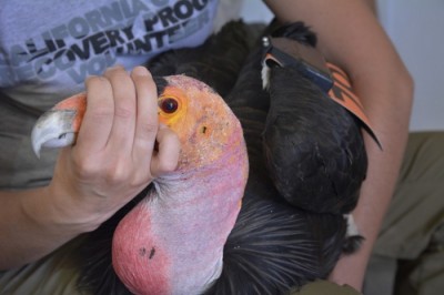
Figure 1. A California condor (Gymnogyps californianus) program volunteer restrains condor #79 (Pitahsi). Photo credit: Jon Myatt of USFWS via Flickr Creative Commons. Click image to enlarge.


1. You are needed

There are many conservation teams that can use your avian veterinary medical skills and experience. Find a project near you or start your own. There are also many international projects that need your support and expertise. Conservation efforts aimed at habitat protection or restoration can also use veterinary consultation. Directly provide your veterinary skills or consult with and teach local veterinarians and students. Provide distance learning and support through email, phone calls, and articles. Provide scholarships for local biologists and veterinarians to travel to your clinic or to conferences where they can acquire the knowledge and skills needed.

To learn more about conservation efforts, visit...

You may also contact your local equivalent of the Environmental Protection Agency, as well as local agencies such as wildlife rehabilitators and sanctuaries, state, county, and city parks, university programs on conservation biology or wildlife management, and national forests, parks, and monuments.


2. Volunteering feels good (or don’t expect to get paid)

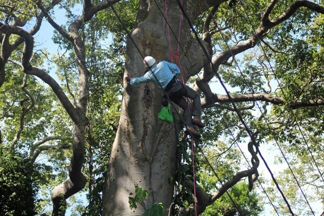
A few projects may be able to pay for veterinary services, some can cover your expenses, but many worthy projects can do neither (Fig 2). Consider volunteering for what may be the experience of your life. Expenses are tax deductible as business expenses or as charitable donations.

climbing nests Ometepe

Figure 2. A volunteer climbing trees to evaluate nests in Ometepe, Nicaragua. Photo credit: Dr. LoraKim Joyner. Click image to enlarge.


3. Go heavily laden; come back empty

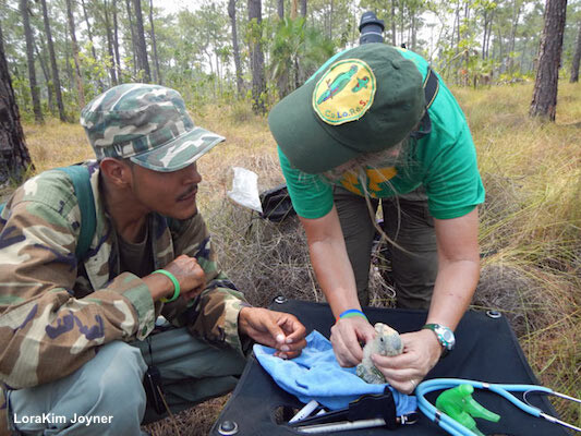
Conservation projects generally do not carry the necessary equipment for physical examination, specimen collection, rudimentary treatment, or record keeping due to limitations in both expertise and funding. When traveling abroad, come “over-prepared”, bringing everything that you may need (see Supplies for the Avian Conservation Medicine Field Kit ) (Fig 3). Valuable time can be spent looking for equivalent pharmaceuticals or equipment in remote locations, only to discover sometimes they are not available at all. Projects that are new or in remote regions often need equipment and supplies, so equipment and supplies that are not used can be left for future use.

Figure 3. Much of the equipment required for a physical exam can be transported abroad. Shown here, demonstrating an avian physical exam in Rio Bravo,  Tamaulipas, Mexico. Photo credit: Dr. LoraKim Joyner. Click image to enlarge.


4. Conservation is for the long haul

In human systems we often overestimate the difference we can make in 5 years and underestimate the difference we can make in 20 years. Conservation projects can take decades to mature and develop. Developing human relations, a funding base, and institutional infrastructure takes time. If possible, plan on a long-term relationship with your project. Stay with the project even when it looks hopeless for the bird’s survival, looks too difficult to navigate human relationships, or looks frustratingly under-supported. Conservation projects include complex ecological and social systems that do change over time if you remain committed to the project and are flexible about your outcome expectations. If you are traveling abroad, stay connected through correspondence throughout the year and plan short visits in subsequent years.


5. Conservation happens all the time

If it is not possible to volunteer your time or donate resources, you can still participate in avian conservation on a daily fashion from your home or clinic.

  • Learn and teach about the conservation status and behavior of species you see in your clinic.
  • Offer resources to colleagues and clients about conservation.
  • “Green” your clinic and home to minimize our negative environmental impact, and decrease your “carbon footprint” in your daily and yearly activities.

Visit Ten Things You Can Do to Promote Avian Conservation for additional information.


6. Avian conservation is about the people

Conservation projects cannot succeed if there is conflict, distrust, burnout, or discontent among the humans. For birds to be saved, the humans require a creative matrix for interdisciplinary synergy. Goals and objectives must be clear, however, even with the best plans and structure, there will be discord. Conflict, in fact, is a part of healthy collaboration. With planning and attention, conflict and tension helps a team develop effectiveness, cohesiveness, focus, and relevancy. Do not be afraid to consult outside of the team, or place on the team someone who understands social systems. Teams and team members can never have too much coaching and support in the human realm.

Ultimately, how you care for others and yourself may be more important than your veterinary expertise. Frontline conservation can be frustrating, depressing, irritating, despairing, physically demanding, and even risky for one’s health. If people aren’t having fun, finding meaning in the work, and contributing creatively, then the team is not doing all it can for the birds and the people in the habitat. Cultivate gratitude for the work itself, regardless of the outcome, and plan for rest and celebration to nurture your well-being. Take time also to mourn and recognize the loss of beauty, lives, habitats, and biodiversity inherent in modern life.


7. There is no right way to “do” avian conservation or medicine—not even yours

The best way to devastate any conservation project is by interjecting “experts” from the outside who tell people the right way to do things without exploring the current status of veterinary care and expertise in the project. People from other cultures, especially those in remote areas with few resources, are sensitive to others that assume other cultures or understandings are flawed, or outdated. People do not work well with others when their hard work is not valued, and such viewpoints can result in an “outsider” versus “insider” dualism.

How can you offer your valuable expertise while also valuing the contribution of others who many not have the skills or experience needed in veterinary matters? Effective conservation veterinarians embrace multiculturalism so that all team voices and experiences are held with respect and honor. As a veterinarian, you can do this in casual conversation and in planned meetings. If possible, consult with a sociologist or who can help your team understand the values and meanings of each other’s actions and words.

Balance offering plenty of time to work with people and unforeseen conditions with working with a sense of urgency. Exigency keeps teams on track and is a responsible response to the alarming threats to birds and their worlds.


8. When working in avian conservation, no clear lines delineate veterinary medicine from biology, ecology, or cognitive ethology.

As part of collecting medical history, performing a physical exam, and suggesting treatment protocols, the veterinarian is in fact taking a history and exam of the entire environment. Therefore veterinarians in conservation familiarize themselves with the natural behavior of a species in relation to its environment and evolution (cognitive ethology), how the species interacts with other species and the environment (ecology), and how the behavior and interspecies interactions produce individual and flock health (biology).

Collecting information for diagnostics and treatment involves the human realm as well. A rudimentary understanding of ethnology and ethno-ornithology helps the veterinarian work with people who are both part of the problem and part of the solution. Ethnology analyzes and compares human cultures, by looking at social structure, language, religion, cultural anthropology, and technology. Ethno-ornithology studies the relationship between people and birds, combining anthropological, cognitive, and linguistic perspectives with natural scientific approaches to the description and interpretation of people’s knowledge and use of birds.

Veterinarians clearly cannot be experts in all these fields, but it is helpful to have a working knowledge of how these fields contribute to the overall knowledge of a species so that the veterinarian can see the complex interactions of birds with their communities of mixed species. Here is an example of how these fields interact with avian medicine. Recently while working in Guatemala determining the health of wild scarlet macaw chicks (Ara macao), investigators found several factors limiting the fledging success rate (Fig 4). Limiting factors included the nesting, feeding and foraging behavior of the parents (biology and ecology), availability of suitable nesting and food trees (ecology), predation (ecology), adaptive behavior of the parents and flocks in different habitats (ethology), and the ability and approaches possible to work with the local biologists, veterinarians, Mayan villages, and drug lords (ethnology and ethno-ornithology).

Poaching of nestlings for the pet trade

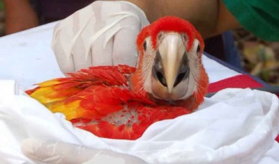
Figure 4. A scarlet macaw chick (Ara macao) in Guatemala. Photo credit:  Dr. LoraKim Joyner. Click image to enlarge.


9. Avian conservation work can be risky to your health.

There are many benefits to working with birds in field conditions, but there are also disadvantages. Working in Central America, for instance, one cannot safely ignore the violence of drug lords, gangs, petty theft and burglary, organized crime, illegal land settlers, and political or military groups (Fig 5). There are also the risks of transportation:  vehicles stranding you in remote areas, car accidents, and the criminal targeting of busses and cars in transportation hubs and roadways. Consult the State Department to discern the threat to your health and safety in each country. Speak to other foreigners who work, live, or travel abroad as well as local people.

parrot patrol

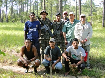
Figure 5. Honduran villagers patrolling local parrot nests. Photo credit:  Dr. LoraKim Joyner. Click image to enlarge.


The physical environment can also pose challenges such as dehydration, unsanitary water supplies, food poisoning, biting and stinging animals and insects, exhaustion, falling debris, and naturally-occurring obstacles, such as sinks, cliffs, falling trees and limbs (Fig 6). Sleeping arrangements may also fall below your usual expectations as you navigate sleeping on the ground, on floors, in cars, and sometimes even on mountainsides. With adequate precaution, practice, support, and equipment these concerns can be minimized.

LoraKim Joyner Bartola River

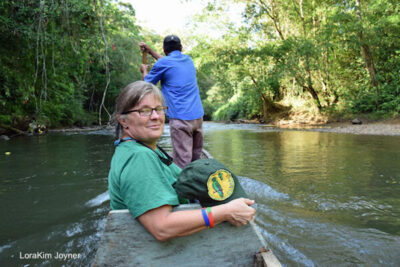
Figure 6. LoraKim traveling up the Bartola River, part of the Indio Maíz Reserve, Nicaragua. Photo credit: Dr. LoraKim Joyner. Click image to enlarge.


10. Avian medicine is not value free.

Each of us comes with preconceived opinions, values, worldviews, ethical norms, and experiences. Much of how we respond to a particular situation works at the emotional, precognitive level and rational discourse may not be helpful, or possible. Despite the reliable data we gather and the scientific method we employ, we must be aware that there are layers upon layers, most of them subconscious, which affect our thinking and our working relationships.

One conservation team, for example, faced the question of whether or not to shoot hawks that were predating an endangered parrot species. Rationally it makes sense to sacrifice some individuals of an abundant species to save an entire other species. However, it can be argued that intentional violence as a means to an end undermines larger values. What an individual or conservation team decides on an issue like this will be based upon complex interactions that will ultimately come down to emotions and previous life experiences that interplay with others in unexpected ways.

What are we to do if so much of how we reason and work with others comes from a precognitive disposition? As veterinarians, we have the training to make sharp critical arguments about animal diagnosis, prognosis, and treatment. The challenge now is to integrate that acumen consciously and effectively with emotional intelligence, moral discernment, empathy, and support for other viewpoints. These are ancient skills, honed by wise women and men in various cultures over millennia. In modernity, we look to reclaim the best of old wisdom while incorporating new scientific and social understanding such as social science, integral ecology, and nonviolent or compassionate communication. Attention to these emerging disciplines will facilitate our service to our beloved birds.


Further reading

Collen C, Bekoff M. Species of Mind: The Philosophy and Biology of Cognitive Ethology. Cambridge, MA: MIT Press; 1999.

Esbjorn-Hargens S, Zimmerman ME. Integral Ecology: Using Multiple Perspectives on the Natural World . Boston: Integral Books; 2009.

Goleman D. Social Intelligence: The New Science of Human Relationships. New York: Bantam Books; 2006.

Joyner L. The socioscientific art of avian medicine. Proc Annu Conf Association of Avian Veterinarians; 2009.

Rosenberg M. Nonviolent Communication: The Language of Life. Encinitas: Puddledancer Press; 2003.

Tinbergen N. On aims and methods in ethology. Zeitschrift für Tierpsychologie 20:410-433, 1963. Available at https://www.esf.edu/biology/faculty/documents/Tinbergen1963onethology.pdf. Accessed April 16, 2024.

Basic Information Sheet: Fennec Fox

Fennec Fox (Vulpes zerda)

Fennec Fox

Photo credit: Yvonne N via Flickr Creative Commons

Natural history


Fennec foxes are highly specialized to desert life and found almost exclusively in arid, sandy regions. Densest populations are found in the central Sahara desert region of North Africa, but the range extends as far north as Morocco, east along the northern Red Sea to Kuwait, and south into northern Nigeria and Chad.

Fennec foxes are nocturnal hunters that dig burrows as shelter to sleep during the day and rear kits.* Scrub vegetation is used to line dens and may be eaten as a source of water.

Fennecs are highly social animals that reside in close family groups usually composed of at least one breeding pair, a litter of immature pups, and some older siblings. A group of foxes may be referred to as a “skulk” or a “leash”.

Fennec foxes are monogamous cooperative breeders that mate for life. Social structure is built around this pairing: each breeding couple have their own territory which is bounded by urine and fecal mounds, which they will vigorously defend.

Social rank among fennecs is communicated mainly through play and visual and tactile communication. Fennec foxes of all ages also make frequent and varied vocalizations such as chatters, whimpers, wails, growls, and shrieks.

*Juvenile fennec fox are general referred to as “kits”, however as canids ‘pups’ is also considered accurate. Youngsters are also referred to as “cubs”.


Sound recording of a fennec fox “singing” Source: Eosin-Y via Wikimedia Commons


Conservation status:


Fennecs once ranged broadly over northern Africa, but sport hunting and human density are shrinking their habitat and numbers and they are considered threatened in the wild.

The IUCN Red List cites fennecs as “data deficient”. The Convention on International Trade in Endangered Species (CITES) places fennecs in Appendix II.

Taxonomy


Class: Mammalia

Order: Carnivora

Family: Canidae

Genus: Vulpes: artic fox, fennec fox, red fox

Formerly, the fennec was classified as a separate genus (Fennecus) due to its rounded skull and weak dentition.


Physical description


Fennec foxes are the smallest canids. Females or “vixens” weigh approximately 0.8 kg (1.8 lb). Adult males or “reynards” reach up to 1.5 kg (3.3 lb) and stand 18 -22 cm (7-8.7 in) at the shoulder.

The most distinctive feature are characteristic large pinnae (15 cm or 5.9 in long), which function to dissipate heat and enhance audition.

A thick buff pelage covers adults, with white along the legs, face, ears and ventrum, in contrast to downy white juveniles. The violet (supracaudal) scent gland, common to all vulpines, is covered in black or dark brown fur, as well as the tail tip. The eyes, rhinal pad, and vibrissae are all black.


Diet


Fennec foxes, like other canids, are omnivores. They commonly hunt and forage for birds, small mammals, reptiles, eggs, carrion, insects, other terrestrial arthropods, leaves, roots, tubers, and fruit. In addition to foraging they commonly cache food. Fennecs obtain much of their food through digging.

Free-ranging fennecs can go without water indefinitely, however fruit, leaves, and roots serve as the sole source of moisture. The captive fennec fox should ALWAYS be offered fresh water.

In captivity, an exotic canine diet (e.g. Mazuri), high-quality dry or canned dog, or cat food is fed. Vegetables, fruits, pinkie mice, rodents, eggs, crickets, mealworms, as well as commercially available raw meat diets are also offered.


Housing


Fennec foxes are adept climbers and can easily escape fenced enclosures. Fennecs should be kenneled while unsupervised; large ferret or cat cages can be suitable. These desert dwellers require low humidity, and good ventilation. Avoid dusty cage substrates.

Fennecs practice site-specific defecation in marking their territories and can thus be litter box trained in a captive environment. Standard clay litter is recommended.

Fennecs can be leash or harness trained, but may be prone to slip out of restraint when startled. Therefore a leash or harness is recommended only for a confined, safe space.


Normal physiologic values


Lifespan: Fennecs can live for up to 10 years in The wild, while captives may survive up to 12 years.
Heart rate (resting) ~118 bpm
Respiratory rate (resting) ~23 bpm
Body temperature 38.2°C (100.8°F)


Anatomy / physiology


Dental formula The fennec fox has the same dental formula as the domestic dog: I3/3, C1/1, PM4/4, M2/3).Compared with other vulpines, their canine and carnassial teeth are reduced.  The teeth are also sharply cuspidate, which may facilitate a partially insectivorous diet.
Special senses Fennecs have highly developed sense of hearing and smell.
Unique physiological
adaptations to desert life:
Fennec fox metabolism is 67% of the rate predicted for an animal of its size.Fennecs will shiver when the ambient temperature drops below 20˚C (68˚F). In hot ambient temperatures the fennec radiates body heat by dilating blood vessels in its feet and large, vascular ears.The enormous ears are able to filter sound through many centimeters of sand, and can detect subtle differences in vocalizations from conspecifics.In contrast to some other canids, the feet are heavily furred to protect the pads from hot desert sand.Fennec foxes are nocturnal hunters. Night vision is enhanced by a reflective tapetum with elliptical pupils.

Fennec foxes allow their body temperature to rise to 40.9˚C (105.6˚F) before beginning to sweat, thereby reducing water loss. Fennecs only lose heat through panting when environmental temperature exceeds 35˚C (95˚F). Panting rates up to 690 breaths per minute have been observed.

Although fennec foxes will drink freely when given the opportunity, laboratory studies suggest that free-ranging fennecs can survive indefinitely without access to free water.

Reproduction Like all foxes, fennecs have three pairs of mammary glands.The breeding season runs from January to February, but vixens remain in estrus for only 1-2 days.Altricial pups (kits) are born after a 50-day gestation in annual litters of two to four pups. For the first 2 weeks pups are cared for in a den by the dam until their eyes open, but full weaning does not occur until nearly 3 months.Parent-raised offspring are weaned by 8-10 weeks of age.Parent-raised offspring are weaned by 8-10 weeks of age, although full weaning may not occur until nearly 3 months.Hand reared kits are sometimes forced weaned although this practice is generally not recommended: Kits are pulled at 10-12 days, and fed a fox milk replacer such as Day One® Formula 35/32 (Fox Valley Animal Nutrition; Lake Zurich, IL). Solid food may be introduced at 1 month in force-weaned animals, and weaning can occur as early as 6 weeks.

Adult size and sexual maturity are reached at 6-9 months of age.

Sexual dimorphism Not pronounced, though males tend to be larger in mass (see physical description above).In general, males (reynards) are a bit more friendly and sociable than are vixens, and may make better pets for a beginning owner. However, they can turn aggressive as they become sexually mature, which makes neutering a very important consideration. One additional benefit is that the males are often simpler to potty train than are the females. However, in addition to the potential aggression, males do have a much stronger odor in general than do females, and they are more likely to “hump” things, particularly as they enter sexual maturity.
Other Like all foxes, paired anal sacs are present. There are also glands located between the toes.


Important medical conditions


Fennec foxes are susceptible to all diseases of domestic dogs
Common clinical presentations include:

  • Obesity
  • Heat stress
  • Dental disease
  • Trauma (bite wounds)
  • Gastrointestinal upset secondary to poor diet and/or stress
  • Fur rings
  • Neoplasia
  • Renal or liver disease
  • Cardiomyopathy
  • Pneumonia
  • Dermatitis (mites, otitis, fleas)
  • Conjunctivitis, corneal lesions (foreign body), glaucoma
  • Neonatal death (nervous mothering)

Fennecs become very nervous and aggressive during breeding and rearing. To prevent neonatal deaths, avoid disturbances until the kits reach 3-4 weeks of age.

Zoonotic potential: Rabies virus, tuberculosis, leishmaniasis

Histoplasmosis and toxoplasmosis have also been described in case reports.


Preventive care


  • Annual to biannual physical examination
  • Fecal parasite testing
  • Killed rabies virus vaccination
  • Recombinant canarypox vector canine distemper virus and canine parvovirus vaccination
  • Flea control
  • Canine heartworm prevention
  • Spay or neuter companion pets, unless these individuals are to be used as part of a breeding program with an experienced fox owner


Restraint


Use same methods of manual or chemical restraint as for canines.

Venipuncture


Jugular vein, cephalic vein, lateral saphenous vein

**Login to view references**

References and further reading