Lymphoid leukosis and Marek’s disease virus are two diseases of the avian leukosis complex. These conditions are commercially important diseases of poultry seen worldwide. Lymphoid leukosis and Marek's disease were the first neoplastic diseases shown to be transmitted and caused by viruses . . .
Pour les vétérinaires. Par les vétérinaires.
Le site Lafervet.com est conçu pour une utilisation par les vétérinaires. Il est ouvert aux vétérinaires diplômés, aux techniciens vétérinaires diplômés, aux animaliers et aux étudiants dans ces domaines.
Créer un compte pour accéder à des articles et des ressources du site.
L'inscription est gratuite.
Para Profesionales Veterinarios. Por Profesionales Veterinarios.
El sitio Lafervet.com es para uso de los profesionales veterinarios. Está abierto a los veterinarios licenciados, técnicos veterinarios licenciados, rehabilitadores licenciados y estudiantes en estos campos.
Cree una cuenta para acceder a los artículos y recursos del sitio.
La registro es gratis.
Already a LafeberVet Member?
Please Login
Veterinary Answers
LafeberVet has teamed with veterinary telemedicine service VETERINARY ANSWERS
Veterinary Answers understands the difficulties you face in your practice. Their goal is to work with you to find the next step in diagnosis or treatment of patients under your care.
Contact Veterinary Answers for:
- Case discussion of challenging cases. Consultations include verbal discussion & written report.
- Development of protocols so you can provide the best standard of care for every species in your practice.
Veterinary Answers consultants in avian & exotic animal medicine include:
- Gwen Flinchum, DVM, MAg, DABVP-Avian
- Douglas Folland, DVM, DABVP-Avian
- Daniel Johnson, DVM, DABVP-Exotic Companion Mammals
- Kenneth Welle, DVM, DABVP-Avian

Fee Schedule:
- Standard consultation $65 ($45 when you mention LafeberVet)
- Extended consultation $75 ($55 when you mention LafeberVet)
- Follow-up consultation $40 ($20 when you mention LafeberVet)
- Std of care protocol consultation $180 ($140 when you mention LafeberVet)
Veterinary Answers Toll Free Phone 877.262.3024
Presenting problem: Paresis, Paralysis, and Tremors in Sugar Gliders
The sugar glider is a small, nocturnal marsupial native to New Guinea and Australia. Sugar gliders are omnivores that eat arthropods and plant products, such as eucalyptus phloem sap, manna, honeydew, nectar, and pollen in the wild. Although there is little medical information available on sugar gliders in captivity, nutritional secondary hyperparathyroidism or metabolic bone disease is recognized as a common problem in this species . . .
Pour les vétérinaires. Par les vétérinaires.
Le site Lafervet.com est conçu pour une utilisation par les vétérinaires. Il est ouvert aux vétérinaires diplômés, aux techniciens vétérinaires diplômés, aux animaliers et aux étudiants dans ces domaines.
Créer un compte pour accéder à des articles et des ressources du site.
L'inscription est gratuite.
Para Profesionales Veterinarios. Por Profesionales Veterinarios.
El sitio Lafervet.com es para uso de los profesionales veterinarios. Está abierto a los veterinarios licenciados, técnicos veterinarios licenciados, rehabilitadores licenciados y estudiantes en estos campos.
Cree una cuenta para acceder a los artículos y recursos del sitio.
La registro es gratis.
Already a LafeberVet Member?
Please Login
Common Chicken Breeds
Chickens are categorized as pure breeds, hybrids, and bantams. A chicken breed is a group of birds with distinctive characteristics. There are more than 500 chicken breeds throughout the world. There are laying breeds, meat breeds, and ornamental breeds. Some breeds are also considered dual purpose or raised for both eggs and meat. Each breed is further subdivided into varieties based on physical characteristic, such as color, comb type, leg feathering, presence of a beard or muffs, or comb type (i.e. single comb white leghorn). Many breeds have a single comb. Rose combs are typically flat and close to . . .
Pour les vétérinaires. Par les vétérinaires.
Le site Lafervet.com est conçu pour une utilisation par les vétérinaires. Il est ouvert aux vétérinaires diplômés, aux techniciens vétérinaires diplômés, aux animaliers et aux étudiants dans ces domaines.
Créer un compte pour accéder à des articles et des ressources du site.
L'inscription est gratuite.
Para Profesionales Veterinarios. Por Profesionales Veterinarios.
El sitio Lafervet.com es para uso de los profesionales veterinarios. Está abierto a los veterinarios licenciados, técnicos veterinarios licenciados, rehabilitadores licenciados y estudiantes en estos campos.
Cree una cuenta para acceder a los artículos y recursos del sitio.
La registro es gratis.
Already a LafeberVet Member?
Please Login
Waterfowl Diseases: A “Cheat Sheet”
Introduction
Although the rare veterinarian routinely deals with large numbers of waterfowl on a regular basis, many avian veterinarians encounter waterfowl only sporadically as wildlife rehabilitation cases, backyard poultry, and/or zoo specimens (Fig 1). When consulting textbooks for help, often a dizzying array of waterfowl diseases are encountered. Some conditions such as “angel wing” and predator trauma are important in captive populations, while infectious diseases like fowl cholera can cause massive die-offs in free-ranging birds. Unless captive populations are exposed to wild birds, the incidence of infectious disease is relatively low, although birds less than 7 weeks of age are at greater risk when compared to adult birds.

Figure 1. Many avian veterinarians encounter waterfowl only sporadically as wildlife rehabilitation cases, backyard poultry, and/or zoo specimens. Photo credit: Getty Images.
Below you will find a collection of differential diagnoses for common clinical problems observed in the anseriform. These abbreviated lists should in no way replace professional judgment when evaluating your patient. This “cheat sheet” is merely designed as an aide or reminder system and should never be used for diagnostic decision-making. Particularly important or common conditions are bolded or linked to disease descriptions (Box 1 through Box 12).
Diseases featured in the ‘Cheat Sheet’
Sudden death
Major rule-outs for acute to peracute death in waterfowl can include:
- Trauma
- Pesticide poisoning
- Chronic renal disease, gout
Other potential causes of sudden death include:
- Aspergillosis (young birds can die peracutely)
- Colibacillosis (ducks)
- Fowl cholera
- Pasteurella anatipestifer
In new duck disease or duck septicemia caused by P. anatipestifer, ducklings usually die within 6-12 hours after the onset of clinical signs. Acute death is seen in older birds.
- Renal coccidiosis (Eimeria truncata)
- Leucocytozoan simondi
- Duck virus enteritis (duck plague)
- Duck virus hepatitis type 1 (death rates up to 100% in ducklings < 1 week)
- Blue-green algal toxins
- Amyloidosis
| Box 1. Fowl Cholera or Avian Cholera |
|---|
| Fowl cholera, or disease caused by Pasteurella multocida, has been reported in many bird species and occurs primarily in free-ranging populations. Dramatic outbreaks, sometimes killing thousands of birds, are reported annually in North America. Although losses can occur any time of the year, outbreaks typically occur during the winter or spring in northern California, Oregon, Nebraska, as well as the Texas panhandle. Bacteria are shed in feces and bodily secretions such as oculonasal discharge and oral discharge. Infection is transmitted by ingestion of contaminated food and water, inhalation, or less commonly inoculation of feet by contaminated debris. In the acute form of disease, some birds may be found dead. Clinical signs can also include:
Death rapidly follows the onset of clinical signs with birds frequently dying within 6 to 12 hours. Chronic fowl cholera can follow an acute outbreak or may arise from a less virulent strain. Clinical signs vary and are related to focal disease like septic arthritis, sinusitis, or respiratory infection. Chronically infected, free-flying birds are the likely source of infection for captive poultry and waterfowl. The classic necropsy finding for the acute form of disease is a bird in good flesh with abundant fat reserves.
Chronic fowl cholera lesions usually involve focal infections with caseous exudate. Important differential diagnoses include duck virus enteritis, erysipelas, E. coli and other bacteremias. Diagnosis relies upon consistent clinical signs, gross necropsy findings, and culture of P. multocida from intestinal fluid or bone marrow. Wright’s stain can also be used to demonstrate bipolar rods in heart blood or liver impression smears. Antimicrobials commonly used against P. multocida include beta-lactam antibiotics, oxytetracycline (50 mg IM followed by 500 g/ton feed x 30d), or enrofloxacin (5 mg/kg PO, IM, SC q12h). Management of disease also relies upon prompt removal and incineration of carcasses as well as cleaning the environment. Pasteurella multocida can live in the environment for up to 3 months, however the organism is easily destroyed by common disinfectants, sunlight, drying, and heat. Therefore good sanitation practices are an important part of disease prevention and control. Additional helpful measures include:
A bacterin is available for valuable flocks with persistent problems. |
| Box 2. Duck Plague or Duck Virus Enteritis (DVE) |
|---|
| Duck plague is an important, acutely contagious viral disease affecting ducks, geese, and swans. The etiologic agent is an alpha-herpesvirus. Duck plague was first documented in the United States in Pekin ducks in New York state. Since then sporadic outbreaks have been documented in commercial operations or park waterfowl. Most outbreaks occur during Apr, May, and June. Anseriforms of all ages can be affected. Highly susceptible species include blue-winged teals (Anas discors), wood ducks (Aix sponsa), and redheads (Aythya americana). The incidence of duck virus enteritis (DVE) is also higher in Muscovy ducks (Cairina moschata), Canada geese (Branta canadensis), and Northern pintails (Anas acuta). Outbreaks can occur in both wild and captive populations, but is disease is relatively rare in free-ranging birds. The herpesvirus that causes DVE is shed in feces and oral secretions. Transmission occurs primarily via ingestion of fecal-contaminated water sources. The virus can survive up to 60 days in water at 4°C (39.2°F) or 30 days at room temperature. Infected birds can be asymptomatic. Clinical disease is usually acute in onset. Both clinical patients and asymptomatic carriers may possess ulcers or cheesy plaques beneath the tongue. Clinical signs can also include:
At necropsy birds are usually in good flesh. Although the severity of lesions can vary, gross lesions often include:
A histopathologist will detect the presence of intranuclear inclusions. Necrosis is identified histologically in the liver, spleen, esophagus, and intestines. There will also be histologic evidence of gastrointestinal hemorrhage and necrosis. Presumptive diagnosis of DVE relies on the presence of consistent clinical signs and gross lesions. Definitive antemortem requires electron microscopy of fecal/oral samples or viral isolation. The liver and spleen are the best tissues for culture. Treatment of the individual patient relies upon aggressive supportive care. Management of an entire flock usually relies upon culling affected birds and disinfecting the premises. Decontaminate the environment by chlorinating water and raising soil pH. Control/prevention measures include:
|
| Box 3. Amyloidosis |
|---|
| Amyloidosis is common in captive waterfowl, but extremely rare in wild birds. In one survey of trumpeter swan (Cygnus buccinator) necropsies, amyloidosis was the primary disease process found. Amyloidosis includes a group of diseases characterized by the abnormal accumulation of amyloid proteins organized into beta-pleated sheets in various tissues. Most avian amyloidosis cases consist of reactive systemic amyloidosis with increased production of serum amyloid A proteins, a normal acute-phase reactant protein. The incidence of amyloidosis increases with chronic inflammatory conditions like pododermatitis, arthritis, and gout as well as aging and social stressors. At necropsy, amyloid deposition is most prevalent in the liver, spleen, and kidney followed by the pancreas and adrenal gland. Affected tissues often appear pale, yellow-brown, firm, and enlarged. |
Non-specific signs of illness
Non-specific signs of illness frequently include weakness, lethargy, reluctance to fly, an unsteady gait, loss of appetite, and fluffed and ruffled feathers. Failure to preen can lead to loss of plumage water repellency. There are a host of potential causes of non-specific signs of illness (Table 1). Important causes in waterfowl include yolk peritonitis, amyloidosis, fowl cholera, renal coccidiosis, traumatic injury, and heavy metal poisoning.
| Table 1. Potential Causes of Non-Specific signs of Illness in Waterfowl | ||
|---|---|---|
| Disease Category | Differential Diagnoses | |
| Metabolic |
|
|
| Nutritional |
|
|
| Inflammatory | ||
| Infectious | Bacterial |
|
| Viral |
|
|
| Protozoal |
|
|
| Toxin |
|
|
| Trauma |
|
|
Lead is a persistent contaminant in the environment, and waterfowl are susceptible to lead poisoning from ingestion of lead pellets and fishing weights. In addition to non-specific signs of illness, clinical signs can include bright green droppings or biliverdinuria, mucoid oral secretions, pallor, and variable neurological deficits, like leg paresis, ataxia, wing droop, and abnormal head position. Waterfowl, particularly Canada geese (Branta canadensis), may also exhibit subcutaneous edema of the head and eyelids.
Conditions affecting the gastrointestinal tract
Foreign body ingestion is a very common problem in waterfowl. Objects such as fish hooks, wire, fishing line, and gunshot are often incidental findings at necropsy but their presence can also be associated with morbidity and mortality.
Potential causes of oropharyngeal plaques include:
- Duck virus enteritis or duck plague
- Derzy’s disease (fibrous material beneath the tongue)
Rule-outs for diarrhea in waterfowl include:
- Fowl cholera
- Avian tuberculosis
- Colibacillosis (ducks)
- Enteritis, colitis caused by Salmonella spp. (most common in young birds)
- Intestinal coccidiosis (occasionally seen in young, wild or captive birds under crowded conditions)
- Acanthocephalans
| Box 4. Acanthocephalans |
|---|
| More than 50 species of thorny-headed worms or Acanthocephalans have been reported in waterfowl. Thorny-headed worms use a hooked proboscis to attach to the intestinal tract setting off an intense inflammatory response. With heavy infestations, the resultant granulomatous, hemorrhagic enteritis leads to malnutrition, morbidity, and even death. |
The appearance of a waterfowl’s stool can sometimes provide a clue to the underlying cause of disease.
- Green or bile-stained watery droppings:
- Lead poisoning
- New duck disease (P. anatipestifer)
- Avian chlamydiosis (domestic ducklings and goslings; rare in the United States but serious outbreaks have been seen in Europe)
- White diarrhea can be due to Derzy’s disease
- Profuse, occasionally bloody diarrhea, may be caused by duck plague
It is possible for Newcastle disease virus to cause watery diarrhea, but most ducks and geese are resistant to clinical disease unless exposed to a virulent strain. It has been theorized that some waterfowl develop chronic infection and may serve as a reservoir of disease.
Emaciation
Potential causes of severe weight loss in waterfowl include:
- Heavy metal poisoning
- Pesticide poisoning
- Oil toxicosis
- Gastrointestinal foreign body ingestion
- Gastrointestinal parasites, such as the nematodes Echinuria uncinata (common proventricular worm), Capillaria contorta, which causes thickening and necrosis of the esophagus, and Amidostomum sp. (ventricular worms)
- Renal coccidiosis (young birds)
- Aspergillosis (young birds)
- Avian tuberculosis
- Colibacillosis (ducks)
- Avian chlamydiosis (domestic ducklings and goslings; rare in the United States but serious outbreaks have been seen in Europe)
Diseases of the juvenile bird
Important diseases of juvenile waterfowl include angel wing and renal coccidiosis caused by Eimeria truncata (Fig 2). Important viral diseases include duck virus hepatitis (affecting ducklings less than 1-2 weeks of age) as well as Derzy’s disease (goslings less than 4-6 weeks). Additional conditions may include yolk sacculitis or omphalitis caused by Gram-negative bacterial overgrowth (e.g. E. coli or Salmonella spp.), Actinobacillus infection, and duck septicemia.
| Box 5. Renal Coccidiosis |
|---|
| Renal coccidiosis caused by Eimeria truncata is a serious disease in domestic ducks and geese. Infection is subclinical in adult birds, but can cause heavy mortality in ducklings and goslings. Flocks housed in crowded surroundings with poor sanitation are at increased risk. Pale, swollen kidneys with multifocal white lesions may be found at necropsy. Oocysts can be identified on renal impression smears and on histopathologic exam. Sulfonamides are the treatment of choice. Control and prevention relies upon strict sanitation and disinfection. It is also helpful to house juvenile birds separate from adults. |
| Box 6. Angel Wing |
|---|
| Angel wing is a conformational abnormality of the wing known by many terms including “airplane wing” and “oar wing”. Angel wing is classically seen in large, juvenile waterfowl as the weight of the growing flight feathers causes the weak muscles of the carpus to rotate the wing outward at the level of the carpus (Fig 2). Known underlying causes include:
The incidence of angel wing decreases when young, growing birds are allowed to swim and dive. The normal waterfowl is in the water as it goes through a molt, which prevents wing dragging from being an important issue. Early treatment of angel wing relies either upon encouraging the bird to swim or bandaging that elevates the wingtip enough to make the bird more comfortable and allow normal feather growth. Tape the wing to itself, and not to the body, for 3 to 5 days. |
Figure 2. Outward rotation of the wing at the level of the carpus or “angel wing”. Photograph by audreyjm529. Click image to enlarge.
Aspergillosis is the most significant fungal disease of young birds. Birds usually become infected by inhalation of A. fumigatus spores in moldy or decaying organic matter.
Lameness
Important causes of lameness include septic arthritis and pododermatitis. Pododermatitis or “bumblefoot” frequently occurs in large-bodied birds such as swans housed on hard, rough surfaces. Obesity and lack of access to swimming facilities can predispose birds to bumblefoot. Most lesions occur on metatarsal pads. Septic arthritis may be caused by Salmonella spp., especially in young birds, and Mycoplasma synoviae.
| Box 7. Mycoplasma synoviae |
|---|
| Mycoplasma synoviae most commonly causes swelling of the hock joint or footpad. A presumptive antemortem diagnosis is based on consistent clinical signs and gross lesions. Definitive diagnosis requires culture or PCR testing. Gross necropsy findings include viscous to caseous exudate within joints and tendon sheaths. Lesions can extend into musculature and air sacs in advanced disease. Antimicrobials used against Mycoplasma spp. may include tylosin, tetracyclines, lincomycin, and spectinomycin. Treatment does NOT eliminate Mycoplasma spp. from a flock. Surgical intervention may be necessary but the prognosis is guarded. It is also important to move the bird to softer ground or grass. Prevention of mycoplasmosis requires that stock be purchased from clean breeder flocks and that strict biosecurity is maintained. |
Other potential causes of lameness include:
- Perosis or medial luxation of the gastrocnemius tendon, enlargement of the hock, bending deformities of tarsal or tarsometatarsal bones
- Arthritis (degenerative joint disease)
- Colibacillosis (ducks)
- Articular gout
- Trauma (bite wound)
- (Avian tuberculosis)
Respiratory signs
Yolk peritonitis is an important cause of respiratory distress in geese. Owners often report a recent history of reproductive activity. Coelomic distension is a common physical exam finding. Delayed molt is also sometimes seen.
Sinusitis caused by Mycoplasma gallisepticum is an important reason for keeping ducks and chickens separate.
There are a variety of other conditions that can cause respiratory signs in waterfowl:
- Duck plague (dyspnea)
- Pox, wet or diphtheritic form (rare, only reported in a mute swan [Cygnus olor])
- Avian influenza (rare sinusitis, domestic ducks seem more susceptible to clinical disease)
- Newcastle’s disease virus (rare, mild respiratory signs)
- Fowl cholera (blood stained nasal d/c, cyanosis)
- New duck disease (occasionally cough, tachypnea, sinusitis)
- Colibacillosis (ducks)
- Avian chlamydiosis (sinusitis in domestic ducklings and goslings; rare in the United States but serious outbreaks have been seen in Europe)
- Aspergillosis (dyspnea in young birds)
- Hemoparasites: Leucocytozoan simondi, Plasmodium spp., Hemoproteus nettionis (dyspnea)
- Nasal leeches (Theromyzon spp.) puddle ducks and swans from northern climates are most susceptible; sneezing, heavy loads can be fatal
- Gape worms or tracheal worms (Syngamus trachea or Cyathostoma sp.) infrequently reported in waterfowl: young, captive birds are most susceptible
- Oil toxicosis
- Botulism (respiratory failure)
- Avian tuberculosis
| Box 8. Avian Influenza |
|---|
| Avian influenza is reported in a variety of bird species including ducks, geese, and swans. The fecal-oral route most commonly transmits the orthomyxovirus causing avian influenza, although virus is also shed in respiratory and ocular secretions. Classic signs of avian influenza can include acute death, respiratory signs like oculonasal discharge and sinusitis, decreased egg production, and occasionally neurological signs. Waterfowl infected with avian influenza are usually asymptomatic although domestic ducks seem more susceptible to clinical disease. When lesions are present, sinusitis is the most common gross necropsy finding in ducks. Additional necropsy lesions can include catarrhal tracheitis, air sacculitis, myocarditis, encephalitis, as well as hemorrhage or congestion in the brain, lung, and gastrointestinal tract. Waterfowl are considered a potential reservoir of avian influenza. Wild waterfowl and free-range commercial ducks are known to play an important role in the spread of disease. Virus can remain stable in lake water for over 200 days. Prevention and control of disease relies upon preventing exposure to wild birds. For instance netting can be set up over pet duck areas. |
Neurologic disease
Important causes of neurologic signs in waterfowl include botulism, lead poisoning, pesticide poisoning, and trauma.
- Exposure to pesticides, such as organochlorines, can cause muscle fasciculations, convulsions, ataxia, and disorientation.
- Central nervous signs seen with lead poisoning can include weakness, drooped wings, erratic flying, leg paresis, ataxia, tremors, convulsions, partial to complete leg paresis, and blindness.
- Blunt force trauma to the head is another potential cause of neurologic deficits. The strong territorial nature of swans may increase the incidence of aggression and subsequent injury.
| Box 9. Botulism |
|---|
| Botulism, also known as “limberneck” or alkali poisoning is caused by exotoxins produced by the Gram-positive, spore-forming anaerobic bacterium Clostridium botulinum. Clostridium botulinum grows best in shallow, alkaline water, especially that overlying limestone with a pH ranging from 5.7-6.2. The organism persists in spore form until environmental temperature reaches 77°F (25°C). Spores can persist in the environment for years, and are resistant to heat and drying. Botulism can occur in wild and captive waterfowl, galliforms, and many other bird species. The distribution of disease is worldwide. In the United States and Canada most outbreaks occur west of the Mississippi river. Clostridium botulinum reproduces and makes toxin within vegetation and dead invertebrates at the water’s edge. Waterfowl then eat invertebrates and vegetation containing C. botulinum exotoxin type C. As waterfowl die, there is a subsequent build-up of fly populations and maggot production within vertebrate and invertebrate carcasses. The maggots concentrate toxin, and are then eaten by additional waterfowl creating a vicious cycle of biomagnification. Major outbreaks are usually associated with summer heat. Environmental conditions that favor bacterial growth and toxin production include:
The toxin affects peripheral nerves, and the clinical signs of botulism are dose-related ascending flaccid paralysis. Birds first exhibit leg paralysis, propelling themselves along with their wings. Paralysis then extends to the wings, nictitans, neck muscles and even the tongue. Birds often die by drowning or cardiopulmonary failure. There are no gross or microscopic lesions in birds with botulism although the stomach is frequently empty. A presumptive diagnosis is based on history, clinical signs, and an absence of obvious gross lesions. Definitive diagnosis relies upon mouse bioassay testing in which botulinum serum is injected into mice. Individual patients can be provided with aggressive supportive care. Gavage birds with charcoal slurry, tube feed as needed, while propping birds up or maintaining them in slings to prevent aspiration. Manage large flocks by providing easy access to fresh water, food, and shade. Antitoxin injections (where available) have also been credited with high rates of recovery of some waterfowl species. Control and prevention relies upon the removal of potential sources of Clostridial organisms and toxin from the environment such as:
|
Additional causes of neurological disease include:
- Vitamin E deficiency (encephalomalacia)
- Erysipelas: ataxia
- Fowl cholera: convulsions, opisthotonus, circular swimming; erratic flight and landings
- New duck disease (P. anatipestifer): central nervous system signs can include ataxia, tremors, abnormal swimming behavior, opisthotonos, and/or seizures
- Pseudomonas/Aeromonas spp. (flaccid paralysis in swans, especially during summer months)
- Salmonella spp: central nervous signs, most common in young birds
- Avian chlamydiosis: ataxia in domestic ducklings and goslings; rare in the United States but serious outbreaks have been seen in Europe)
- Avian influenza (very rare cause of neurologic signs)
- Derzy’s disease
- Duck plague: CNS signs – difficulty standing, ataxia, opisthotonos, convulsions
- Duck virus hepatitis (DVH Type 1): loss of balance, ataxia, opisthotonus
- Eastern equine encephalitis: neurologic deficits in imported species like Pekin ducklings
- Newcastle’s disease virus (rare cause of ataxia)
- Aspergillosis: ataxia or torticollis
- Blue-green algal toxins caused by summer algal blooms (Anabaena flosaquae): opisthotonos, convulsions
| Box 10. Duck Virus Hepatitis |
|---|
| Duck viral hepatitis or DVH type 1 is a worldwide disease of waterfowl caused by a picornavirus. Ducklings less than 1 week of age are particularly susceptible with mortality rates reaching 100%. Up to 50% of 1-3 week old ducklings die, while the death rate is negligible in ducklings over 4-5 weeks. An exception to this rule of thumb is the Muscovy duck (Cairina moschata), which is particularly prone to DVH with high mortality rates up to 6 weeks of age. Disease is transmitted by the fecal/oral route, aerosol, vertically, and via fomites. Survivors can excrete virus for up to 8 weeks post-infection. The clinical picture typically involves acute onset, rapid spread of clinical signs and death within 3-4 days. Ducklings may display non-specific signs of illness such as lethargy and separation from the flock as well as central nervous system signs such as opisthotonus and ataxia. Antemortem diagnosis relies upon antibody titers. Gross necropsy findings often include hepatomegaly with punctate or ecchymotic hemorrhages. Occasional findings include splenomegaly and mottling of the kidneys. The most consistent histologic finding is severe hepatic necrosis. Additional diagnostic tests include viral isolation and direct fluorescent antibody testing of the liver. Management of DVH typically relies upon strict quarantine (8 weeks minimum) and supportive care. Intramuscular injections of DVH Type 1 antiserum have also been found to be effective in susceptible ducklings in the face of an outbreak. Control and prevention relies upon strict isolation of young ducklings and vaccination of breeder ducks and/or vaccination of ducklings. |
When faced with a waterfowl suffering from presumptive neurologic disease, two additional rule-outs must be considered.
- Capture myopathy, causing leg paralysis and death, has been described in large birds like the Canada goose.
- Unfortunately neurologic signs can also herald imminent death in waterfowl as terminal convulsions and opisthotonos occur with many disease processes.
Ocular disease
- New duck disease (P. anatipestifer): serous to purulent ocular discharge, conjunctivitis
- Colibacillosis (ducks)
- Avian chlamydiosis: conjunctivitis, tearing in domestic ducklings and goslings; rare in US but serious outbreaks have been seen in Europe
- Nasal leeches (Theromyzon spp.): conjunctivitis, corneal damage, even blindness; puddle ducks and swans in northern climates are most susceptible
- Duck plague: oculonasal discharge, conjunctivitis
- Derzy’s disease: oculonasal discharge, swollen eyelids, conjunctivitis
| Box 11. Derzy's Disease |
|---|
| Derzy’s disease is caused by a highly resistant goose parvovirus that infects goslings (< 4-6 weeks) and ducklings (< 4 weeks). Derzy’s disease affects all domestic goose breeds, Canada geese, and snow geese (Chen caerulescens) as well as Muscovy ducks. Disease is primarily seen in Europe and Asia. Virus is transmitted by fecal-oral and vertical routes. Clinical signs vary with age.
Survivors are persistently infected and resistant to clinical disease. Available antemortem diagnostic tests include antibody titers and fecal electron microscopy. Definitive diagnosis relies upon viral isolation in goose or Muscovy duck eggs or virus neutralization testing. Gross necropsy findings in acute disease can include cardiomegaly, a pale myocardium, hepatomegaly with gray-white areas of necrosis, as well as a congested spleen and pancreas. Chronic goose parvovirus lesions can include serofibrinous pericarditis, perihepatitis, ascites, pulmonary edema, liver dystrophy, and/or catarrhal enteritis. The most significant histologic finding are intranuclear inclusions within the liver, spleen, and intestines. Management of Derzy’s disease relies upon supportive care. Prevention relies upon elimination of adult carrier geese and Muscovy ducks as well as vaccination of adult breeder birds. The virus is susceptible to bleach, stabilized chlorine dioxide, and glutaraldehyde. |
Dermatologic lesions
Feather loss
The mating process in ducks frequently results in a bald neck on the female. Some breeds like Khaki Campbell drakes are particularly active and can cause significant feather loss when there is only one or two ducks per drake.
Frostbite
Frostbite can be observed in species from tropical or subtropical regions like whistling ducks. Lesions begin as pale areas in webbing or on the knob that can progress to dry gangrene.
Loss of waterproofing
Waterproofing of the plumage is maintained through careful and frequent preening. Anything that disrupts this process will interfere with waterproofing, leading to a soggy appearance known as “wet feather”.
Common causes include:
- Lice leading to overpreening
- Chemical contamination (e.g., oil)
- Mold spores, particularly from willow trees, can fall on the plumage
Scratching at face
Nasal leeches (Theromyzon spp.) are a classic cause of head shaking and scratching at the face. Puddle ducks and swans in northern climates are particularly susceptible to infestation.
Additional causes of dermatologic lesions include:
- Avipoxvirus, dry form: wart-like nodules on featherless areas, usually legs and feet (reported only occasionally in wild birds)
- Derzy’s disease
- Erysipelas
- Fire ants
- Maggots
| Box 12. Erysipelas |
|---|
| Erysipelothrix rhusiopathiae is a small, Gram-positive facultative anaerobic bacillus resistant to many chemical and environmental factors. Erysipelas primarily affects turkey of all ages, but outbreaks have been reported in other fowl like ducks and geese. Bacteria are introduced through breaks in the skin or mucosa. Contaminated soil or carrier birds may serve as sources of infection. Some outbreaks have been associated with poultry housed in areas previously occupied by swine and sheep. Clinical signs include non-specific signs of illness, ataxia, skin lesions, and acute death. Gross necropsy lesions consistent with septicemia can include:
A presumptive diagnosis is supported by demonstrating beaded, pleomorphic, Gram-positive rod-shaped bacteria in smears of liver, spleen, heart blood, or bone marrow. Definitive diagnosis relies upon culture. Effective antimicrobials include first-generation beta-lactams or macrolide antibiotics like erythromycin and azithromycin. Control relies upon cleaning and disinfection with 1-2% sodium hypochlorite. |
Prolapse of the phallus
Important differentials for prolapse of the phallus include:
- Duck virus enteritis has been associated with prolapse of the phallus.
- Frostbite
- Excessive sexual stimulation
- Trauma, including over exuberant vent sexing
Prolapse of the duck penis is a common problem, particularly in light breeds like the Indian runner duck. Unless the problem is recognized early, the phallus ulcerates and becomes necrotic. Surgical amputation is often required.
Summary
Medical conditions commonly seen in captive waterfowl include pododermatitis, foreign body ingestion, toxicoses, nutritional disorders, exposure, and predator trauma. Unless captive populations are exposed to wild birds, the incidence of infectious disease is relatively low. Hatchlings are at greater risk for infectious disease when compared to adult birds.
References
Physical Examination of the Avian Patient
Be prepared for your next bird patient. Review the basic approach to the avian physical examination, including history, review of signalment, and visual examination. Key parts of the exam will vary, but generally include a body weight in grams, the oropharynx, crop, sternum, coelom, and vent. The fundus should be routinely evaluated in trauma patients . . .
Pour les vétérinaires. Par les vétérinaires.
Le site Lafervet.com est conçu pour une utilisation par les vétérinaires. Il est ouvert aux vétérinaires diplômés, aux techniciens vétérinaires diplômés, aux animaliers et aux étudiants dans ces domaines.
Créer un compte pour accéder à des articles et des ressources du site.
L'inscription est gratuite.
Para Profesionales Veterinarios. Por Profesionales Veterinarios.
El sitio Lafervet.com es para uso de los profesionales veterinarios. Está abierto a los veterinarios licenciados, técnicos veterinarios licenciados, rehabilitadores licenciados y estudiantes en estos campos.
Cree una cuenta para acceder a los artículos y recursos del sitio.
La registro es gratis.
Already a LafeberVet Member?
Please Login
Presenting problem: Retained Spectacles in Reptiles
Like other shedding problems, retained spectacles or "eye caps" are a sign of an underlying problem related to patient health or husbandry. If retained spectacles are not removed, they can interfere with vision, damage the eye, and/or serve as a source of infection . . .
Pour les vétérinaires. Par les vétérinaires.
Le site Lafervet.com est conçu pour une utilisation par les vétérinaires. Il est ouvert aux vétérinaires diplômés, aux techniciens vétérinaires diplômés, aux animaliers et aux étudiants dans ces domaines.
Créer un compte pour accéder à des articles et des ressources du site.
L'inscription est gratuite.
Para Profesionales Veterinarios. Por Profesionales Veterinarios.
El sitio Lafervet.com es para uso de los profesionales veterinarios. Está abierto a los veterinarios licenciados, técnicos veterinarios licenciados, rehabilitadores licenciados y estudiantes en estos campos.
Cree una cuenta para acceder a los artículos y recursos del sitio.
La registro es gratis.
Already a LafeberVet Member?
Please Login
Estimating Energy Requirements
Introduction
Exotic animal veterinarians are well aware of the differences between energy expenditure in small and large species. Larger animals use more energy overall, however when energy expenditure is divided by body weight (kcal/kg/day), large animals use relatively less energy than small animals (Fascetti 2012) (Fig 1).

Figure 1. Proportionally the energy requirements of a large species like the ostrich (Struthio camelus) (left) (Photo by S. Nygaard) are much lower than that of a tiny species like the marsh wren (Cistothorus palustris) (right) (Photo by USFWS)
Basal metabolic rate
Definition
The basal metabolic rate or BMR, also known as basal energy expenditure or basal energy requirement (BER), is defined as the amount energy expended by a healthy animal under thermoneutral conditions 12 hours after a meal. The animal is conscious but not exercising.
Practical applications
The basal metabolic rate applies primarily to human nutrition because the conditions needed to measure BMR are so stringent that it is virtually impossible to do so in animals.
Resting energy rate
Definition
The resting energy rate (RER) or resting metabolic rate is defined as the number of calories required for an animal to maintain homeostasis while resting quietly. The resting metabolic rate is similar to BMR but includes energy expended for recovery from physical activity and feeding.
Practical applications
The RER is a parameter commonly used in animal nutrition.
There are multiple ways to calculate RER, but each method is based on the following equation in which a pre-determined K value is multiplied by body weight in kilograms: K (Wkg)0.75 = kcal/day
In dogs and cats, one method is based on body weight (Table 1).
| Body weight | Formulas used to calculate recommended kcal/day |
| < 2 kg | 70 (BWkg)0.75 |
| 2-45 kg | 30 (BWkg) + 70 |
Faster metabolic rates translate into higher K factors for exotic animal patients (Table 2 2).
BER = K (Wkg)0.75 = kcal/day
| Taxonomic group | K value |
| Psittacine birds | 175 |
| Passerine birds | 175 |
| Aquatic non-passerine birds | 150 |
| Terrestrial non-passerine birds | 125 |
Alternatively, RER can also be calculated as 1.25 x BER. Traditionally RER was then multiplied by an “illness factor” between 1.0 and 1.5 to account for increases in metabolism associated with different conditions and injuries however this practice has fallen out of favor (see overfeeding below). HOWEVER current recommendations place much less emphasis on these subjective “illness factors”. Instead RER is used as an initial estimate or starting point of a critically ill animal’s energy requirements. The patient’s response is then monitored closely, and adjustments in nutritional support are made as needed.
Overfeeding
Use of “illness factors” (see above) put the patient at risk for overfeeding. Potential sequelae to overfeeding include vomiting, diarrhea, cramping, nausea, electrolyte disturbances, hyperglycemia, hepatic dysfunction, and even respiratory distress.
Consequences of overfeeding
- Vomiting, diarrhea, cramping, nausea
- Electrolyte disturbances
- Hyperglycemia
- Hepatic dysfunction
- Dyspnea
Maintenance energy requirement
The maintenance energy requirement is defined as the energy required for a moderately active adult in a thermoneutral environment. This includes the amount of energy needed to maintain an animal at its current weight and body condition.
Activities such as flying and swimming require large amounts of energy. These activities generally burn up five to ten times the energy of BER.
Caloric content of select diets
There are a number of reputable products available for enteral feeding of exotic animal patients (Box 3).
Box 3. Caloric content of select diets
| Manufacturer | Product | kcal per gram | kcal per ml* |
| EmerAid LLC | EmerAid Intensive Care Carnivore | 4.98 | 1.60 |
| EmerAid LLC | EmerAid IC Herbivore | 2.95 | 1.32 |
| EmerAid LLC | EmerAid Sustain Herbivore | 3.04 | 1.00 |
| Emeraid LLC | EmerAid IC Omnivore | 4.09 | 1.86 |
| Harrison’s Bird Foods | Recovery Formula | 3.92 | n/a |
| Kaytee Exact | Hand-Feeding Formula | 3.840 | n/a |
| Kaytee Exact | Macaw Hand-Feeding Formula | 3.980 | n/a |
| Roudybush | Formula AA | 3.450 | n/a |
| Kilocalories (kcal) milliliters (ml) information not available (n/a). *When prepared as directed. |
|||
References
Care of the Senior Rabbit
With proper care, pet rabbits can live long, happy lives. In this client education handout, we explore the aging changes that can be expected in the senior house rabbit including common health problems. Veterinary screening as well as home care of the geriatric rabbit are also explored.
Veterinary Answers Case Study: 8-year Old Iguana with Ascending Tail Necrosis
The goal of this case study is to reinforce and highlight common concepts, situations, and presentations that reptile veterinarians encounter on a regular basis, while also expanding knowledge by including content not entirely available in textbooks. This case study is based on a report prepared by Veterinary Answers consultant Dan Johnson, DVM, DABVP (Exotic Companion Mammal). An 8-year old ovariectomized green iguana presents today for gradual onset of ascending tail necrosis (gangrene) over weeks to months . . .
Pour les vétérinaires. Par les vétérinaires.
Le site Lafervet.com est conçu pour une utilisation par les vétérinaires. Il est ouvert aux vétérinaires diplômés, aux techniciens vétérinaires diplômés, aux animaliers et aux étudiants dans ces domaines.
Créer un compte pour accéder à des articles et des ressources du site.
L'inscription est gratuite.
Para Profesionales Veterinarios. Por Profesionales Veterinarios.
El sitio Lafervet.com es para uso de los profesionales veterinarios. Está abierto a los veterinarios licenciados, técnicos veterinarios licenciados, rehabilitadores licenciados y estudiantes en estos campos.
Cree una cuenta para acceder a los artículos y recursos del sitio.
La registro es gratis.
Already a LafeberVet Member?
Please Login
Rabbit Introductions
Does my rabbit really need a companion? Many experts on house rabbit care agree that most individuals are not meant to live in solitude, away from members of their own kind. This client education handout discusses house rabbit companionship and the challenging process of rabbit introductions.
Download the Rabbit Introductions PDF Handout with color photos or the black and white version.
Aggression in Rabbits
To the uninitiated, rabbits have a reputation for being docile, passive creatures. Any aggressive actions from a house rabbit can be surprising–even alarming–to new owners. In this client education handout, fights between rabbits as well as rabbit aggression towards people are discussed.
Ariana Finkelstein, DVM

Ariana Finkelstein DVM, was born and raised in the Tri-state area. She graduated as a George H. Cook Scholar with a Bachelor of Science Degree in Animal Science from Cook College, Rutgers University. She then attended The Ohio State University, College of Veterinary Medicine. After working with companion and exotic animals for 5 years in private practice, Dr. Finkelstein completed an internship in avian, exotic and zoological medicine at Oklahoma State University. Upon completion of the internship, Dr. Finkelstein worked at the San Antonio Zoo. After 2 years at the zoo, Dr. Finkelstein returned to small animal practice. She now practices at VCA Florida Veterinary League where she is available for exotic animals exclusively one day a week.
Dr. Finkelstein has contributed many images to LafeberVet.
Understanding the Avian Egg: From Outside to In
Introduction
The bird egg, sometimes called a “miracle of packaging”, can be intimidating for the avian veterinarian. This amazing structure comes with a plethora of new vocabulary terms, complex anatomy, and intricate physiology (Fig 1). LafeberVet’s “Understanding the Avian Egg” describes the basics of egg anatomy from the eggshell to the embryo with a brief description of hatch.

Figure 1. Schematic illustrating egg anatomy by de:Benutzer: Horst Frank via Wikimedia Commons. (1) Eggshell (2) Outer shell membrane (3) Inner membrane (4) Chalaza (5) Outer thin albumen (6) Inner thick albumen (7) Vitelline membrane (8) Nucleus of pander (9) Germinal disc (blastoderm) (10) Yellow yolk (11) White yolk (12) Internal albumen (13) Chalaza (14) Air cell (15) Cuticula. Click image to enlarge
Cuticle
The outermost component of the eggshell is called the “cuticle”. Composed of protein, polysaccharide, and lipid, the thin, waxy cuticle protects the egg from water evaporation as well as microbial invasion. The cuticle is found on most, but not all, bird eggs (Johnson 1999).
Testa
The testa is the largest portion of the eggshell consisting primarily of calcite, the crystalline form of calcium carbonate. The testa can be further divided into external, spongy, and mammillary layers.
The external layer, also known as the ‘outermost crystal layer’ is present in many, but not all birds. This dense crystalline layer measures anywhere from 3-8 μm in thickness and serves as a transitional zone to the outer surface of the egg.
The spongy or palisade layer makes up approximately two-thirds of the shell. Approximately 200 μm thick, the palisade layer contains calcified crystals intermixed with an organic matrix, that consists of a series of protein layers and mucopolysaccharides.
The thin, inner cone or mammillary layer consists of organic cores and cone-shaped knobs embedded within the outer shell membrane below. Cores are made primarily of protein, but contain carbohydrates and mucopolysaccharides. Deposition of calcium carbonate crystals begins upon these organic cores (Johnson 1999).
Normal areas of incomplete crystallization create thousands of funnel-shaped openings or pores within the eggshell. The number of pores varies with the species and has been shown to correlate with metabolic demands. The number of pores increases as egg weight decreases. The diameter of the pores ranges from 0.3-0.9 μm. The pores serve as a major site of gas exchange (water and carbon dioxide) as well as water vapor transport across the eggshell. The exchange of gases and water occurs primarily by passive diffusion.
When eggs are pigmented, variable amounts of blue-green biliverdin and/or red-brown porphyrin crystals are deposited within the inorganic portion of the testa and sometimes the cuticle (Fig 2).

Figure 2. Robin (Turdus migratorius) eggs are a beautiful blue color. Photo credit: Soltrcy. Click to enlarge.
Shell membranes
If you’ve ever peeled a boiled egg, you’ve noted the presence of membranes beneath the eggshell. Semipermeable shell membranes serve as a barrier against microbial invasion and they also play a minor role in egg respiration (Nys 2007).
The outer shell membrane is in contact with the mammillary layer of the testa, while the inner shell membrane is in direct contact with albumen (see below). The inner and outer shell membranes are firmly attached except in a small region, usually at the blunt end of the egg.
Air cell
The air cell, located at the blunt end of the egg, is created by separation of the inner and outer shell membranes. The air cell is created as the egg cools after oviposition or egg laying. Creation of the air cell is a dangerous time in egg development, as this is the most likely time for microbes to be drawn into the egg.
Go to Hatch below for more information on the air cell.
Chorion
The chorion is a highly vascular outer embryonic membrane that is found in not
only birds, but also reptiles and mammals. The chorion is another
semipermeable membrane that participates in gaseous exchange.
Amnion
The amnion is a fluid filled sac that surrounds and cushions the embryo. The amnion serves as a source of protein, water, and minerals throughout embryological development. Albumen drains into amnion, allowing this structure to serve as a protein source.
There are four distinct layers of albumen (Johnson 1999):
- Outer thin (fluid) layer
- Outer thick layer
- Inner thin (liquid) layer
- Inner thick chalaziferous layer
Chalazae
The inner thick layer of albumen or the chalaziferous layer, consists of a dense,
gel-like albumen that holds the yolk within the center of the egg. Chalaze
consists of a double strand at the sharp end of the egg and a single
strand at the rounded end.
Allantois
The allantois is another vascular fetal membrane of reptiles, birds, and mammals that forms as a pouch from the hindgut.
Eggs need to be turned regularly, and the reason for this relates to the existence of the allantois and amnion. Regular turning stirs nutrients and waste material within the egg, while preventing fetal membranes from adhering to the embryo. Psittacine eggs pulled for artificial incubation are typically turned every 1 to 2 hours. Inadequate turning rates have an adverse effect on embryonic development and are associated with increased mortality rates.
Yolk
The yolk membrane, or vitelline membrane, consists of four membranes that separate the yolk sac from albumen. Yolk is another important nutrient, providing protein and lipids in late development and during the first few days of life.
Not all yolk is the same. Yellow yolk is higher in fat, while white yolk is higher in protein. The latebra is a region of white yolk that can be seen during candling. The latebra consists of small round center, a neck, and a cone-shaped disc that lies beneath the germinal disc.
Candling is a technique used to evaluate the egg (Box 1). Candling is used to look for evidence for:
- Fertility or infertility
- Cracks
- Shell thickness
- Membrane and vessel integrity
- Air cell size and shape
- Yolk size
- Chick size, position, and/or movement
Visit the University of California Division of Agriculture and Natural Resources Publication 8134: Egg Candling and Breakout Analysis for additional information and illustrative color photographs.
| Box 1. Distinguishing fertile and infertile eggs during candling | ||
|---|---|---|
| Appearance of… | Fertile egg | Infertile egg |
| Blood vessels | Radiate out like a spider | Blood ring (blood vessels disintegrate and blood diffuses into embryonic membranes |
| Germinal disc | Blastoderm: White spot on yolk consisting of an area opaca (distinct ring) surrounded by the area pellucida (clear region) | Blastodisc: Lacks organization, looks like a tuft of cotton |
Fetal position
The normal chicken embryo is positioned with the head closer to the blunt end of the egg and the air cell. The beak normally sits beneath the right wing tip (Fig 3). Because of their shorter, thicker necks, the psittacine beak merely lies close to the wing.
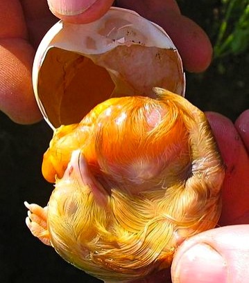
Figure 3. Chick embryo. Photo credit: Crispin Semmens. Click to enlarge.
For more information on malpositions, go to Chick Embryo Malpositions by the University of Florida’s IFAS Extension.
Hatch
The process of hatch begins with drawdown when the air cell expands and extends down the side of the egg. Drawdown occurs approximately 24 to 48 hours prior to internal pip.
Internal pip is caused by rising carbon dioxide (CO2) levels within the egg. When CO2 levels reach a critical level, the overdeveloped muscles on the back of the (otherwise) scrawny chick’s neck begin to twitch. Repeated twitching allows the egg tooth to penetrate the inner shell membrane, allowing the chick’s head to pops into the air cell. This is a crucial moment as the chick’s respiration now changes from chorioallantoic to pulmonary. Once internal pip has occurred, the egg no longer needs to be turned and the chick can actually be heard peeping within the egg.
Eventually the chick uses up the small amount of oxygen present within the air cell. As CO2 levels begin to rise again, muscular twitching causes the egg tooth to penetrate the eggshell. Twitching of abdominal muscles also causes the yolk sac to be pulled within the coelom. Also the time frame is species-specific, external pip or breakout generally occurs within 36-72 hour of internal pip. Assisted hatch or ovotomy should never be initiated prematurely as there are a host of possible complications including bacterial infection, iatrogenic damage to the chick, and unabsorbed yolk sac. If associated vasculature has not receded the chick can also suffer fatal hemorrhage.
References
Nebulization of Avian Patients
Nebulization is used in avian medicine to deliver certain medications, usually antibiotics or antifungals, directly to the respiratory tract. Nebulization can provide hydration to the mucous membranes as well as provide an expectorant effect to help clear debris from the respiratory tract . . .
Pour les vétérinaires. Par les vétérinaires.
Le site Lafervet.com est conçu pour une utilisation par les vétérinaires. Il est ouvert aux vétérinaires diplômés, aux techniciens vétérinaires diplômés, aux animaliers et aux étudiants dans ces domaines.
Créer un compte pour accéder à des articles et des ressources du site.
L'inscription est gratuite.
Para Profesionales Veterinarios. Por Profesionales Veterinarios.
El sitio Lafervet.com es para uso de los profesionales veterinarios. Está abierto a los veterinarios licenciados, técnicos veterinarios licenciados, rehabilitadores licenciados y estudiantes en estos campos.
Cree una cuenta para acceder a los artículos y recursos del sitio.
La registro es gratis.
Already a LafeberVet Member?
Please Login
Kimberly Mickley, DVM, DABVP (Avian Practice)
Dr. Kimberly Mickley is board-certified in avian medicine and surgery. She received an Associates of Science Degree in Veterinary Technology from Central Carolina Community College in North Carolina. After several years working as a licensed veterinary technician, she decided to pursue her dream of becoming a veterinarian. She received her degree from Iowa State University, College of Veterinary Medicine. After graduation, she accepted a small animal medicine-surgery internship at Angell Animal Medical Center in Boston, MA where she also continued to pursue her passion for exotic animals. She then completed a residency in avian and exotic medicine at Angell. Dr. Mickley currently practices at Lehigh Valley Animal Hospital in Allentown, PA.
Prevent Problems with Boas and Pythons
According to the Humane Society of the United States,17 deaths and many more injuries have been related to large constrictors since 1978. Given the tens of thousands of large boas and pythons sold, the incidence of fatalities and injuries is relatively low, however every incident—including the death of four babies in their cribs and three additional children—is particularly tragic since these cases are completely preventable. In this client education handout, safety tips involving snake feeding, housing, and behavior are discussed.
Breeder Checklist
…or How to Question the Source of Your Future Lifelong Companion:
By the time an owner is ready to select the breeder or shop to purchase their companion parrot, they have hopefully done their homework.
Many of the behavioral problems commonly seen in pet birds can be traced back to difficulties encountered during early development like forced weaning and poor socialization. It is essential that the prospective companion parrot owner identifies a breeder or shop that is sincerely concerned with the physical and psychological development of their chicks. How will the prospective owner know? This checklist can be used as an aid when interviewing a breeder or shop.
Download the PDF version of this client education handout, or modify the DOCX version for your veterinary hospital.
Aquatic and Semi-Aquatic Turtles, Care of
Aquatic turtles are personable, popular pets, however their upkeep can be labor intensive. This educational handout will help your client understand how to care for and maintain semi-aquatic turtle species such as sliders (Trachemys spp.), painted turtles (Chrysemys), pond turtles . . .
Pour les vétérinaires. Par les vétérinaires.
Le site Lafervet.com est conçu pour une utilisation par les vétérinaires. Il est ouvert aux vétérinaires diplômés, aux techniciens vétérinaires diplômés, aux animaliers et aux étudiants dans ces domaines.
Créer un compte pour accéder à des articles et des ressources du site.
L'inscription est gratuite.
Para Profesionales Veterinarios. Por Profesionales Veterinarios.
El sitio Lafervet.com es para uso de los profesionales veterinarios. Está abierto a los veterinarios licenciados, técnicos veterinarios licenciados, rehabilitadores licenciados y estudiantes en estos campos.
Cree una cuenta para acceder a los artículos y recursos del sitio.
La registro es gratis.
Already a LafeberVet Member?
Please Login
Ear Abscess in Turtles
Ear or aural abscess is extremely common in box turtles and aquatic or semi-aquatic turtles like red-eared sliders. This educational handout will help your client understand this clinical problem, the veterinary treatment commonly required as well as follow-up care and monitoring . . .
Pour les vétérinaires. Par les vétérinaires.
Le site Lafervet.com est conçu pour une utilisation par les vétérinaires. Il est ouvert aux vétérinaires diplômés, aux techniciens vétérinaires diplômés, aux animaliers et aux étudiants dans ces domaines.
Créer un compte pour accéder à des articles et des ressources du site.
L'inscription est gratuite.
Para Profesionales Veterinarios. Por Profesionales Veterinarios.
El sitio Lafervet.com es para uso de los profesionales veterinarios. Está abierto a los veterinarios licenciados, técnicos veterinarios licenciados, rehabilitadores licenciados y estudiantes en estos campos.
Cree una cuenta para acceder a los artículos y recursos del sitio.
La registro es gratis.
Already a LafeberVet Member?
Please Login
Body Condition Scoring the Rabbit
Body condition scoring is a technique used to assess body condition in many species. Although no official scoring system exists for rabbits, evaluation of rabbit body condition can be adapted from methods used in cats, dogs, and large animals. This brief article describes specific examination techniques, before reviewing obesity in rabbits and client education . . .
Pour les vétérinaires. Par les vétérinaires.
Le site Lafervet.com est conçu pour une utilisation par les vétérinaires. Il est ouvert aux vétérinaires diplômés, aux techniciens vétérinaires diplômés, aux animaliers et aux étudiants dans ces domaines.
Créer un compte pour accéder à des articles et des ressources du site.
L'inscription est gratuite.
Para Profesionales Veterinarios. Por Profesionales Veterinarios.
El sitio Lafervet.com es para uso de los profesionales veterinarios. Está abierto a los veterinarios licenciados, técnicos veterinarios licenciados, rehabilitadores licenciados y estudiantes en estos campos.
Cree una cuenta para acceder a los artículos y recursos del sitio.
La registro es gratis.
Already a LafeberVet Member?
Please Login
Presenting problem: Shell Fractures in Chelonians
Shell fractures are one of the more common presentations of turtles and tortoises to the private exotic animal practitioner. Shell fractures are frequently caused by vehicular trauma, lawn mowers, predation by dogs and raccoons, or drops from balconies or porches. This presenting problem article reviews the key points of urgent care for this traumatic injury as well as the principles of case management . . .
Pour les vétérinaires. Par les vétérinaires.
Le site Lafervet.com est conçu pour une utilisation par les vétérinaires. Il est ouvert aux vétérinaires diplômés, aux techniciens vétérinaires diplômés, aux animaliers et aux étudiants dans ces domaines.
Créer un compte pour accéder à des articles et des ressources du site.
L'inscription est gratuite.
Para Profesionales Veterinarios. Por Profesionales Veterinarios.
El sitio Lafervet.com es para uso de los profesionales veterinarios. Está abierto a los veterinarios licenciados, técnicos veterinarios licenciados, rehabilitadores licenciados y estudiantes en estos campos.
Cree una cuenta para acceder a los artículos y recursos del sitio.
La registro es gratis.
Already a LafeberVet Member?
Please Login
Box Turtle, Care of the
Box turtles are one of the most common reptile pets in the United States. There are many subspecies of the box turtle, with the Eastern box turtle and three-toed box turtle being most commonly kept as pets. This educational handout will help your client understand how to care for and maintain this species in captivity. Recommendations for pet turtle diet and housing, as well as common clinical problems seen in veterinary practice are described . . .
Pour les vétérinaires. Par les vétérinaires.
Le site Lafervet.com est conçu pour une utilisation par les vétérinaires. Il est ouvert aux vétérinaires diplômés, aux techniciens vétérinaires diplômés, aux animaliers et aux étudiants dans ces domaines.
Créer un compte pour accéder à des articles et des ressources du site.
L'inscription est gratuite.
Para Profesionales Veterinarios. Por Profesionales Veterinarios.
El sitio Lafervet.com es para uso de los profesionales veterinarios. Está abierto a los veterinarios licenciados, técnicos veterinarios licenciados, rehabilitadores licenciados y estudiantes en estos campos.
Cree una cuenta para acceder a los artículos y recursos del sitio.
La registro es gratis.
Already a LafeberVet Member?
Please Login
Basic Information Sheet: White’s Tree Frog
White’s Tree Frog (Pelodryas caerulea)
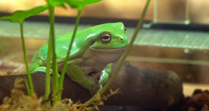
Photo credit: Dara_Kero via Flickr Creative Commons
Natural history
The White’s tree frog is indigenous to Australia and Indonesia. Also known as the dumpy tree frog or the Australian giant green tree frog. This species is captive bred in large numbers. Wild-caught frogs from Indonesia are also still in the pet trade.
Taxonomy
Class: Amphibia
Order: Anura
Family: Hylidae (tree frogs)
Color and Size
This medium to large tree frog reaches 2-4.5 in (5-11 cm) in length. Most adults weigh 50-90 grams. Males are smaller than females. The dorsum is jade green to olive brown and the lips are greenish.
Diet
Feed a variety of gut-loaded appropriately-sized invertebrates, primarily insects. Dust food items with a vitamin/mineral supplement twice weekly for juveniles and once weekly for adults. Feed juveniles daily and adults two to three times weekly.
Husbandry
| Temperature | Maintain a temperature gradient of 70-90°F (24-32°C) with a basking spot that reaches 95°F (35°C). Cage temperature may drop to 65-70°F (18-21°C) at night. |
| Humidity/water | Maintain humidity at 60-70%. Mist the enclosure daily and provide a sizable, but shallow, water dish. Between mistings the substrate should remain damp, but not water logged. Potential water sources include:
|
| Cage size and design | This quiet frog does not require much space. One to four frogs can thrive in a 20-gallon (75-L) terrarium. A woodland terrarium is ideal, but White’s tree frogs also do well with a simple set up. |
| Cage furniture/supplies | Dry paper towels can suffice as a cage substrate for a patient but care must be taken to maintain adequate humidity. Provide a large tree limb at least the diameter of the body or a clay pot to serve as a perching spot. Provide low-intensity full-spectrum lighting for optimal absorption of dietary calcium. Provide plants for visual security. |
| Social structure | Communal |
Lifespan
20+ years
White’s tree frogs often reach sexual maturity during their second year of life.
Anatomy/ physiology
| Dermatologic: | Frog skin is a dynamic organ involved in water and electrolyte balance, chemical synthesis and secretion, and immunity. Poor husbandry may result in skin disease that can quickly kill the patient. Frogs periodically shed and eat their skin. The skin will appear cloudy beforehand. |
| Gastrointestinal: | A short, simple gastrointestinal tract empties into a cloaca |
| Cardiac: | A three-chambered heart is encased within the pectoral girdle. |
| Lymphatics: | All amphibians possess lymph hearts, which beat in synchrony independently of the heart. |
| Urogenital: | A renal portal system is present |
| Miscellaneous: | Frogs possess coelomic and inguinal fat bodies. |
| Sexual dimorphism: | Adult males are about 30-50% smaller than adult females. Males also call during the breeding season and they also develop small brown nuptial pads on each thumb. |
Restraint
Venipuncture
Ventral abdominal vein
Preventive medicine
Perform a visual exam by placing the frog in a clear plastic box or tub.
Physical examination prn (as needed).
Fecal parasite testing
Important medical conditions
- Chytridiomycosis
- Endoparasitism
- Nutritional secondary hyperparathyroidism or “metabolic bone disease” (Download the client handout: Metabolic Bone Disease in Reptiles and Amphibians ).
- Obesity
- “Red leg” bacterial syndrome
**Login to view references**
References
Basic Information Sheet: Veiled Chameleon
Veiled Chameleon (Chamaeleo calyptratus)
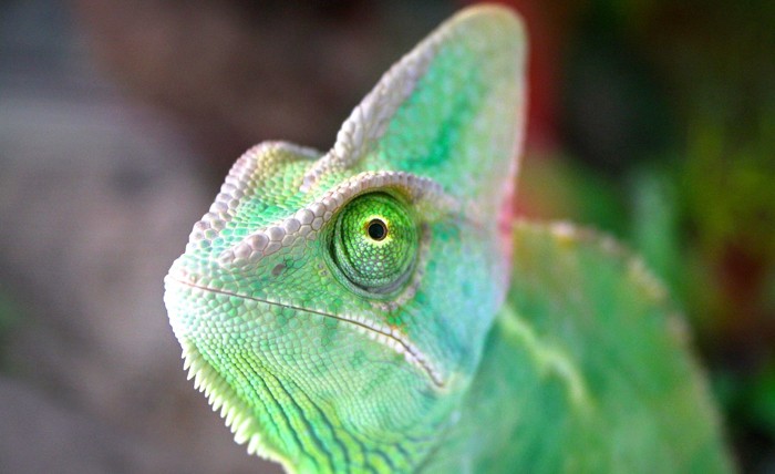
Photo credit: Mrs. Logic via Flickr Creative Commons
Natural history
Taxonomy
Class: Reptilia
Order: Squamata
Family: Chamaeleonidae
Color and Size
Males are brightly colored, ranging from blue-green to green or yellow in appearance.
Males are 18-24 inches (45-60 cm) long, while female “veileds” typically measure up to 14 in (35 cm) long.
Diet
Veiled chameleons are typically fed a variety of gut-loaded insects such as crickets, mealworms, superworms, waxworms, grasshoppers, silkworms, and Madagascar roaches of appropriate size. Veiled chameleons also enjoy blossoms and leaves such as hibiscus, dandelions, ficus, romaine, and escarole.
Dust the non-breeding adult’s diet with a calcium carbonate or calcium gluconate supplement once weekly. Calcium supplements should be devoid or low in phosphorus with a minimum Ca:P ratio of 2:1. Avoid products containing Vitamin D as this can lead to toxicity. A general vitamin/mineral supplement may also be offered once weekly.
For more information, download the client handout: Feeding Insect Eating Reptiles.
Husbandry
| Temperature | Maintain a temperature gradient of 70-95°F (20-35°C) using an overhead radiant heat source. Provide a 10-15°F (5-8°C) drop in temperature at night. |
| Humidity/water | Maintain 40-60% relative humidity. Offer water either by misting the plants every 4-8 hours or with an automatic watering system. |
| Cage size and design | Minimum cage size is 2 x 2 x 3 feet (0.6 x 0.6 x 0.9 m) but much larger is recommended. Plastic-coated wire-welded mesh enclosures serve well. House adults in a large, vertical wire enclosure. |
| Cage furniture/supplies | Provide multiple branches or twigs for climbing, potted plants (e.g. Ficus benjamina or hibiscus) to provide visual security, and a full-spectrum light source for normal absorption of dietary calcium. |
| Social structure | A mixed pair or two females can usually coexist well in a large cage with visual barriers. |
Lifespan
Females typically live 3-5 years; the average male lifespan is 4-6 years. may live up to 6-7 years.
Sexual maturity is reached between 4-8 months.
Anatomy/ physiology
Dermatologic:Some chameleon species, including the Veiled, have “chromatophores” or specialized cells in the skin that allow color change.
Musculoskeletal:Chameleons are didactyl: five toes are fused into groups of two laterally and three medially giving the foot a mitten-like appearance.Gastrointestinal:
- The tongue is a complicated structure that sits within a structure at the base of the oral cavity. The tip of the tongue is normally darker where the taste buds are found.
- Acrodont dentition: Teeth are not set in sockets, but instead are weakly attached to the jawbone surface.
- Lizards have incomplete tracheal rings.
Ophthalmic:The upper and lower eyelids are fused with only a pinhole opening for the pupil. The eyes can rotate and focus separately.Urogenital:
- A renal portal system is present.
- Like many lizards, the chameleon has a thin-walled bladder.
- The male copulatory organ is the hemipenes.
Sexual dimorphism:Males are larger than females (see Size above).
The prominent casque on top of the head is also taller in males, and males have distinct calcars or heel spurs that are apparent at birth.
Chameleons are most comfortable when allowed to perch on a wooden dowel or finger. When manual restraint is necessary, place one hand underneath to allow the chameleon to grip with its feet. Place the palm of your other hand over the chameleon’s back. Grasp the head behind the eyes with the thumb and index finger.
Venipuncture
Ventral tail (coccygeal) vein
(The ventral abdominal vein is not easily found).
Preventive medicine
Regular physical examination
Fecal parasite testing
Quarantine
Use ivermectin with caution; toxicity has been reported. Avoid use in debilitated animals.
Important medical conditions
- Egg binding
- Nutritional secondary hyperparathyroidism (metabolic bone disease)
- Ophthalmic disease
- Stomatitis, periodontal disease
**Login to view references**
References
Basic Information Sheet: Uromastyx
Uromastyx (Uromastyx spp.)
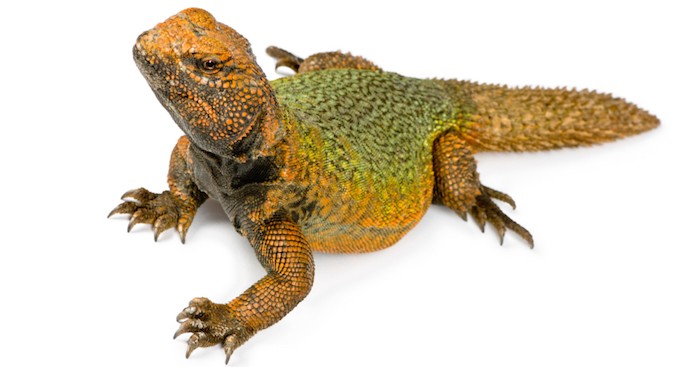
Natural history
Uromastyx spp. are also known as dabb lizards or spiny-tailed lizards. This latter name comes from its thick, short tail covered with large, spiny scales. The Moroccans spiny-tailed lizard or agama is native to the deserts of northern Africa. Colorful specimens of the pet trade are often captured from Morocco, Algeria, Tunisia, and Mauritania. The range of the ornate spiny-tailed agama is restricted to the Sinai Peninsula in Egypt.
Wild-caught animals are more common than captive bred in the pet trade, this is particularly true for the ornate spiny-tailed agama.
Taxonomy
Class: Reptilia
Order: Squamata
Family: Agamidae – bearded dragons, frilled lizards, water dragons
Uromastyx acanthinurus: Moroccoan spiny-tailed lizard
Uromastyx aegyptis: Egyptian spiny-tailed lizard
U. ocellatus ornatus: ornate spiny-tailed agama
Color and Size
- Moroccan spiny-tailed agamas are red, orange, yellow, bright green, or a combination of these colors. The colors of this lizard will intensify with age. Adults may reach 15-17 in (38-43 cm) in length.
- The ornate spiny-tailed agama has a tan ground color with yellow and turquoise crossbands and a turquoise head. Adults are 10-14 in (25-35 cm) long.
- Egyptian spiny-tailed lizards are the largest members of the genus. Total length may equal 30 in (76 cm). The juvenile is light gray-brown with yellow bars on spots on the back. The adult turns from black to white or yellow as it warms up.
Diet
- Feed dark, leafy greens and grasses such as bok choy and spring salad mix, as the bulk (~70%) of the diet. Mix salad greens with vegetables like corn, squash, carrots, sweet potato, cucumber, zucchini, green peppers, parsley, peas, and beans as well as hay and birdseed. Dried lentils, dried and fresh peas are often favorite foods. Uromastyx may be fed insects occasionally. Fruit like melons or berries and blossoms such as hibiscus leaves, flowers, and mulberry leaves can also be an occasional treat.
- Dust the non-breeding adult’s diet with a calcium carbonate or calcium gluconate supplement once weekly. Calcium supplements should be devoid or low in phosphorus with a minimum Ca:P ratio of 2:1. A general vitamin/mineral supplement may also be offered once weekly.
- Feed juveniles daily and adults every 1-3 days
- For more information, download the client handout: Feeding Insect Eating Reptiles.
Husbandry
| Temperature | Maintain a temperature gradient of 80-100°F (27-38°C). Offer a basking spot of 120°F (49°C). During the winter months the temperature of this basking area should be lowered to around 100-110°F (38-43°C). Provide a 10-15°F (5-8°C) drop in temperature at night. These high temperatures are essential for the health of Uromastyx spp. and are achieved by utilizing heaters and powerful light bulbs, which often create fire hazards in the home. |
| Humidity/water | The humidity level should be from 10-40%. A dehumidifier may be necessary in many regions. Although it may not be used, a water dish should be offered as long as its presence does not raise humidity levels. Most Uromastyx species come from areas with less than 2 inches (5 cm) of rainfall annually. The majority of the water needed is obtained from the foods they consume. |
| Cage size and design |
|
| Cage furniture/supplies | Provide a full-spectrum light source for normal absorption of dietary calcium and hide boxes. |
| Social structure | One male may be housed with one to three females year round. Females can also exhibit territorial behavior so monitor groups carefully for signs of aggression. |
Lifespan
Approximately 15-20 years
Sexually mature Spiny-tailed lizards are over 18-24 months of age.
Anatomy/ physiology
| Dermatologic: | Unlike snakes, lizards normally exhibit a patchy shed or “ecdysis”. |
| Respiratory: | Lizards have incomplete tracheal rings. |
| Urogenital: | A renal portal system is present. Agamas possess a thin-walled bladder. |
| Miscellaneous: |
|
| Sexual dimorphism: |
|
Restraint
Uromastyx are usually mild tempered, friendly lizards but they are capable of inflicting a painful bite and the well-armored tail may be used as a defensive weapons. Although these lizards do not possess tail autotomy, never hold a Uromastyx by the tail.
Venipuncture
Ventral tail (coccygeal) vein
Ventral abdominal vein
Preventive medicine
Regular physical examination
Fecal parasite testing
Quarantine
Important medical conditions
- Endoparasitism
- Nutritional secondary hyperparathyroidism or metabolic bone disease
- Cachexia due to poor food intake caused by lack of hot enough temperatures.
- Failure of fresh imports to thrive, probably due to excess chilling, dehydration and starvation, and parasitism.
**Login to view references**
References
Basic Information Sheet: Savannah Monitor
Savannah Monitor (Varanus exanthematicus)
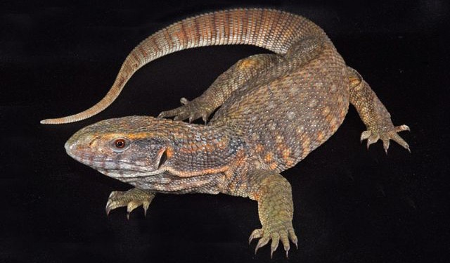
Juvenile savannah monitor. Photo from General Exotics.
Natural history
Savannah monitors in the pet trade are either wild-caught or captive-raised.
[/ezcol_2third_end]
Taxonomy
Class: Reptilia
Order: Squamata
Family: Varanidae– monitors
Color and size
Savannah monitors are tan to gray with a lighter pattern on the back, sides, and anterior tail.
Adult size can be quite variable. Some individuals reach 2.5 ft (0.8 m) while others exceed 4.5 ft (1.4 m) and can even reach 6 ft (1.8 m) or more.
Diet
Much controversy surrounds the feeding of Savannah and other grassland monitors in captivity, and clients often bring questions about appropriate foods for their pet.
- The Savannah monitor is a carnivore. Offer gut-loaded insects such as large crickets, superworms, king mealworms, silkworms, grasshoppers, cockroaches, as well as crayfish and other low-fat foods like cooked egg whites or Egg beaters®. Mice or rats may be offered, but only occasionally to reduce the risk of obesity.
- Dust the non-breeding adult’s diet with a calcium carbonate or calcium gluconate supplement once weekly. Calcium supplements should be devoid or low in phosphorus with a minimum Ca:P ratio of 2:1. Avoid products containing Vitamin D as this can lead to toxicity. A general vitamin/mineral supplement may also be offered once weekly.
- Adults may be fed two to three times weekly.
- For more information, download the client handout: Feeding Insect Eating Reptiles.
Husbandry
Lifespan
5-10+ years
Anatomy/ physiology
| Dermatologic: | Unlike snakes, lizards normally exhibit a patchy shed or “ecdysis”. |
| Fat stores: | Fat pads are present with the caudoventral coelom. |
| Urogenital: | Monitors possess a thin-walled bladder. |
| Sexual dimorphism: | Adult males are larger and more robust with prominent femoral pores and a hemipenal bulge. The hemipenis is the copulatory organ of the male. The hemipenis will appear as radiodense opacities in some individuals. Femoral pores are the opening through which glands produce a thick, waxy secretion. This secretion plays a role in scent marking and other pheromone-based communication. |
Savannahs are the most mild mannered of the monitors, however all monitor lizards are capable of delivering a painful bite. Monitors also have strong talons.
Venipuncture
Ventral tail (coccygeal) vein
Ventral abdominal vein
Preventive medicine
Regular physical examination
Fecal parasite testing
Important medical conditions
- Coelomic neoplasia
- Egg yolk coelomitis
- Hepatic lipidosis
- Nutritional secondary hyperparathyroidism or metabolic bone disease (Download the client handout: Metabolic Bone Disease in Reptiles and Amphibians ).
- Obesity (rodent-based diet)
- Starvation (poor husbandry, parasitism, cold temperatures, under-feeding)
**Login to view references**
References
Basic Information Sheet: Red-Eared Slider
Red-eared sliders are native to the eastern and central United States river valleys. Most pet sliders are captive bred and hatched. Red-eared sliders are hardy and outgoing. Although pretty and personable as pets, red-eared sliders occupy a niche of dark history in herpetoculture, first as transmitters of Salmonella bacteria to small children, second as an invasive species that have disturbed ecosystems throughout the waterways of the world. The former problem is the result of husbandry and marketing practices of large-scale commercial producers; the latter due to illegal release of unwanted pets . . .
Pour les vétérinaires. Par les vétérinaires.
Le site Lafervet.com est conçu pour une utilisation par les vétérinaires. Il est ouvert aux vétérinaires diplômés, aux techniciens vétérinaires diplômés, aux animaliers et aux étudiants dans ces domaines.
Créer un compte pour accéder à des articles et des ressources du site.
L'inscription est gratuite.
Para Profesionales Veterinarios. Por Profesionales Veterinarios.
El sitio Lafervet.com es para uso de los profesionales veterinarios. Está abierto a los veterinarios licenciados, técnicos veterinarios licenciados, rehabilitadores licenciados y estudiantes en estos campos.
Cree una cuenta para acceder a los artículos y recursos del sitio.
La registro es gratis.
Already a LafeberVet Member?
Please Login
Basic Information Sheet: Panther Chameleon
Panther Chameleon (Furcifer or Chamaeleo pardalis)
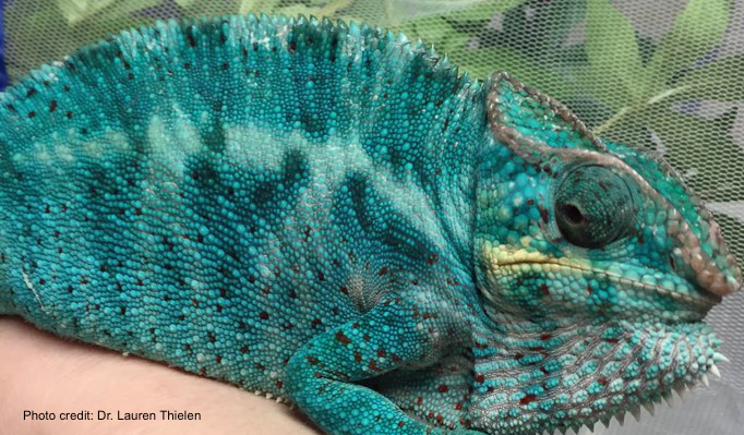
Natural history
The panther chameleon is indigenous to Madagascar.
Most specimens in the pet trade used to be wild caught, however with changes in Madagascar’s export regulations and better understanding of the care of these exquisite lizards, most are now captive-bred.
Taxonomy
Class: Reptilia
Order: Squamata
Family: Chamaeleonidae
Color and size
Males may reach 20 in (50 cm) long, while females typically measure about 14 in (35 cm) long. By 2-2.5 years of age, adult males typically weigh 200- 220 grams while females weight 140-160 grams.
The panther chameleon is variably colored in green, brown, red, orange, or turquoise.
Chameleons change color as a result of complex internal and external cues involving such factors as recognition of nearby conspecifics, adjustments to environmental temperature, breeding behaviors, and basking in sunlight. Color change is generally not used as camouflage. Instead, chameleons utilize their stillness and halting gait, and to a lesser degree their coloration, as protection from predator detection.
Diet
Feed a variety of gut-loaded insects such as crickets, mealworms, waxworms, superworms, grasshoppers, silkworms, and Madagascar roaches of appropriate size.
Dust the non-breeding adult’s diet with a calcium carbonate or calcium gluconate supplement once weekly, perhaps more often if exposure to UVB light is limited. Calcium supplements should be devoid or low in phosphorus with a minimum Ca:P ratio of 2:1.
A general vitamin/mineral supplement may also be offered once weekly. The panther chameleon may be relatively sensitive to over-supplementation of vitamin A so use caution.
For more information, download the client handout: Feeding Insect Eating Reptiles.
Husbandry
Lifespan
2-5 years
Chameleons typically reach sexual maturity between 6-9 months.
Anatomy/ physiology
| Dermatologic: | “Chromatophores” or specialized cells in the skin allow color change. |
| Respiratory: | Lizards have incomplete tracheal rings. |
| Musculoskeletal: | Chameleons are didactyl: five toes are fused into groups of two laterally and three medially giving the foot a mitten-like appearance. |
| Gastrointestinal: | The tongue is a complicated structure that sits within a structure at the base of the oral cavity. The tip of the tongue is normally darker where the taste buds are found. Acrodont dentition: Teeth are not set in sockets, but instead are weakly attached to the jawbone surface. |
| Ophthalmic: | The upper and lower eyelids are fused with only a pinhole opening for the pupil. The eyes can rotate and focus separately. |
| Urogenital: | A renal portal system is present. Like many lizards, the chameleon has a thin-walled bladder. The male copulatory organ is the hemipenes. |
| Sexual dimorphism: | Males are larger than females (see Size above). |
Restraint
Chameleons are most comfortable when allowed to perch on a wooden dowel or finger. When manual restraint is necessary, place one hand underneath to allow the chameleon to grip with its feet. Place the palm of your other hand over the chameleon’s back. Grasp the head behind the eyes with the thumb and index finger.
Venipuncture
Ventral tail (coccygeal) vein
(The ventral abdominal vein is not easily found).
Preventive medicine
- Regular physical examination
- Fecal parasite testing
- Quarantine
- Use ivermectin with caution; toxicity has been reported. Avoid use in debilitated animals.
Important medical conditions
The panther chameleon is a relatively hardy species as long as its somewhat extensive and complex husbandry needs are met. Conditions that may be seen include:
- Endoparasitism
- Nutritional secondary hyperparathyroidism (metabolic bone disease)
- Stomatitis, periodontal disease
- Egg binding or dystocia
- Ophthalmic disease
**Login to view references**
References
Basic Information Sheet: Leopard Gecko
Leopard Gecko (Eublepharis macularius)
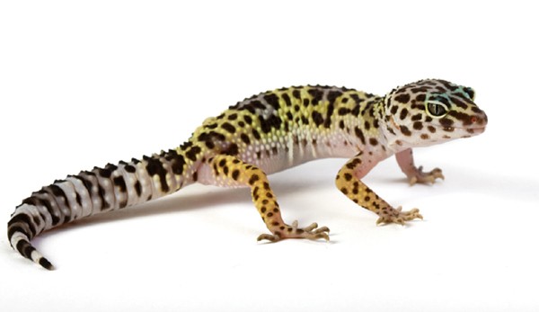
Natural history
Leopard geckos are originally from the grassland and desert regions of India and Pakistan. The vast majority of pets are captive bred and hatched. Leopard geckos are nocturnal, terrestrial, long-lived lizards that make excellent pets.
Taxonomy
Class: Reptilia
Order: Squamata
Family: Gekkonidae
Color and Size
The wild or standard type leopard gecko is yellow with a variable pattern of dark pigment that is banded in juveniles and reticulated in adults. There are also many other color and pattern morphs available including albinos, leucistic (completely pattern-free with only faint traces of yellow color), striped, and “high” yellow.
Adults are typically 16.5-20 cm (6.5-8 inches) long. Mature male geckos weigh between 60-90 grams. Females may be as light as 45 grams. At birth, leopard geckos weigh 6-8 grams.
Diet
- Feed leopard geckos a variety of gut-loaded or dusted insects of appropriate size such as crickets, Phoenix worms, mealworms, and superworms. May be fed the occasional pinky mouse.
- Dust the non-breeding adult’s diet with a calcium carbonate or calcium gluconate supplement once weekly, more often if the gecko is actively breeding. Calcium supplements should be devoid or low in phosphorus with a minimum Ca:P ratio of 2:1. A general vitamin/mineral supplement may also be offered once weekly.
- Feed adult geckos once dailyand offer prey in late afternoon because these lizards are primarily nocturnal.
- For more information, download the client handout: Feeding Insect Eating Reptiles.
Husbandry
| Temperature | Provide a temperature gradient of 70-80°F (21-27°C) and a warm area that reaches 90°F (32°C). Drop night temperatures to the 70s (21-23°C). |
| Humidity/water | Strive for a relative humidity of approximately 40-60%. Provide a shallow water dish and a moist hide area, but make sure the remainder of the habitat is dry. |
| Cage size and design | A single male requires a long, 10-15 gallon (38-57 L) aquarium at minimum. |
| Cage furniture/supplies | Offer leopard geckos hiding places to provide visual security. Also provide low-intensity full spectrum lighting during the day for optimal absorption of dietary calcium. Although leopard geckos are nocturnal exposure to UV-B light seems to be helpful. Offer rocks and logs as climbing structures for exercise. |
| Social structure | Males will fight each other, but can coexist well with one or more females. |
Lifespan
Leopard geckos can live 25-30 years. These species typically reach sexual maturity between 9-18 months.
Anatomy/ physiology
| Dermatologic: | Unlike snakes, lizards normally exhibit a patchy shed or “ecdysis”. |
| Miscellaneous: |
|
| Urogenital: | Geckos also possess a thin-walled bladder. |
| Sexual dimorphism: | Secondary sex characteristics of the male include larger or more distinct femoral pores and the presence of a hemipenal bulge. The hemipenis is male copulatory organ. Femoral pores are the opening through which glands produce a thick, waxy secretion. This secretion plays a role in scent marking and other pheromone-based communication. |
Restraint
Leopard geckos utilize tail autotomy, a defense mechanism that utilizes tail loss. A vertical fracture plane of fibroconnective tissue and cartilage runs through the body and part of the neural arch of each caudal vertebrae. The tail can fall off when grasped, sometimes when very little pressure is applied. The tail will regrow as a cartilaginous rod.
Venipuncture
Ventral tail (coccygeal) vein (see Restraint above)
Ventral abdominal vessel
Preventive medicine
Quarantine
Important medical conditions
Leopard geckos are relatively hardy creatures, however common presenting problems include:
- Cryptosporidiosis
- Cystic calculi
- Dysecdysis (difficult shed on toes)
- Nutritional secondary hyperparathyroidism (metabolic bone disease)
References
Basic Information Sheet: Kingsnakes
Kingsnakes (Lampropeltis spp.)
Natural history
There are several species of kingsnake and their habitat and range vary. The Eastern kingsnake is found in humid forests of the eastern seaboard states and as far west as the Appalachians and Alabama. The California kingsnake is native to desert, arid grassland, and rocky hillsides of Baja Mexico and the western United States.
Pets may be bred in captivity or wild-caught. Kingsnakes are fairly docile, medium-sized, hardy reptiles, that make good “starter” snake pets.
Taxonomy
Class: Reptilia
Order: Squamata
Family: Colubridae – corn snake, rat snake
Lampropeltis getula californiae: California kingsnake
L. getula getula: Eastern kingsnake
Color and size
Hatchlings are gray with black dorsal saddles. Although an array of color morphs are available, adult Eastern kingsnakes are dark chocolate or black with white, cream, or yellow cross-bands.
This species is usually between 4-6 ft (1.2-1.8 m) in length, while California kingsnakes are usually 3-4 ft (0.9-1.2 m) long. The ground color of the California kingsnake is black or brown with a pattern of white, cream, or yellow stripes or bands.
Diet
Free-ranging kingsnakes prey upon rodents, lizards, and frogs and especially upon other snakes. Captive specimens will eat mice and small rats. An adult may be fed 1-3 mice once weekly. For more information, download the client handout: Feeding Snakes.
Husbandry
| Temperature | Provide a temperature gradient of 70-85°F (21-28°C) with a 10-15°F (2-5°C) drop at night. A winter cool-down or “brumation” is also recommended for healthy specimens. |
| Humidity/water | Provide a water bowl large enough for the snake to completely immerse its body, and heavy enough that the animal will not tip it over. Shedding may also be helped by the presence of a humidity box” or a hiding place filled halfway with damp sphagnum moss. Place the container half on and half off the heat. |
| Cage size and design | At minimum, adults require a long, 20-gallon (75-L) aquarium or terrarium. |
| Cage furniture/supplies | Provide visual security in the form of multiple hide boxes. Although kingsnakes are active, these terrestrial snakes do not require a climbing branch. Commercial substrate such as aspen bedding is recommended. |
| Social structure | Kingsnakes are solitary and will kill and eat other snakes. Similarly sized adults can be housed together for breeding, but are otherwise maintained separately in most cases. |
Lifespan
Most kingsnakes live 15+ years in captivity.
Sexually maturity is reached at approximately 200 g (7 oz) in males or 225 g (8 oz) in females. Kingsnakes are typically 18 months of age by this time.
Anatomy/ physiology
| Dermatologic: | Snakes lack movable eyelids; instead the eyes are protected by a clear “spectacle”, which is an embryonic fusion of the eyelids. The scaled skin is normally shed in one piece in a process called “ecdysis”. |
| Gastrointestinal: | The teeth are arranged in six rows with double rows present in the upper jaw. Snake teeth are shed through life, and are attached to the bone’s surface instead of roots. |
| Cardiopulmonary: | The snake’s three-chambered heart is mobile within the ribcage. Snakes possess incomplete tracheal rings. The right lung joins with a large air sac that may extend to the caudal coelom. The left lung is absent or vestigial. |
| Gastrointestinal: | The tongue is a complicated structure that sits within a pouch at the base of the oral cavity. The tip of the tongue is normally darker where the taste buds are found. Acrodont dentition: Teeth are not set in sockets, but instead are weakly attached to the jawbone surface. |
| Urogenital: | Snakes possess a renal portal system. Kingsnakes are “oviparous” or egg layers. |
| Sexual dimorphism: | The copulatory organs of the snake are “hemipenes”. Hemipenes lie within pouches at the ventral tail base. Probing of the male pouches will be deeper when compared to females. |
Restraint
Kingsnakes are relatively docile and rarely bite.
When handling a snake, take care not to smell like snake food (i.e. rodents or rabbits). Wash your hands thoroughly beforehand.
Support the head, neck, and body. Make sure the snake’s weight is not borne by its single occipital condyle and the cervical spine.
Tail vessel
Cardiac puncture
Injections
Give intramuscular injections between the scales in the epaxial muscles. Use the cranial half of the body to avoid the renal portal system.
Preventive medicine
Regular physical examination
Fecal parasite testing
Quarantine
Important medical conditions
Kingsnakes are relatively hardy species, however problems seen in captivity can include:
- Anorexia (juveniles of some species may refuse mice in preference of snakes or lizards)
- Dysecdysis, retained spectacles
- Egg binding or dystocia
- Endoparasitism (wild-caught)
- Pustular dermatitis or “blister disease”
**Login to view references**
References
Metabolic Bone Disease in Reptiles and Amphibians
What is metabolic bone disease or nutritional secondary hyperparathyroidism? This educational handout will help the reptile and amphibian owner understand this common problem from the underlying cause and signs of illness to the testing and treatment commonly recommended by your veterinarian.
Download the Metabolic Bone Disease in Reptiles and Amphibians PDF Handout
Basic Information Sheet: Jackson’s Chameleon
Jackson’s or Three-horned chameleon (Chamaeleo jacksoni xantholophus)
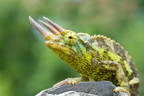
Natural history
Jackson’s chameleons comprise three distinct subspecies native to the montane regions of Kenya. Large feral populations are found in Hawaii where specimens are often caught for the pet trade. Small numbers are also captive-bred.
Taxonomy
Class: Reptilia
Order: Squamata
Family: Chamaeleonidae
Color and Size
Adults may reach 12-15 in (30.5-38.1cm) in total (snout to tail) length, with a body length of about 5 in (12.7 cm).
Adults can reach 0.9-1.8 kg but most weigh less.
Diet
- Feed a variety of gut-loaded insects such as crickets, mealworms, grasshoppers, roaches, superworms, silkworms, and wax worms as well as pesticide-free snails, slugs, caterpillars, sowbugs, spiders, and flies of appropriate size.
- Dust the adult non-breeding diet with a calcium carbonate or calcium gluconate supplement once weekly. Calcium supplements should be devoid or low in phosphorus with a minimum Ca:P ratio of 2:1. Avoid products containing high levels of Vitamin D as this can lead to toxicity.
- A general vitamin/mineral supplement may be offered once weekly. Jackson’s chameleon may also be more sensitive to over-supplementation of vitamin A than some other lizards so use caution.
- For more information, download the client handout: Feeding Insect Eating Reptiles
Husbandry
| Temperature | Maintain a temperature gradient of 70-80°F (21-27°C) with a basking spot that reaches 82-85°F (28-29°C). Never maintain a Jackson’s chameleon above 84°F (29°C) for long periods of time. Provide a drop in temperature at night that ranges from 55°F to the low to mid 70s (13-24°C). |
| Humidity/water | Maintain 60-100% relative humidity. Jackson’s chameleons only drink water droplets from leaves and other surfaces. Offer water either by misting the plants every 4-8 hours or with an automatic watering system. Use of a cool-mist humidifier may also be necessary. |
| Cage size and design | House adults in a large, vertical wire enclosure. Plastic-coated wire-welded mesh enclosures serve well. The minimum cage size for an adult pair is 2 x 2 x 3 ft (0.6 x 0.6 x 0.9 m). |
| Cage furniture/supplies | Provide multiple branches or twigs for climbing, potted plants (e.g. Ficus benjamina or hibiscus) to provide visual security, and a full-spectrum light source for normal absorption of dietary calcium. |
| Social structure | May be housed as a mixed pair or one male may coexist with one or two females in a large cage with many visual barriers. |
Lifespan
3-8+ years
Chameleons typically reach sexual maturity between 6-9 months of age.
Anatomy/ physiology
| Dermatologic: | Some chameleon species, including the Jackson’s chameleon, have “chromatophores” or specialized cells in the skin that allow color change. |
| Respiratory: | Lizards have incomplete tracheal rings. |
| Musculoskeletal: | Chameleons are didactyl: five toes are fused into groups of two laterally and three medially giving the foot a mitten-like appearance. |
| Gastrointestinal: | The tongue is a complicated structure that sits within a structure at the base of the oral cavity. The tip of the tongue is normally darker where the taste buds are found. |
| Acrodont dentition: | Teeth are not set in sockets, but instead are weakly attached to the jawbone surface. |
| Ophthalmic: | The upper and lower eyelids are fused with only a pinhole opening for the pupil. The eyes can rotate and focus separately. |
| Urogenital: | A renal portal system is present. Like many lizards, the chameleon has a thin-walled bladder. The male copulatory organ is the hemipenes. |
| Sexual dimorphism: | Males have three distinctive rostral horns and a parietal crest. |
Restraint
Chameleons are most comfortable when allowed to perch on a wooden dowel or finger. When manual restraint is necessary, place one hand underneath to allow the chameleon to grip with its feet. Place the palm of your other hand over the chameleon’s back. Grasp the head behind the eyes with the thumb and index finger.
Venipuncture
(The ventral abdominal vein is not easily found).
Preventive medicine
Regular physical examination
Fecal parasite testing
Use ivermectin with caution; toxicity has been reported. Avoid use in debilitated animals.
Important medical conditions
The Jackson’s chameleon is a relatively hardy species.
- Dehdyration from housing at high temperatures or failure to provide adequate drinking water as droplets.
- Egg binding
- Endoparasitism
- Nutritional secondary hyperparathyroidism or metabolic bone disease
- Ophthalmic disease
- Stomatitis, periodontal disease
**Login to view references**
References
Basic Information Sheet: Green Iguana
Green or Common Iguana (Iguana iguana)
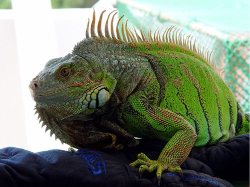
Photo credit: Cy via Flickr Creative Commons
Download the Care of the Green Iguana client handout PDF.
Natural history
The green iguana is native to the tropical rain forests of Latin America. Most pet iguanas are raised on farms in El Salvador and Costa Rica although wild-caught imports and domestically captive-bred lizards are also available.
Taxonomy
Class: Reptilia
Order: Squamata
Family: Iguanidae
Iguanas, chuckwallas
Color and Size
Juveniles should be a bright green color although color becomes less vivid as the lizard ages. All ages have dark vertical bands on the tail. Breeding males may also have an orange tinge. Red and albino color morphs have also been developed.
Including the tail, adults can reach 1.8 m (6 feet) or more.
Diet
Green iguanas are herbivores.
- Dark, leafy greens should make up the majority of the adult diet. Offer a variety including collard greens, kale, romaine, dandelion greens, turnip greens, mustard greens, beet greens, bok choy, Swiss chard, spinach, chicory, and escarole.
- Mix greens with other chopped or grated vegetables like squash, zucchini, sweet potato, broccoli, peas, and carrots.
- Fruit should make up no more than 5% of the diet and should include nutrient dense items such as papaya, melon, and banana.
- Occasional treats may consist of non-toxic flower blossoms such as hibiscus.
Dust the non-breeding adult’s diet with a calcium carbonate or calcium gluconate supplement once weekly. Calcium supplements should be devoid or low in phosphorus with a minimum Ca:P ratio of 2:1. A general vitamin/mineral supplement can also be offered once weekly.
Husbandry
| Temperature | The green iguana has a preferred optimal temperature zone of 80-88°F (27-31°C). The basking spot should reach 92-95°F (33-35°C). |
| Humidity/water | Green iguanas will often soak in their water bowl and can defecate in their water. Drinking and soaking bowls should be cleaned at least daily. Iguanas should be misted daily with warm water. |
| Cage size and design | Adults require very large arboreal enclosures (i.e. 6 x 3 x 6 ft or 2 x 1 x 2 m) so custom built cages are often needed. Screening is best for adequate air circulation although more effort will be needed to keep humidity levels high. |
| Cage furniture/supplies | Provide a full-spectrum light source for normal absorption of dietary calcium. Iguanas are arboreal, and are most comfortable when provided with climbing structures. Offer sturdy branches to support heavy-bodied adults. |
| Social structure | Iguanas are solitary and territorial. |
Lifespan
Green iguanas can live up to 15-20 years when cared for properly.
Anatomy/ physiology
| Gastrointestinal: | The tip of the tongue is normally a dark red color. |
| Dermatologic: | Adult iguanas have a dewlap, or flap of skin, beneath the mandible.Unlike snakes, lizards normally exhibit a patchy shed or “ecdysis”. |
| Cardiopulmonary: | Unlike many lizards, the iguana heart is located just caudal to the thoracic inlet. Lizards possess incomplete tracheal rings. |
| Urogenital: | The paired kidneys are found within the pelvic canal. The green iguana has a thin-walled bladder. In the Western hemisphere, the breeding season is typically between October to June. |
| Sexual dimorphism: | Male iguanas have a hemipenal bulge. The hemipenes is the male copulatory organ. Males also tend to be larger in size with larger dorsal spines, dewlap, and femoral pores when compared to the female iguana. Femoral pores are the opening through which glands produce a thick, waxy secretion. This secretion plays a role in scent marking and other pheromone-based communication. |
| Miscellaneous: | Nasal salt glands excrete excess sodium. Iguanas “sneeze” clear, high sodium fluid, which later dries as a fine, white powder.Two or more fat bodies are found in the caudal abdomen. |
Restraint
Never grasp iguanas by the tail. Green iguanas utilize tail autotomy, a defense mechanism that utilizes tail loss. A vertical fracture plane of fibroconnective tissue and cartilage runs through the body and part of the neural arch of each caudal vertebrae. The tail can fall off when grasped, sometimes when very little pressure is applied. The tail will regrow as a cartilaginous rod.
Iguanas can also go on the “offensive”. They can bite and twist, utilize their sharp claws to scratch the handler, and larger specimens use their tails as a whip and will direct tail lashes at human eyes.
Venipuncture
Ventral tail (coccygeal) vein (see Restraint above)
Ventral abdominal vein
(Jugular vein)
Preventive medicine
Regular physical examination
Consider elective castration and feeding high levels of plant phytoestrogens to reduce aggression in males.
Important medical conditions
- Abscesses
- Cystic calculi
- Egg binding or dystocia
- Foreign body ingestion
- Nutritional secondary hyperparathyroidism or metabolic bone disease*
*(Download the client handout: Metabolic Bone Disease in Reptiles and Amphibians). - Renal disease
**Login to view references**
References
Basic Information Sheet: Ornate Horned Frog
Ornate Horned Frog (Ceratophrys ornata)
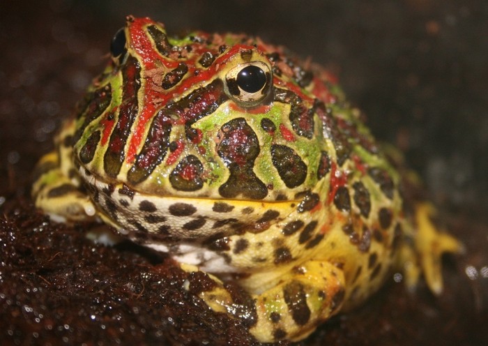
Photo credit: ‘her wings’ via Flickr Creative Commons
Natural history
The ornate horned frog is found in the tropical and montane rain forests of South America. This frog’s camouflaged coloration allows it to hide as it lies half buried in leaf litter on the forest floor. As soon as prey passes by, the horned frog grabs and swallows its prey whole in one or two gulps. This is why this species is also known as the “Pac-Man frog”.
The pet trade is made up primarily of captive bred specimens.
Taxonomy
Class: Amphibia
Order: Anura
Family: Ceratophryidae – horned frogs, fantasy frog, Budgett’s frog
Color and Size
Ornate horned frogs are large, brown or green frogs. Adults may be 3-6 in (8-15 cm) long and are often just as wide. Large females may weigh up to 480 g (1 lb).
Diet
Gut-loaded insects such as crickets, superworms, and earthworms as well as vertebrates like appropriately-sized fish and mice can be offered.
Dust invertebrate foods with a vitamin/mineral supplement two to three times weekly for juveniles and once weekly for adults. Ornate horned frogs have a voracious appetite. Feed adults with forceps to avoid injury.
Husbandry
| Temperature | Maintain the gradient between 75-82°F (24-27°C) during the day. Temperature may drop to 65°F (18°C) at night. Do not use a radiant heat source as these tend to dry amphibian environments. |
| Humidity/water | Strive for 80% relative humidity. Lightly mist the enclosure regularly. Between misting, the substrate should remain damp, but not water logged. Potential water sources include: |
- Aged water: Allow chlorinated water to sit in an open container for 24-48 hours so chlorine can dissipate
- Bottled spring water
- Filtered tap water: run through a sediment and activated charcoal filter
Cage size and designThis inactive frog can survive in a 10-gallon terrarium, but will thrive in larger habitats.Cage furniture/suppliesProvide low-intensity full-spectrum lighting for optimal absorption of dietary calcium. Provide hiding places at each end of the cage (i.e. live or artificial plants). Unmilled sphagnum or non-aromatic mulch can serve as the cage substrate. Alternatively, frogs may be housed in a bare tank containing 0.5 in (1 cm) of water that cleaned daily or more often.Social structureSolitary
Lifespan
16+ years
Sexual maturity is reached at 18-24 months of age.
Anatomy/ physiology
| Gastrointestinal: | A short, simple gastrointestinal tract empties into a cloaca |
| Cardiovascular: | A three-chambered heart is encased within the pectoral girdle. |
| Lymphatics: | All amphibians possess lymph hearts, which beat in synchrony independently of the heart. |
| Dermatologic: | Horned frogs periodically shed and eat their skin at regular intervals. The skin will appear cloudy beforehand. The old skin is pushed off with the hind legs and the skin peels off from the back end. The skin should come off in one piece and is normally eaten by the frog. The skin is pushed forwards using its legs towards the mouth. |
| Miscellaneous: | If temperatures fall below recommended level, then the Ornate horned frog may estivate or enter a period of “partial hibernation”. Frogs possess coelomic and inguinal fat bodies. |
| Urogenital: | A renal portal system is present |
| Sexual dimorphism: | Females are usually larger than males with a more rounded body shape.Males will call during the mating season. They will also develop nuptial pads on each thumb and tend to have darker throats. |
Restraint
Amphibian skin is very sensitive so handle the ornate horned frog as little as possible. To manually restrain the frog, moisten your hands with water or wear moistened surgical gloves. The ornate horned frog is known for its readiness to bite, not because of an aggressive nature, but because it always seem ready to eat! So be prepared for this tendency. Although frogs lack true teeth, boney outgrowths of the jawbone called “vomerine teeth” can deliver a painful bite.
Venipuncture
Ventral midline vein (midline abdominal vein)
Preventive medicine
Perform a visual exam by placing the frog in a clear plastic box or tub.
Physical examination prn (as needed).
Important medical conditions
- Corneal lipidosis (rodent-based diet)
- Foreign body ingestion
- Nutritional secondary hyperparathyroidism or “metabolic bone disease”
- Obesity (rodent-based diet)
- Trauma (rostral abrasions)
**Login to view references**
References
Basic Information Sheet: Poison Dart Frog
Dart Frogs (Dendrobatidae spp.)

Photo credit: kakissel via Flickr Creative Commons
Natural history
Native to the tropical rain forests of Central and South America, tiny poison dart frogs secrete lipophilic alkaloid toxins through their skin. These toxins serve as a chemical defense against predation. Some native tribes are renowned for dipping arrow tips in the toxins from these frogs.
These beautiful, active frogs are outgoing and diurnal, making excellent “look but don’t touch” pets. Many but not all specimens in the pet trade today are captive bred. Frogs bred for several generations in captivity fail to synthesize skin alkaloid toxins because of differences in diet. However all specimens should be considered to be wearing noxious alkaloids on their skin until demonstrated otherwise.
Taxonomy
Class: Amphibia
Order: Anura
Family: Dendrobatidae
Dendrobates auratus: green and black poison frog
D. azureus: blue poison frog
D. leucomelas: yellow and black poison frog
Color and Size
These vividly colored frogs are tiny, typically reaching 1.5 in (3.8 cm) in length. Adult poison frogs typically weigh between 2-6 grams.
Diet
These small frogs have big appetites! Feed poison frogs small insects like fruit flies, termites, aphids, and pinhead crickets. Dust foods for juveniles with a vitamin/mineral supplement twice weekly. Dust food items for adults once weekly.
Husbandry
| Temperature | Provide a temperature gradient of 72-80°F (22-27°C) during the day. At night the temperature may fall to 65-70°F (18-21°C). Do not use a radiant heat source as these tend to dry amphibian environments. |
| Humidity/ water | Strive for a relative humidity of 80-100%. Provide a shallow water dish and lightly mist the enclosure regularly. Between misting the substrate should remain damp, but not water logged. Potential water sources include:
|
| Cage size and design | House one to four frogs in a heavily planted 20-gallon (75-L) terrarium. |
| Cage furniture/supplies | Provide plants and other visual barriers, as well as one or two tree limbs arranged diagonally. Provide low-intensity full-spectrum lighting for optimal absorption of dietary calcium. Dart frogs particularly enjoy commercially available huts made from coconut and bromeliads for climbing. |
| Social structure | Communal |
Lifespan
7-15 years (Green and black poison frog, yellow and black poison frog)
2-8 years (Blue poison frog)
Anatomy/ physiology
| Gastrointestinal: | A short, simple gastrointestinal tract empties into a cloaca |
| Dermatologic: | Dart frogs have the fragile, sensitive skin characteristic of all amphibians, plus are known for the skin secretions of toxic alkaloids. To protect the patient and clinician, moistened gloves should be worn for examination and treatment. |
| Cardiac: | A three-chambered heart is encased within the pectoral girdle. |
| Lymphatics: | All amphibians possess lymph hearts, which beat in synchrony independently of the heart. |
| Urogenital: | A renal portal system is present |
| Sexual dimorphism: | Males will call during the mating season. They will also develop nuptial pads on each thumb and tend to have darker throats. Females are usually larger than males with a more rounded body shape. | Miscellaneous: | Frogs possess coelomic and inguinal fat bodies. |
Restraint
Amphibian skin is very sensitive so handle the poison frog as little as possible. Perform a visual exam by placing the frog in a clear plastic box or tub.
To manually restrain the frog, moisten your hands with water or wear moistened latex gloves.
Important medical conditions
- Chytridiomycosis
- Spindly leg syndrome
**Login to view references**
References
Feeding Snakes
Care of the captive snake requires proper nutrition. All snakes are strict carnivores. Depending on the species of interest, preferred prey items may include appropriately sized rodents, rabbits, fish, birds, reptiles, and even other snakes. This client education handout strives to answer many of the questions that can arise related to feeding snakes and the snake diet: When should my snake be fed? Should I feed fresh or frozen prey? How often should I feed my snakes? What size prey should be fed? What if my snake won’t eat?
Download the Feeding Snakes PDF handout
Basic Information Sheet: Corn Snake
Corn Snakes (Pantherophis guttatus guttatus or Elaphe guttata)
Photo credit: Lauren Mitchell via Flickr Creative Commons
Natural History
The corn snake is also known as the red rat snake. Corn snakes are found in woodland and forest regions of the southeastern and central United States as far west as Kentucky and Louisiana and as far south as Mexico.
Pets may be wild-caught but most are captive bred and many color morphs are now available in the pet trade.
Taxonomy
Class: Reptilia
Order: Squamata
Family: Colubridae
Corn snake, rat snake,
Color and Size
The ground color of the normal or wild type corn snake ranges from orange to gray. There are orange, brown, or reddish patterns with black borders on the back and sides. The belly is checkered black and white, and the underside of the tail is usually striped. There are also albinos and many color morphs produced in captivity.
Adults reach 2.5-5 ft (0.8-1.5 m) in length.
Diet
Free-ranging corn snakes eat small mammals, birds, and eggs. In captivity they thrive on rats and mice.
For more information, download the client handout: Feeding Snakes.
Husbandry
Lifespan
Corn snakes may live up to 15-20 years in captivity.
Anatomy/ physiology
| Dermatologic: | Snakes lack movable eyelids; instead the eyes are protected by a clear “ spectacle”, which is an embryonic fusion of the eyelids. |
The scaled skin is normally shed in one piece in a process called “ecdysis”.Gastrointestinal:The teeth are arranged in six rows with double rows present in the upper jaw. Snake teeth are shed through life, and are attached to the bone’s surface instead of roots.Cardiopulmonary:The snake’s three-chambered heart is mobile within the ribcage.
Snakes possess incomplete tracheal rings.
The right lung joins with a large air sac that may extend to the caudal coelom.
The left lung is absent or vestigial.Urogenital:Snakes possess a renal portal system.Sexual dimorphism:The copulatory organs of the snake are “hemipenes”. Hemipenes lie within pouches at the ventral tail base. Probing of the male pouches will be deeper when compared to females and snakes are usually sexed by this technique. Stainless steel probes for this purpose are sold in the pet trade.
Restraint
Corn snakes are relatively docile and rarely bite.
When handling a snake, take care not to smell like snake food (i.e. rodents or rabbits). Wash your hands thoroughly beforehand.
Support the head, neck, and body. Make sure the snake’s weight is not borne by its single occipital condyle and the cervical spine.
Cardiac puncture
Tail vessel
Injections
Give intramuscular injections between the scales in the epaxial muscles. Use the cranial half of the body to avoid the renal portal system.
Preventive medicine
Regular physical examination
Fecal parasite testing
Important medical conditions
Corn snakes are relatively hardy. Problems seen in captivity can include:
- Dysecdysis
- Endoparasitism (wild-caught)
- Pustular dermatitis or “blister disease”
**Login to view references**
References
Basic Information Sheet: Chinese or Green Water Dragons
Chinese or Green Water Dragon (Physignathus cocincinus)
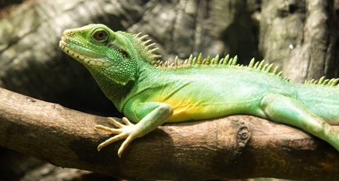
Download the Water Dragon Client Handout PDF.
Natural history
Green water dragons are native to tropical forests or areas near the river’s edge in southeast Asia.
Pets are primarily wild-caught and wild-caught adults can have a difficult time adjusting to captivity. Water dragons are beautiful lizards that make stunning display animals in naturalistic vivaria, however these lizards are inexpensive and imported in large numbers. These flighty lizards then end up in private homes where they may be housed poorly and rapidly become stressed and ill.
Taxonomy
Class: Reptilia
Order: Squamata
Family: Agamidae
Water dragons, bearded dragons, frilled lizards, agamas
Color and Size
- Variable shades of green or brown with dark tail bands and sometimes light body bands. Coloring is often brightest during the breeding season.
- Including the tail, adult males may reach 24-30 in (0.6-0.8 m) in length. More than half of this length is tail.
- Females are somewhat smaller.
Diet
- Feed Water dragons a variety of gut-loaded insects such as crickets, king mealworms, silkworms, and waxworms, as well as grasshoppers, roaches, and earthworms. Pinky mice and small fish may also be fed occasionally. As water dragons age, they become more omnivorous. Offer chopped fruits, greens, and other vegetables.
- Dust the non-breeding adult’s diet with a calcium carbonate or calcium gluconate supplement just prior to feeding once weekly. Calcium supplements should be devoid or low in phosphorus with a minimum Ca:P ratio of 2:1. A general vitamin/mineral supplement may be offered once weekly
- Feed juveniles daily. Feed adults once or twice weekly, more often if they are active and in a large habitat.
- For more information, download the client handout: Feeding Insect Eating Reptiles.
Husbandry
| Temperature | Maintain a temperature gradient of 80-86°F (27-30°C) with a basking spot that reaches 95°F (35°C). Provide a 10-15°F (5-8°C) drop in temperature at night to 70-77°F (21-25°C). |
| Humidity/water | Provide a tropical environment with relative humidity ranging from 60-90%. This lizard requires a large bathing area. |
| Cage size and design | Water dragons require large, spacious enclosures. At minimum, provide this active lizard with a 75-gallon (283-L) terrarium. House one to three lizards in an enclosure at least 6 x 3 x 7 ft (1.8 x 1 x 2 m). |
| Cage furniture/supplies | Provide a full-spectrum light source for normal absorption of dietary calcium. This arboreal species requires branches for climbing. Provide hiding places in sufficient numbers and in various locations so that all animals have opportunities to hide from other lizards and humans. |
| Social structure | Water dragons may be housed alone, in pairs, or as trios. |
Lifespan
When properly cared for, Water dragons may live 12-15 years. Sexual maturity occurs when dragons exceed 16 in (40 cm) in length when Dragons are usually 2-3 years of age.
Anatomy/ physiology
| Respiratory: | Lizards have incomplete tracheal rings. |
| Gastrointestinal: | Acrodont dentition: Teeth are not set in sockets, but instead are ankylosed to the jawbone surface. |
| Miscellaneous: | Two or more fat bodies are found in the caudal abdomen. |
| Sexual dimorphism: | Males are somewhat larger than and they posses a hemipenal bulge. The hemipenes are the male copulatory organ. Males also tend to be larger than females with larger heads, jowls, and crest, as well as larger femoral pores. Femoral pores are openings on the ventral thighs where a waxy secretion is produced. Femoral pores are the opening through which glands produce a thick, waxy secretion. This secretion plays a role in scent marking and other pheromone-based communication. |
Restraint
Never grasp a lizard by the tail. Water dragons utilize tail autotomy, a defense mechanism that utilizes tail loss. A vertical fracture plane of fibroconnective tissue and cartilage runs through the body and part of the neural arch of each caudal vertebrae. Therefore the tail can fall off when grasped sometimes when very little pressure applied. The tail will regrow as a cartilaginous rod.
Ventral tail (coccygeal) vein (see Restraint above)
Ventral abdominal vein
Preventive medicine
- Regular physical examination
- Fecal parasite testing
- Quarantine
Important medical conditions
- Abscesses
- Ectoparasites
- Egg binding
- Endoparasitism
- Nutritional secondary hyperparathyroidism or metabolic bone disease
- Obesity
- Respiratory infection
- Rostral abrasions (snout rubbing)
- Stomatitis, gingivitis, periodontal disease
**Login to view references**
References
Feeding Insect-Eating Reptiles
An insectivore diet consists completely or partially of insects. Amphibians like frogs and toads, some turtles and snakes, and many lizards are all insectivores. This client education handout explores many practical matters related to feeding insect prey from types (like mealworms and waxworms), size, and number, as well as toxic insects that should never be fed. Techniques used to improve calcium content from gut-loading to dusting are also discussed.
Water Bottles in Birds
Proper pet bird care requires constant access to fresh water. Some bird owners prefer water bottles because open water bowls are vulnerable to contamination from droppings, food, and other debris. This educational handout will help your client understand the pros and cons of this husbandry option. Selection of appropriate equipment, mounting, and proper bottle sanitation and maintenance are discussed. Training is also essential and the basics of bottle introduction are also reviewed.
Basic Information Sheet: Giant African Bullfrog
Giant African Bullfrog (Pyxicephalus adspersus)
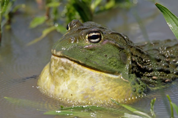
Natural history
The African bullfrog lives in a variety of arid and semiarid habitats in central and southern Africa. This frog has long ridges on the skin of its back and a huge head.
The pet trade is supplied by captive-bred and wild-caught specimens. African bullfrogs are often called “pixie” frogs, derived from their genus, not their size. It is theorized that a number of different subspecies or species are now sold in the trade as African bullfrogs.
Taxonomy
Class: Amphibia
Order: Anura
Family: Ranidae
Bullfrogs, leopard frogs, rice paddy frogs
Color and Size
African bullfrogs are olive green in color. The males have a yellow or orange throat, while female throats are cream-colored. Females may also retain the dorsal yellow stripe seen in the juvenile.
African bullfrogs are large, often weighing more than 2 kg (4.4 lbs). Males may reach 20-25 cm (8-10 in) in length. Females are typically half this size.
Diet
Feed adult bullfrogs insects such as locusts, crickets, cockroaches, mealworms, moths, beetles and waxworms, as well as fish, earthworms, and mice. Bullfrogs may also be fed an occasional mouse. Bullfrogs have a voracious appetite and sometimes jump quite high. Since they can mistake fingers for food, they should be fed with tongs.
Dust invertebrate prey regularly with a vitamin/mineral supplement.
Husbandry
| Temperature | Provide a temperature gradient of 77- 82°F (25-28°C). Do not use a radiant heat source as these tend to dry amphibian environments. |
| Water | Offer a large, shallow water dish, in which the bullfrog will spend much of its time, and lightly mist the enclosure regularly. Between mistings the substrate should remain damp, but not water logged.Potential water sources include: |
- Aged water: Allow chlorinated water to sit in an open container for 24-48 hours so chlorine can dissipate
- Bottled spring water
- Filtered tap water: run through a sediment and activated charcoal filter
Cage size and designAlthough bullfrogs are very large, they are not very active. One adult may be kept in a long, 20-gallon (75 liter) terrarium.Cage furniture/suppliesProvide low-intensity full-spectrum lighting for optimal absorption of dietary calcium. Provide hiding places at each end of the cage (i.e. live or artificial plants). Consider sandy soil as a cage substrate for burrowing. Alternatively, frogs may be housed in a bare tank containing 1-2 in (2.5-5 cm) of water and a styrofoam platform, which can serve as a haul-out region. Frequent cleaning of the tank is necessary because of the prodigious amount of feces bullfrogs can pass.Social structureHouse African bullfrogs separately.
Lifespan
Up to 20 years is common, however African bullfrogs may live 45+ years.
Mature by 1.5-2 years.
Anatomy/ physiology
| Dermatologic: |
|
| Gastrointestinal: | A short, simple gastrointestinal tract empties into a cloaca. |
| Cardiac: | A three-chambered heart is encased within the pectoral girdle. |
| Lymphatics: | All amphibians possess lymph hearts, which beat in synchrony independently of the heart. |
| Miscellaneous: | If temperatures fall below recommended levels, then the African bullfrog may estivate or enter a period of “partial hibernation”.Frogs possess coelomic and inguinal fat bodies. |
| Urogenital: | A renal portal system is present |
| Sexual dimorphism: | Unlike most other species of frog, the male is larger than the female. Females are usually a lot smaller than males with rounder body shape.During the breeding season, male bullfrogs will call. They will also develop nuptial pads on each thumb as well as yellow or orange throats. |
Restraint
Their ridged skin means that the African bullfrog is one of the few amphibians that can be handled, although handling still should not be done too regularly or for prolonged amounts of time. Like all amphibians, bullfrogs possess relatively delicate, absorbent skin so handle the frog with clean, wet hands or with moistened surgical gloves.
Use caution since adult African bullfrogs can inflict a painful bite.
Venipuncture
Ventral midline vein
Preventive medicine
Perform a visual exam by placing the frog in a clear plastic box or tub.
Physical examination prn (as needed).
Important medical conditions
- Foreign body ingestion
- Nutritional secondary hyperparathyroidism or metabolic bone disease (Download the client handout:
Metabolic Bone Disease in Reptiles and Amphibians). - Obesity
**Login to view references**
References and further reading
Basic Information Sheet: Box Turtle
Box turtles are indigenous to North America. Free-ranging box turtles spend much of their time in woodland and grassy habitats, near streams or other water sources. Most box turtles offered for sale in the pet trade are . . .
Pour les vétérinaires. Par les vétérinaires.
Le site Lafervet.com est conçu pour une utilisation par les vétérinaires. Il est ouvert aux vétérinaires diplômés, aux techniciens vétérinaires diplômés, aux animaliers et aux étudiants dans ces domaines.
Créer un compte pour accéder à des articles et des ressources du site.
L'inscription est gratuite.
Para Profesionales Veterinarios. Por Profesionales Veterinarios.
El sitio Lafervet.com es para uso de los profesionales veterinarios. Está abierto a los veterinarios licenciados, técnicos veterinarios licenciados, rehabilitadores licenciados y estudiantes en estos campos.
Cree una cuenta para acceder a los artículos y recursos del sitio.
La registro es gratis.
Already a LafeberVet Member?
Please Login
Lories and Lorikeets, Care of
Lories and lorikeets are some of the most colorful members of the parrot family. Native to Australia, Tasmania, and the South Pacific islands, many species are threatened or endangered in the wild due to habitat loss or trapping. This educational handout will help your client understand how to care for and maintain these beautiful birds in captivity. Recommendations for diet, housing, and bathing are described as well as common problems seen in the pet lory and lorikeet.
Download the Care of Lories and Lorikeets PDF Handout (129 KB)
Basic Information Sheet: Boas and Pythons
Boas and Pythons

Photo credit: ‘Dept’ by Flickr Creative Common
Natural history
The group of snakes called “boas” consists of over 40 species. Most popular pet boas are New World species, however boas are also found in Africa and Asia. Most boas live in environments that range from tropical rain forest to dry woodland and scrub forest. Sand boas (Gongylophis spp.) may be found in desert and savannah land and Rosy boas (Lichanura spp.) live in dry, rocky habitats.
Pythons are found in Africa, Australia, and Asia under conditions that range from rain forest to desert. Some species are primarily ground dwellers, but most can be found in bushes and trees. The larger species are often found near water and are strong swimmers.
Pet boas and pythons may be farm-bred, captive-born, or wild-caught imports. Popular companion snakes include the Boa constrictor (Boa constrictor imperator), Rosy boa, and Ball python (Python regius). Ball pythons are docile snakes that coil into a tight ball with the head buried within when frightened. Captive-bred ball pythons are much preferred over wild-caught specimens since the latter can fast for long periods and can be stubborn feeders.
In January 2012, the U.S. government banned the importation of snakes and eggs of four species, Burmese python, northern and southern African pythons, and yellow anacondas, in reaction to the python problem in southern Florida.
[/ezcol_2third_end]
Taxonomy
Class: Reptilia
Order: Squamata
Family: Pythonidae-pythons
Family: Boidae-boas, anacondas
Color and size
 Photograph by gjofilli. Click to enlarge. |
Boa constrictors usually reach 7-8 feet (2 meters) in captivity, but wild specimens may reach more than 12 ft (4 m). |
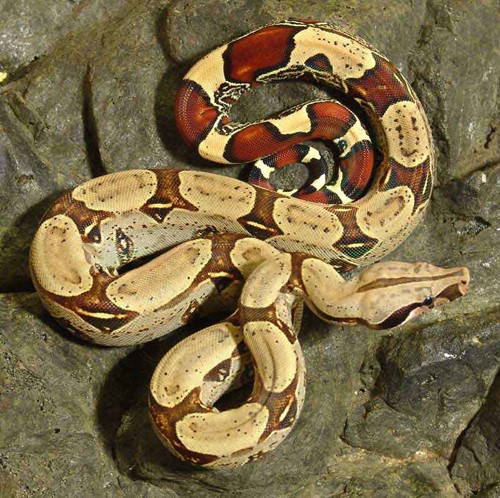 Peruvian red-tailed boa from petinfospot.com. Click to enlarge. |
Red-tailed boas (Boa constrictor constrictor) can reach 6-12 ft (2-4 m) |
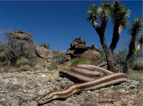 Image from itsnature.org. Click to enlarge. |
Rosy boas have a blunt tail and smooth scales. The ground color ranges from tan to silver with deep chocolate to orange stripes. Rosy boas are relatively small snakes that rarely exceed 3 ft (0.9 m). |
 Photograph by Brian Gratwicke. Click to enlarge. |
Pythons include some of the world’s largest snakes. Giant snakes like the Burmese python (Python molurus bivitattus) and reticulated python (Python reticulatus) can reach 20 feet (6.1 m) or more and are not recommended for casual hobbyists. However the popular ball python (Python regius) [shown left] rarely exceeds 5 ft (1.5 m) and is usually about 4 ft (1 m) in length. |
Diet
Boas and pythons feed on rodents of appropriate size, primarily mice and rats predominately although gerbils may be preferred by recently imported adult ball pythons. Larger snakes may also be fed rabbits and chickens.
Snakes should be fed every 1-4 weeks depending on the size of the individual. Smaller snakes should be fed more frequently, and larger snakes less frequently. Pre-killed food is preferable since live prey can inflict serious bites and scratches.
Download the client handout: Feeding Snakes for more information.
Husbandry
| Temperature | Provide a daytime temperature gradient of 80-85°F (27-29°C). At night, the temperature may be reduced to 70-75°F (21-24°C). A winter cool-down or “brumation” is important for captive breeding of some species like the rosy boa. |
| Humidity/water | Most (BUT NOT ALL) species do well with a relative humidity of 50-70%.Provide a water bowl large enough for the snake to completely immerse its body, and heavy enough that the animal will not tip it over.Provide ball pythons with a humidity box” or a hiding place filled halfway with damp sphagnum moss. Place the container half on and half off the heat. |
| Cage size | Snakes up to 4 ft (1.2 m) in length can be maintained in at least a 20-gallon (75 L) aquarium or similar sized cage but ideally the habitat should be as long as the snakes. Larger animals need larger cages. For example, breeders often keep ball pythons in cages (or Sterilite tubs) that are 4 ft long. |
| Social structure | Boas and pythons are solitary. |
| Cage furniture |
|
Lifespan
Reported life span ranges:
Common boa constrictor: 20+ years
Ball python: 20+ years
Burmese python: 12-20 years
Red-tailed boa: 20+ years
Rosy boa: 15+ years
Anatomy/ physiology
| Dermatologic: | The scaled skin is normally shed in one piece in a process called “ecdysis”. Snakes lack movable eyelids. Instead a clear scale called the “spectacle”, which is an embryonic fusion of the eyelids, protects the eyes. |
| Cardiopulmonary: |
|
| Gastrointestinal: | The teeth are arranged in six rows with double rows present in the upper jaw. Snake teeth are shed through life, and are attached to the bone’s surface instead of roots. |
| Urogenital: | Snakes possess a renal portal system. Pythons lay eggs (“oviparous”). Boas bear live young, and are “ovoviviparous”. Live young are hatched from eggs within the reproductive tract. |
| Sexual dimorphism: | Both boas and pythons have external or cloacal spurs. These sharp, keratin-covered, bony structures are remnants of the vestigial pelvic girdle. Spurs are better developed in males, and are very small or absent in females. The copulatory organs of the snake are “hemipenes”. Hemipenes lie within pouches at the ventral tail base. Probing of the male pouches will be deeper when compared to females. |
Restraint
- When handling a snake, take care not to smell like snake food (i.e. rodents or rabbits). Wash your hands thoroughly beforehand.
- Support the head, neck, and body. Make sure the body weight is not borne by the snake’s single occipital condyle and the cervical spine.
- A towel is useful for aggressive snakes.
- Never carry a constrictor draped around the neck.
- Another rule of thumb for safety’s sake is to utilize one handler per 3-4 ft (0.9-1.2 m) of snake. Giant constrictors require special precautions to ensure safety of its handlers.
Tail vessel
Cardiac puncture
Injections
Give intramuscular injections between the scales in the epaxial muscles. Use the cranial half of the body to avoid the renal portal system.
Preventive medicine
- Regular physical examination
- Determine gender
- Fecal parasite testing
Important medical conditions
-
- Boid inclusion body disease (IBD) virus
- Dysecdysis, retained spectacles
- Pneumonia
- Stomatitis
**Login to view references**
References
Basic Information Sheet: Northern Blue-Tongued Skink
Northern Blue-Tongued Skink (Tiliqua spp.)
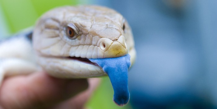
Photo credit: Conor Lawless via Flickr Creative Commons
Download the Skink Client Handout
Natural history
The blue-tongued skinks are represented by a variety of species, subspecies and races native to Australia, New Guinea and Indonesia. Two of the more common varieties in the pet trade are the northern, Tiliqua scincoides intermedia, and Irian Jaya, an undescribed T. scincoides. These ground-dwelling diurnal skinks have tiny legs and feet, heavy bodies, and a large, blue tongue that can be bared as a warning to potential enemies.
Some blue-tongued skinks are domestically bred, however many are imported from Indonesia and New Guinea.
Taxonomy
Class: Reptilia
Order: Squamata
Family: Scincidae-skinks, sandfish
Color and size
This large, heavy-bodied skink has ridged or “keeled” scales and dark crossbands. Pink to orange coloring is often found on the sides.
Adults reach 12-15 in (30-38 cm) in length.
Diet
Blue-tongued skinks are omnivores. They needs are best met by feeding a wide variety of foods:
- The bulk of the diet (45-60%) should consist of greens. Feed dark, leafy greens like mustard, kale, dandelion and collard greens. Mix vegetables with the salad such as peas, green beans, corn, squash, carrots, sweet potato, cucumber, zucchini, green peppers, and parsley.
- Also offer a protein source such as gut-loaded crickets, mealworms, and kingworms, snails, and the occasional pinky mouse. Some skinks may also accept earthworms. Juveniles eat more prey items than adults. Download the client handout: Feeding Insect Eating Reptiles for additional information.
- Fruit and blossoms may be offered as treats. Dust the non-breeding adult’s diet with a calcium carbonate or calcium gluconate supplement once weekly. Calcium supplements should be devoid or low in phosphorus with a minimum Ca:P ratio of 2:1. A general vitamin/mineral supplement may also be offered once weekly.
Feed adults every 1-2 days.
Husbandry
| Temperature | Provide a temperature gradient with a cool end of 70-85°F (21-29°C) and a basking spot of 92-100°F (33-38°C) at the opposite end. |
| Humidity/water | Maintain approximately 30% relative humidity Fresh drinking water should always be available. The bowl or tub should be large enough for the skink to easily access. |
| Cage size and design | At minimum, provide adults with a 40-55 gallon aquarium (or a cage of equivalent size), however these lizards have wide ranges in the wild and they will do best with much larger enclosures. |
| Cage furniture/supplies |
|
| Social structure | Must be housed alone; even juveniles will fight savagely. |
Lifespan
20+ years
Blue-tongued skinks are usually sexually mature by 2-3 years of age.
Anatomy/ physiology
Restraint
Blue-tongue skinks are considered to be very docile lizards, however always wash your hands before handling a skink to minimize the risk of smelling like one of their food items. Rawhide gloves help prevent scratches and give skinks a toe-hold while being held.
Never grasp a skink by the tail. Skinks possess tail autotomy, a defense mechanism that utilizes tail loss. A vertical fracture plane of fibroconnective tissue and cartilage runs through the body and part of the neural arch of each caudal vertebrae. The tail can fall off and will regrow as a cartilaginous rod, however skinks do not lose their tails easily.
Venipuncture
Preventive medicine
- Regular physical examinations
- Fecal parasite testing
- Quarantine
Important medical conditions
- Bite wounds (group housing)
- Nutritional secondary hyperparathyroidism or metabolic bone disease
- Obesity
- Cachexia from improper husbandry
- Blue-tongued skinks are relatively hardy, however common problems include:
**Login to view references**
References
Basic Information Sheet: Inland Bearded Dragon
Inland Bearded Dragon (Pogona vitticeps)
Download the Bearded Dragon Client Handout PDF
Natural history
Native to the central deserts of Australia, the Bearded dragon’s name comes from its practice of extending the flap of skin under the jaw or “beard” in a defensive posture.
Taxonomy
Class: Reptilia
Order: Squamata
Family: Agamidae
bearded dragon, frilled dragon, water dragon
Color and size
The wild type or standard bearded dragon is tan to yellow color on its dorsum with irregular blotches and a pale belly. There are numerous other color variations in captive-bred specimens.
Including the tail, bearded dragons commonly attain a length of up to 2 ft (0.6 m). Juveniles weigh about 2-3 grams and adults weigh at least 250 g. Males tend to grow larger than females and have larger heads.
Diet
Bearded dragons are omnivores, however juveniles require a diet of almost 100% live, appropriately sized prey.
- Animal protein should make up at least 25% (by volume) of the adult dragon diet. Offer appropriately sized, gut-loaded insects such as crickets, superworms, and waxworms as well as grasshoppers, locusts, and the occasional pinky mouse. Download the client handout: Feeding Insect Eating Reptiles for additional information.
- Dark, leafy greens such as should make up 50-55% (by volume) of the adult dragon diet. Offer a variety of greens including collard greens, kale, romaine, dandelion, turnip greens, mustard greens, beet greens, bok choy, Swiss chard, spinach, chicory, and escarole.
- Other chopped or grated vegetables may comprise up to 20% of the diet (squash, zucchini, sweet potato, broccoli, peas, carrot, beans, okra, bean sprouts, tofu).
- Fruit should make up no more than 5% of the diet and should include nutrient dense items such as papaya, melon, and banana.
- Occasional treats may consist of non-toxic flower blossoms such as hibiscus.
Dust the non-breeding adult’s diet with a calcium carbonate or calcium gluconate supplement just prior to feeding once or twice weekly, more often if actively breeding. Dust every meal for hatchlings. Calcium supplements should be devoid or low in phosphorus with a minimum Ca:P ratio of 2:1. A general vitamin/mineral supplement may be offered once weekly. This supplement should contain vitamin D, and bearded dragons should have exposure to ultraviolet light (specifically UVB rays via specialized bulbs or direct sunlight).
Feed adults every one to two days. Hatchlings should be fed twice daily.
Husbandry
| Temperature | The bearded dragon has a preferred optimal temperature zone of 80-88°F (27-31°C). The basking spot should reach 92-100°F (33-38°C). Bearded dragons heat up quickly but cool down very slowly, putting them at risk for heat stroke. Never place a bearded dragon in direct sunlight when housed in a glass tank. |
| Humidity/water | Although bearded dragons thrive in low humidity, drinking water should always be provided in a shallow bowl or saucer. Dragons will often soak in their water bowl and may defecate in their water. Drinking and soaking bowls should be cleaned at least daily. |
| Lighting | Bearded dragons require bright light for adequate food intake and normal behaviors. Ceramic heaters, red bulbs, and low wattage bulbs are inadequate. Provide daily sunlight directly or a fluorescent full-spectrum light source. If a fluorescent full-spectrum bulb is used, also provide a bright spotlight at one end of the habitat. |
| Cage size | As a rule, bearded dragons require housing at least three times as long as their snout-tail length. At minimum, the single adult requires a 75-gallon (283-L) aquarium although larger enclosures are recommended. |
| Cage furniture | Bearded dragons require hiding areas (rock cave, plant pot, cardboard box, etc.) as well as thick branches upon which to climb and bask. Branches must be sturdy enough to support heavy-bodied adults. Provide a full-spectrum light source for normal absorption of dietary calcium. |
| Social structure | Bearded dragons may be housed in small groups of one male and a few females. Breeders may set up groups of two males and three to five females in large habitats. |
Lifespan
Bearded dragons typically live 8-10 years.
Bearded dragons reach sexual maturity between 8-18 months, although sexual activity may begin before 12 months of age.
Anatomy/ physiology
| Dermatologic: | The Bearded dragon has a gular “beard” or flap of skin beneath its chin that is covered with large, conical scales. Unlike snakes, lizards normally exhibit a patchy shed or “ecdysis”. |
| Gastrointestinal: | The mucous membranes of the oral cavity are normally yellow. Acrodont dentition: Teeth are not set in sockets, but instead are weakly attached to the jawbone surface. |
| Sexual dimorphism: | Males tend to grow larger than females and have larger heads. Males also have hemipenal bulges on the ventral side of the tail base and larger, more prominent femoral pores. Femoral pores are the opening through which glands produce a thick, waxy secretion. This secretion plays a role in scent marking and other pheromone-based communication. |
Bearded dragons tend to be relatively mild-mannered lizards.
Venipuncture
Preventive medicine
- Regular physical examinations
- Fecal parasite testing
- Quarantine
Important medical conditions
- Agamid adenovirus 1
- Coccidiosis (Isospora amphiboluri)
- Follicular stasis
- Nutritional secondary hyperparathyroidism or metabolic bone disease
- Periodontal disease
- Renal disease
**Login to view references**
References
Do’s and Don’ts of Avian First Aid
First aid is the initial treatment given to an injured or ill bird during an emergency. The goal of first aid is to stabilize the bird until veterinary medical care can be provided. Donated by Dr. Greg Burkett, this client educational handout helps owners prepare for emergencies. Topics covered include the creation of a home hospital cage, stocking a first aid kit, the basics of owner supportive care as well as specific advice for emergencies like bite wounds, bleeding, and head trauma.
Basic Information Sheet: Sugar Glider
Sugar Glider (Petaurus breviceps)
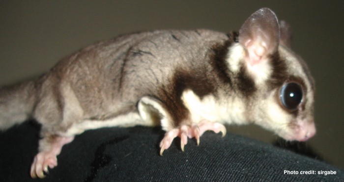
Natural history
The sugar glider is native to northern and eastern Australia, New Guinea, and surrounding islands. This arboreal, nocturnal creature spends its days in leaf-lined nests in tree hollows. Sugar gliders are extremely social and vocal.
Taxonomy
Class: Mammalia
Interclass: Marsupialia
Order: Diprotodontia-kangaroos, koala
Family: Petauridae – possums
Diet
Free-ranging gliders feed on insects, larvae, arachnids, and small vertebrates during the spring and summer. Plant products such as sap, blossoms, and nectar make up the bulk of the diet during the autumn and the winter wet season.
There are a number of captive diets recommended for gliders. The commonly recommended diet listed below comes from the Taronga Zoo. The following recipe feeds two animals:
- Apple (3 g)
- Banana/corn (3 g)
- Grapes/kiwi (3 g)
- Pear (2 g)
- Melon (2 g)
- Orange with skin (4 g)
- Sweet potato (3 g)
- Dog kibble (1.5 g)
- Fly pupae (1 tsp)
- Leadbeater’s mixture (2 tsp)
- Day-old chick, when available (once weekly)
- Large insects, mealworms once weekly
Leadbeater’s Mix:
- 150 ml warm water
- 150 ml honey
- 1 shelled boiled egg
- 25g high protein baby cereal
- 1 tsp vitamin/mineral supplement
- 100mg calcium carbonate
Mix water and honey. In a separate container, blend egg until homogenized, then gradually add vitamin/mineral powder, then baby cereal, blending after each addition until smooth. Keep refrigerated. The mixture can be frozen in ice cube trays.
Husbandry
Sugar gliders are social animals that normally live in groups of five to twelve. These groups are exclusive and territorial, and introduced gliders may be attacked.
Solitary gliders require at least 1-2 hours of human attention daily.
Provide as large a cage as possible like a tall aviary wire cage. Wire spacing should be no more than 1.0 x 0.5 in (2.5 x 1.3 cm). Cage furniture should include items for visual security (i.e. hide box, sleeping pouch) as well as exercise and enrichment (i.e. branches, shelves, solid running wheel, swings, and bird toys). Provide supplemental heat during the winter months.
Normal physiologic values
| Cloacal temperature | 89.6°F | 32°C |
| Pulse | 200-300 bpm | |
| Respiration | 16-40 bpm | |
| Body weight | 80-160g (female is smaller) | |
| Mean life span | 12-14y captivity | 4-5y wild |
| Sexual maturity | females (8-12 mo) males (12-14 mo) | |
| Gestation | 15-17 days (young stay in the pouch 70-74d) | |
| Birth weight | 30-50 grams | |
| Litter size | 1-2 (usually 2) | |
| Weaning age | 110-120 days | |
| Target environmental temperature: | 65-90°F | 18.3-32.2°C |
- Gliders possess a large gliding membrane (patagium) which extends from the 5th digit of the forepaws to the ankles.
- The tail is weakly prehensile and it also serves as a rudder during gliding.
- Dental formula: I (3/2) C (1/0) PM (3/3) M (4/4) = 40
- The incisors are specialized for gouging tree bark.
- An enlarged cecum assists in digesting gum from the acacia tree.
- The bifurcated penis splits about halfway down the protrusion.
- Testicles are permanently descended into pendulous, pre-penile scrotal sacs.
- Males possess large prostate and Cowper’s glands.
- Females have two uteri and two long, thin lateral vaginae
- Females are polyestrous, cycling every 29 days.
- Scent glands: male (frontal, sternal, paracloacal) female (within pouch)
- Gliders may enter torpor during the winter; torpor may last up to 16h/day.
Restraint
Options for restraint of the sugar glider includes:
- Grasp the head between the thumb and middle finger.
- Scruff
- Restrain through a small cotton bag or pouch
- Inhalant anesthesia
- Larger volumes: Jugular vein, cranial vena cava +/- tibial artery
- Smaller volumes: Lateral saphenous, lateral coccygeal, cephalic, and femoral veins
Preventive medicine
Important medical conditions
- Alopecia, stress-related
- Bacterial enteritis
- Cataracts
- Cloacal or rectal prolapse
- Dental disease
- Encephalomalacia, hypovitaminosis E
- “Ick“, is an emerging disease in sugar gliders
- Malnutrition
- Nutritional secondary hyperparathyroidism
- Obesity
- Pouch infection, mastitis
- Self-mutilation of the penis and scrotum
- Traumatic injury
- Urinary tract infection, urolithiasis
**Login to view references**
References
Basic Information Sheet: Rat
Rat (Rattus norvegicus)
Natural history
The Norwegian or brown rat (Rattus norvegicus) is originally from Asia where it lived in burrows on the plains of northern China and Mongolia. The rat arrived in Europe in the early 18th century and had reached America by the late 18th century. The brown rat was the first species to be domesticated for scientific purposes.
Taxonomy
Class: Mammalia
Order: Rodentia
Suborder: Myomorpha – gerbils, hamsters, mice
Family: Muridae – The largest and most diverse family of mammals including Old World rats and mice, gerbils
Breeds
A variety of breeds, colors, and markings are recognized.
Diet
Rats are omnivores. The bulk of the diet should consist of a rodent block or pellet containing a minimum 16% crude protein and 4-5% fat.
Seed-based diets promote obesity and should be avoided. Longevity is increased by feeding a low fat, vegetable protein-based diet.
Husbandry
Rats are social creatures, although females are more likely to fight.
Rats are also primarily nocturnal, but they will have cycles of activity during the day and night. Avoid bright lighting in albino rats.
Adult brown rats tolerate cold well when allowed to acclimate.
Provide rats with items and materials for burrowing, climbing, and chewing.
Normal physiologic values
| Temperature | 99.9°F | 37.7°C |
| Pulse | 300-500 bpm | |
| Respiration | 70-150 bpm | |
| Body weight | Adult male | 267-500 g |
| Adult female | 225-325 g | |
| Mean life span | 2.5-3.5y | Female rats typically live longer. |
| Sexual maturity | 37-75 days | Puberty occurs earlier in female rats. |
| Gestation | 21-23 days | |
| Birth weight | 5-6 g | |
| Litter size | 6 to 13 | |
| Weaning age | 21 days | |
| Target environmental temperature | 50-68°F | 18-26°C |
| Target environmental humidity | 40-70% | |
| Water intake | 22-33 ml/day |
Anatomy / physiology
|
Restraint
Rats are generally docile and are easy to handle. For manual restraint, grasp the rat over the back and place a thumb in the axilla pushing a foreleg up towards the chin.
Venipuncture
Collect blood samples from the lateral tail vein, ventral tail artery, femoral vein, jugular vein, or lateral saphenous vein. Warm the tail first to promote vasodilation.
Preventive medicine
Annual physical examination
Weight control may reduce the risk of mammary gland tumor development.
Studies also suggest that ovariectomy before 5-7 months of age can inhibit the development of spontaneous mammary tumors.
**Login to view references**
References and further reading
Basic Information Sheet: Mouse
House Mouse (Mus musculus)
Natural history
The most common pet mouse is the standard white laboratory mouse, although pet mice are not as inbred as some strains of lab mice.
Taxonomy
Class: Mammalia
Order: Rodentia
Suborder: Myomorpha
Family: Cricetidae-gerbils, lemmings, muskrats, New World rats and mice, voles
Breeds
The standard white lab mouse is most commonly seen, but different
colors and varieties are entering the pet trade such as satin or spotted.
Diet
The bulk of the diet should consist of a rodent block or pellet that contains a minimum crude protein content of 16% and 4-5% fat. Seed-based diets promote obesity and should be avoided.
Husbandry
Mice are nocturnal and crepuscular. Females or mixed pairs do well together, but male mice tend to fight.
Provide the largest cage possible made of stainless steel, durable plastic, or wire. Provide ample nesting material and deep bedding such as aspen shavings or recycled paper product. Provide enrichment in the form of tunnels, exercise wheels, nest boxes, and items to chew on old paper towel rolls or toilet paper tubes.
Normal physiologic values
| Temperature | 98.8°F | 37.1°C | |
| Pulse | 400-700 bpm | ||
| Respiration | 90-200 bpm | ||
| Body weight | Adult male | 20-40 g | The average mouse weighs about 30 grams. |
| Adult female | 18-35 g | Male mice are often twice the size of females. | |
| Mean life span | 1-3 years | ||
| Sexual maturity | 50-60 days | varies among strains | |
| Gestation | 19-21 days | ||
| Birth weight | 1.0-1.5 g | ||
| Litter size | 10-12 | ||
| Weaning age | 18-21 days | ||
| Target environmental temperature: | 50-68°F | or 10-20°C | |
| Target environmental humidity: | 45-55% | ||
| Water intake: | 5-8 ml/day |
Anatomy / physiology
Dental formula: Incisor 1/1 Canine 0/0 Premolar 0/0 Molar 3/3
The incisors are open-rooted and continuously growing.
The stomach consists of a non-glandular forestomach and a glandular stomach.
Restraint
Although mice rarely bite normally, they may bite when handled.
Mice are also quick to jump. Manually restrain mice by scruffing the neck.
The lateral tail vein may be used to collect small volumes of blood.
Preventive medicine
Important medical conditions
- Mammary adenocarcinoma
- Mange
- Mycoplasmosis
- Obesity
- Sendai virus
**Login to view references**
References
Basic Information Sheet: Hamster
Hamster
Natural history
Hamsters are short, stocky rodents with an abundance of loose skin. Today’s pet or laboratory hamster, known as the Syrian hamster (Mesocricetus auratus), was bred from a small group of hamsters removed from a burrow near Syria in 1930 as well as 11 more collected in 1971 and three in 1978.
Taxonomy
Class: Mammalia
Order: Rodentia
Suborder: Myomorpha
Family: Cricetidae-gerbils, lemmings, muskrats, New World rats and mice, voles
Breeds
The Syrian hamster is also known as the Golden hamster because the colony were reddish-gold, however many varieties exist today. Dwarf hamsters may also be seen in practice.
Diet
Hamsters can do very well on a commercial rodent chow (24% protein) supplemented with vegetables and a small amount of fruit. Hamsters fed seed-based diets are very sensitive to vitamin E deficiency.
Housing
Hamsters are nocturnal. As desert animals, the Syrian hamster instinctively burrows and hoards food. Adult Syrian and Chinese hamsters are fiercely territorial and should be housed singly. Winter white Russian and Russian Campbell hamsters are social, however it is still ill advised to introduce new individuals to a group.
Hamsters can enter torpor if temperatures fall below 41°F (5°C), particularly when exposed to short days.
Normal physiologic values
| Temperature | 97-102.3°F | 36.1-38.9°C | |
| Pulse | 250-500 bpm | ||
| Respiration | 34-114 bpm | ||
| Body weight | Adult male | 85-110g | |
| Adult female | 95-130 g | ||
| Mean life span | 2-3y | Maximum 4y | 18-36 months |
| Sexual maturity | 6-8 weeks | 6 wks (F) 8 wks (M) | For optimal breeding, males should be 10-12 weeks old and females 8-10 weeks. |
| Gestation | 15-18 days | ||
| Birth weight | 2-3 g | ||
| Litter size | 4 to 12 | Maximum 17 | |
| Weaning age | 3-4 weeks | ||
| Target environmental temperature: | 68-75°F | 20-24°C | |
| Target environmental humidity: | 40-60% | ||
| Water Intake: | 9-12 ml/day | ||
| Russian hamsters live 1.5-2 years. |
Anatomy / physiology
| Derm: | Sebaceous flank glands are present in both genders, but are better developed with darker pigmentation in males. |
| Gastrointestinal: |
|
| Renal: | Syrian hamster urine is normally creamy and turbid. |
| Reproductive: | Females pass a thick, creamy white post-ovulatory discharge. Each uterine horn opens into a separate cervix. |
| Physiology: | Syrian hamsters will hibernate if kept in a cool area with limited food.Syrian hamsters possess renal and respiratory water conservation mechanisms, however unlike some desert rodents they cannot survive on metabolic water. |
Restraint
Syrian hamsters tend to bite, especially when awakened from a deep sleep or put through excessive or prolonged restraint.
To pick up, cup hamsters in both hands. To restrain, grasp the loose skin over the thorax.
Roborovskii hamsters are gentle and docile, but very fast.
Venipuncture is challenging in the hamster. The jugular and saphenous veins may be tried. The Syrian hamster has a rapid coagulation time so it may help to heparinize needles.
Important medical conditions
- Adrenocortical hyperplasia and adenomas
- Cheek pouch impaction
- Ocular proptosis
- Proliferative ileitis or “wet tail”
**Login to view references**
References
Basic Information Sheet: Guinea Pig
Guinea Pig (Cavia porcellus)
Natural history
The guinea pig is a native of Peru, Brazil, and Argentina that was domesticated by the Inca Indians. Guinea pigs are also known as “cavies”, a term derived from the South American vernacular. Male guinea pigs are called “boars” and females are “sows”.
Taxonomy
Class: Mammalia
Order: Rodentia
Suborder: Hystricomorpha
Family: Caviidae
Breeds
Over 20 breeds of guinea pig are recognized with the three most popular being:
| Peruvian: | Long hair |
| American or English: | Short hair |
| Abyssinian: | Whorled or rough hair |
Diet
Guinea pigs are strict herbivores. A sound diet consists of high-quality hay, fresh foods, and water. Offer unlimited grass hay, as guinea pigs eat small portions throughout the day. Alfalfa or clover hay should only be given to juvenile animals as the high calcium content can predispose adults to renal calcification. Fresh food can consist of grasses, herbs, leafy greens, peppers, and other vegetables. Avoid or limit fruits in the diet. Too many simple carbohydrates can lead to diarrhea and/or dysbiosis.
Guinea pigs of all ages require a dietary source of vitamin C (10-30 mg/kg per day). Fresh foods rich in vitamin C can provide sufficient levels of this nutrient. Additionally, commercial guinea pig pellets often contain vitamin C and small amounts of pellets (about ¼ cup a day) can be offered alongside hay and fresh food. Pellets should be fresh and relatively high in fiber, low in sugars and calcium, and grain free. The stability of vitamin C decreases over time and the vitamin C content of pellets is also reduced by exposure to dampness, heat, and light.
Husbandry
|
Normal physiologic values
| Temperature | 37.2-38.6ºC | 99-101.5ºF |
| Pulse | 230-380 bpm | |
| Respiration | 40-100 bpm | |
| Body weight | Adult male | 900-1200 g |
| Adult female | 700-900 g | |
| Mean life span | 6-8 y | |
| Sexual maturity | 2-4 months Males will exhibit mounting behavior as early as 1 month of age. Females can become pregnant as early as 4-5 weeks of age. |
2 months (F) 3 months (M) |
| Breeding maturity | Male: 3-4 months Female: 6-8 months |
|
| Estrous cycle | 16-17 d, polyestrous | |
| Gestation | 59-72 days (average 68 days) | |
| Birth weight | 45-115 g (inversely related to litter size) 70-100 g |
|
| Litter size | 1 to 13 (2 to 4 is the usual) | |
| Weaning age | 21 days (180 g) | |
| Target environmental temperature: | 18-26ºC Avoid temperatures above 28°C (82°F) |
65-79ºF |
Anatomy / physiology
| Musculoskeletal: | Rodents have a distinctive masseter muscle. The zygomatic arch of the skull and the dental anatomy is related to their feeding habits.
Females must be bred before 6-9 months of age. Immature male s and females have a fibrocartilaginous pelvic symphysis fusion which becomes ossified over 1 year of age. The symphysis remains cartilaginous in primiparous sows so the pelvic canal can dilate allowing passage of the large fetus. |
| Dental formula: | I 1/1 C 0/0 PM 1/1 M 3/3
All teeth are open-rooted and show lifelong growth. The lower incisors are normally 3X longer than the upper incisors. |
| Special Senses: | Large ears with thin-walled pinnae and well-developed bullae. |
| Urogenital: | Males have lateral scrotal swellings on each side of the anus.
Accessory glands include a prostate, coagulating gland, bulbourethral gland, and seminal vesicles. Seminal vesicles are large, yellow-white tubular structures which lie dorsal to the bladder. The seminal vesicles can be confused with the uterus. |
| Respiratory: | All rodents are obligate nasal breathers. |
Restraint
Most pet guinea pigs are calm, gentle animals that rarely bite. To pick up a guinea pig, cup one hand gently under the thorax and use the other hand to support the hind end. Hold the rump lower than the rest of the body.
Large volumes: Jugular vein, cranial vena cava (only under sedation/anesthesia)
Small volumes: Cephalic vein or lateral saphenous vein
Fluid therapy
Administer warm fluids: 38-39°C (100-102°F).
Maintenance fluids are estimated as 50-75 ml/kg/day.
- Dehydration (%) x BW (kg) x 1000 ml/L = fluid deficit (ml)
- Correct fluid deficits over 12-24 hours
Subcutaneous (SC) fluids are indicated only for animals with a stable circulatory system and mild dehydration (max 5%).
- Administer SC fluids up to three times per day (give one-third of calculated fluid volume at a time)
- Infuse up to 150 ml/kg/day (average 40-60 ml/kg)
Intravenous or intraosseous fluid therapy is indicated in critical patients. Intravenous (IV) catheters are placed with growing frequency in guinea pigs, but when IV catheterization fails or when veins are too small or too fragile, an intraosseous or IO catheter can be an excellent option.
| Catheter type | Recommended sites | Comments |
|---|---|---|
| Intravenous catheter | Cephalic or lateral saphenous vein | Slow bolus: 5-10 ml/kg IV every 8 hours |
| Intraosseus catheter | Proximal humerus, proximal tibia, proximal femur | 20-22 gauge (1-1.5 inch) spinal needle Local anesthesia with lidocaine Use survey radiographs to confirm placement |
Important medical conditions
- Bacterial pneumonia
- Dystocia
- Fur mites
- Hypovitaminosis C or scurvy (can reportedly occur within 4 days of dietary decrease)
- Malocclusion
- Ovarian cysts
- Pododermatitis
- Pregnancy toxemia, preeclampsia
Antibiotics to avoid
Avoid antimicrobials that attack only gram-positive bacteria such as beta-lactams.
P.L.A.C.E.:
- Penicillin
- Lincosamide, lincomycin
- Amoxicillin, ampicillin
- Cephalosporins, clindamycin
- Erythromycin
References
Basic Information Sheet: Gerbil
Gerbil
Photo credit: MDL.hu via Flickr Creative Commons
Natural history
The Mongolian gerbil or “jird” is a native of the Gobi desert, originating from Mongolia and Northern China.
Taxonomy
Class: Mammalia
Order: Rodentia
Suborder: Myomorpha
Family: Muridae-The largest and most diverse family of mammals including Old World rats and mice, and gerbils.
There are 14 genera of gerbils.
There are 87 known species of gerbil including the Mongolian gerbil (Meriones unguiculatus).
Physical description
Including the tail, the Mongolian gerbil is 12-14 cm long. The head is broad and short, and the tail is covered in fur with a small tuft at the end.
Diet
Gerbils are omnivores. In the wild the diet consists of grasses and seeds with small amounts of insects. A captive diet consists of a high-quality pellet supplemented with small amounts of fruits and vegetables.
Husbandry
Gerbils are nocturnal, but some daytime activity normally occurs. Gerbils are sociable and generally do not fight when raised together.
Breeding pairs of gerbils are usually housed together for life.
Normal physiologic values
| Temperature | 99.3-100.8 F | 37.4-38.2 C | |
| Pulse | 260-600 | ||
| Respiration | 85-160 | ||
| Body weight | Adult male | 46-131 g | |
| Adult female | 50-55 g | ||
| Mean life span | 2-4y | ||
| Sexual maturity | 10 weeks | 9-18 wks (M) | 9-12 wks (F) |
| Gestation | 22-26 days | ||
| Litter size | 3 to 8 | ||
| Weaning age | 21-28 days | ||
| Target environmental temperature: | 50-68 F | 18-22 C | |
| Target environmental humidity: | 45-55% |
Anatomy / physiology
|
Restraint
|
Small amounts of blood may be collected from the lateral saphenous vein.
Important medical conditions
Ovarian cysts, tumors
Squamous cell carcinoma
**Login to view references**
References
Basic Information Sheet: Ferret
Ferret (Mustela putorius furo)
Natural history
The domestic ferret is probably derived from the European polecat (M. putorius putorius) (Chitty 2009). Ferrets serve as working animals (in the age-old tradition of “ferreting”), pets, and laboratory animals. In the United States, ferrets are raised on ferret farms where they are spayed or neutered at 6 weeks of age. After each procedure, a tattoo is placed on the ear pinna. Male ferrets are called “hobs”, females are “jills”, and juveniles are “kits”.
Taxonomy
Class: Mammalia
Order: Carnivora
Family: Mustelidae – weasels, skunks, stoats, otters, badgers
Colors
Ferrets come in a variety of colors with albino and sable or fitch being the two original ferret colors. Other color standards listed by The American Ferret Association lists include black, black sable, champagne, and chocolate. Pattern standards include panda, dark-eyed white, roan, and color point or Siamese.
Ferrets with strong white features on the head have a genetic disposition for deafness (Fehr and Sassenburg 2015).
Diet
Ferrets are obligate carnivores. Free-ranging ferrets feed on fresh whole carcasses of their prey (Chitty 2009). In the US, pet ferrets should be fed a high-quality ferret food. Cat food that contains animal-based protein can be used if no ferret food is available (Bell 1999). Crude protein should be 30-35% and fat content should be 15-20%. Avoid kitten food as this contains higher fat levels than is necessary.
In the UK and Europe, pets are sometimes offered high-quality, fresh carcass instead of ferret food (Chitty 2009). Vitamin supplements may be necessary when feeding defrosted carcasses (Chitty 2009).
Taste preferences develop in the first months, therefore different food should be offered to ferrets in a young age (Quesenberry & Carpenter 2012).
Husbandry
Although technically nocturnal, ferrets easily adjust their schedule to human activity (Ball 2002). House ferrets in multilevel cages with solid-bottom flooring. Provide toweling and other items for burrowing and hiding. Ferrets are also commonly litter pan trained (Boyce 2001).
Never allow ferrets free roam of the home. Instead supervised play should be limited to a ferret-proofed room or region of the home.
Normal physiologic values
| Temperature | 100-103 F | 37.8-39.4 C |
| Pulse | 180-250 bpm | |
| Respiration | 30-40 bpm | |
| Body weight | 600-2000g | Males are larger |
| Mean life span | 6-10 y | |
| Sexual maturity | 9-12 months | |
| Gestation | 41-42 days | |
| Litter size | 8 | |
| Birth weight | 6-12 g | |
| Teeth first erupt | 3 weeks | |
| Eyes open | 32-34 days | |
| Weaning age | 6-8 weeks | |
| Daily water intake | 75-100 ml | |
| Target environmental temperature: | 65-70 F | |
| Target environmental humidity: | 40-65% |
Anatomy / physiology
| Integumentary | Seasonal molts occur, especially in ferrets living outdoors, coats lighten during the summer and darken during the winter. Even when descented and castrated, sebaceous skin glands convey a musky odor (Chitty 2009). A lack of sweat glands makes ferrets vulnerable to heat stress. |
| Dental formula Gastrointestinal | I3/3 C1/1 PM3/3 M1/2 Short, simple digestive tract with no cecum or ileocolic valve. |
| Musculoskeletal | Ferrets have a slender and elongated body shape with a very flexible spine. The vertebral formula is C. 7, T. 15, L. 5-7, S. 3 (fused), Cd. 18. The thorax is very long with 15 paired ribs.
The extremities are relatively short, and there are five toes on each paw (Chitty 2009). Ferrets possess powerful jaw tone due their very strong masticatory muscles (Chitty 2009). |
| Special Senses | Small pinnae and well-developed bullae. Olfactory sense highly developed; visual sense moderately developed (Chitty 2009). |
| Urogenital | Renal cysts are common incidental findings. The male ferret possesses a J-shaped os penis. Females are induced ovulators. |
| Cardiovascular | The heart lies more caudal in the chest than in similarly sized dogs and cats. |
| Respiratory | A very narrow ventral space in the nasal conchae makes passing a nasogastric tube difficult. |
| Hematopoietic | Blood types have not been identified in ferrets and transfusion can be performed (sans cross-matching) with little risk (Marini 2014). |
| Endocrine | Early neutering and a lack of natural photoperiod may predispose ferrets to adrenocortical disease. |
Ferrets are sweet natured, gregarious animals that may be minimally
restrained. Ferrets may be manually restrained:
- Scruff and stretch. Instead of holding the rear limbs as in a cat, grasp the pelvis in one hand.
- Roll the ferret up in a thin towel to create a ferret burrito.
The total blood volume in ferrets is 40-60 ml; up to 10% can be withdrawn in healthy individuals.
Large volumes: Jugular vein, cranial vena cava
Small volumes: Cephalic vein or lateral saphenous vein
The ferret jugular vein is located more lateral than in a cat.
There is thickened skin over the skin of the neck in hobs.
Preventive medicine
Annual examinations are recommended until ferrets are 3-4 years old, then biannual exams are recommended. Almost all pet ferrets in the United States are descented and neutered before they enter the market. As induced ovulators, all female ferrets should be spayed to prevent the risk of persistent estrus and potentially fatal anemia.
Surgically sterilized females also have a high risk for the development of hyperadrenocorticism. Chemical castration with a Deslorelin implant reduces the risk of adrenal disease (Schoemaker et al 2018).
Vaccinate against rabies virus and canine distemper virus (CDV). Ferrets are exquisitely sensitive to CDV and postvaccinal distemper infection has been reported after use of vaccines for dogs. Therefore ferrets should never be vaccinated with products intended for use in dogs (Meredith 2009).
Important medical conditions
**Login to view references**
References
Basic Information Sheet: Degu
Degu (Octodon degus)
Natural history
Degus, also known as brush-tailed or trumpet-tailed rats, are natives of central Chilean open scrubland where they are routinely exposed to droughts. Degus survive on very poor diets in the wild.
Taxonomy
Class: Mammalia
Order: Rodentia
Suborder: Hystricomorpha
Family: Octodontidae
Diet
Wild degus feed on grasses, seeds, cactus fruits, tubers, and local crops. The captive diet should consist of rodent chow low in sugar (devoid of cane molasses) such as guinea pig or chinchilla pellets supplemented with green vegetables, and free-choice grass hay. Avoid fresh fruit and other sugar-rich foods such as corn, peas, and potatoes. Degus normally drink very little water.
Husbandry
| Cage furniture/Enrichment: | Degus are very active. Provide a wheel, PCV tubing for burrowing, and a hide box. Provide small Nylabones, paper towel rolls, and pine cones for chewing. Hang small, sturdy branches across the cage for climbing and chewing. Also provide ladders and ramps for climbing. Provide a dust bath at least twice a week. |
| Numbers: | Degus are social animals that may be housed in same sex pairs. Singly housed individuals may develop stereotypies, depression, or barbering without a lot of owner attention. |
| Temperature: | Degus easily succumb to heat stress at temperatures > 86 F. |
Normal physiologic values
| Temperature | 100.9 F | 38.3°C |
| Body weight | Adult male | 275 g |
| Adult female | 250 g | |
| Mean life span | 5-9 years | (Should live 8+ years) |
| Sexual maturity | 75 days | |
| Gestation | 87-93 days | |
| Litter size | 3-11 (5-6) | |
| Weaning age | 4-6 weeks |
Anatomy / physiology
| Dental formula Gastrointestinal
|
I (1/1) C (0/0) PM (1/1) M (3/3) Incisors are yellow to orange in color due to iron pigments. Molars are deeply enfolded giving them a “figure 8” appearance. |
| Urogenital | As in chinchillas, the vaginal is normally closed with a thin membrane. Both sexes have a cone appendage or genital papilla used for urination. In females, the distance between the anus and urethral cone is very short. The testicles are intra-abdominal. |
| Special Senses | Degus possess a good sense of sight, hearing, and smell. |
Restraint
Most degus can be easily handled by encircling the middle with one hand and the tail base with the other. NEVER pick a degu up by the tail as they can shed their tails.
Preventive medicine
To minimize the risk of diabetes mellitus, avoid fruit and high-carbohydrate vegetables, and maintain a healthy body weight (no more than 250 g for females, 275 g for males).
Important medical conditions
- Cataracts
- Diabetes mellitus
- Obesity
Antibiotics to Avoid
Avoid antimicrobials that attack only gram-positive bacteria such as beta-lactams.
P.L.A.C.E.:
- Penicillin
- Lincosamide, lincomycin
- Amoxicillin, ampicillin
- Cephalosporins, clindamycin
- Erythromycin
**Login to view references**
Resources
Basic Information Sheet: Chinchilla
Chinchilla (Chinchilla lanigera)
Photo credit: Darekp via Wikimedia Commons
Natural history
The chinchilla is a small mammal native to South America. Originally found in the Andes Mountains of Peru, Bolivia, Chile, and Argentina, chinchillas were hunted and trapped for their pelts to near extinction in the early 1900s.22 The last free-living chinchillas are found in the mountains of northern Chile.13 Chinchillas are perfectly adapted to the rough conditions of their natural habitat: wind, cold, and dryness.22
Taxonomy
Class: Mammalia
Order: Rodentia
Suborder: Hystricomorpha
Family: Chinchillidae
Genus: Chinchilla
Chinchilla lanigera, also known as the long-tailed chinchilla (formerly Chinchilla velligera) has a smaller body, large ears, and a long tail.
Chinchilla brevicaudata, or the short-tailed chinchilla (formerly C. chinchilla chinchilla), has a relatively large body, small ears, and a short tail.
Conservation status
The International Union for Conservation of Nature lists both long-tailed and short-tailed chinchillas as endangered due to loss of natural habitat and hunting and trapping of animals.21
Color
The normal wild-type chinchilla is a smoky blue-gray (standard gray).17 The are many other colors including black velvet, beige, white, ebony, violet, and sapphire. These colors serve as the basis for color mutations such as tan, brown velvet, mosaic, pink-white, etc.12
Diet
The natural diet of chinchillas consists of grasses, cactus fruits, dry roots and tubers, as well as the bark and leaves of small shrubs and bushes. Almost all food items are high in dietary fiber with little fat, sugar, or protein.28 The digestive system of chinchillas is perfectly adapted to these nourishments, the long intestine guarantees an optimal utilization of the sparse food.14,27
Captive animals should be fed a high-fiber diet, similarly low in protein, sugar, and fat. The bulk of the diet should consist of high-quality grass hay. The hay must always be freely available, dry, and free of odor, mold, or dust.28 Dried herbs and high-fiber chinchilla or rabbit pellets, approximately 15-30 ml (1-2 teaspoons) per animal per day, can be offered. Small quantities of fresh vegetables can also be fed (see important medical conditions below).
The digestive system of the chinchilla is perfectly adapted to extract energy and nutrients from a barren food supply, which puts captive animals at risk for obesity and hepatic lipidosis. Do not feed high-fat foods, like nuts and seeds, or foods rich in sugar, like fruits (e.g. raisins).
Water has to be freely available. In the wild, chinchillas nibble rain drops from leaves or stones, either water bowls or water bottles are suitable and have different advantages. While water intake is larger from bowls, which may reduce the risk for urolithiasis 11, water in bottles stays clean longer.19
Husbandry
Chinchillas are crepuscular to nocturnal, although they can be active during the day.
| Caging | Chinchillas require a great deal of space with multiple levels for jumping and climbing. The minimum dimensions should be 50 cm (20 in) wide and 150 cm (60 in) high with a volume of at least 3 m3 (800 gallon). For each additional animal at least 0.5 m3 are required. The cage floor should ideally be solid and covered with aspen shavings or recycled paper product. Select small-gauge wire (15 mm x 15 mm) to prevent limb injury.1
The best enclosure for chinchillas is a chinchilla room with branches and wooden platforms (S. Mayr, written communication). A latrine area can be cleaned daily, but the remainder of the room can be cleaned once a week. The floor is covered with recycled paper bedding or aspen wood shavings. |
| Cage furniture | Provide visual security, such as PVC piping, cork caves or sleeping cottages with a minimum size of 30 x 20 x 20 cm. Provide a separate hiding box to each animal. The hiding boxes should be placed on different levels in the cage, however the highest hide box will probably be the preferred sleeping place.
Provide free choice access to dust baths, whenever possible. Use commercially available chinchilla dust. Clean or sieve sand regularly (see important medical conditions below). Chinchillas also require material for gnawing or chewing to reduce the risk of dental overgrowth. Branches of unsprayed deciduous trees (e.g. hazelnut, willow, birch) are suitable; do not offer branches from stone fruit trees or conifer trees. |
| Enrichment | If not kept in a room especially set up for chinchillas, animals require at least 30 minutes of exercise or play outside of their cage daily.
“Flying saucer” exercise wheels are preferable over “regular” exercise wheels to minimize the risk of back and limb problems. The larger “flying saucer” wheels do not force the animal into an abnormal hyperextended back position, but individuals can still develop limb problems due to abnormal loading of joints and muscles. |
| Numbers | Chinchillas are social animals. Pairs, trios, and harems as well as single-sex groups can live together peacefully.18 The best period to socialize animal is before sexual maturity. |
| Temperature | Chinchillas easily succumb to heat stress at temperatures exceeding 26.7°C (80°F). Temperatures between 18.3-26.7°C (59°F-70°F) are best for holding and ambient temperature.19 One source describes optimal temperatures as low as 10°C (50°F).26 |
| “Neighbors” | Rodents should not be housed near rabbits. Even healthy rabbits can harbor a variety of bacteria that can cause morbidity or mortality. |
Normal physiologic values
Note: Physiologic values reported are for C. lanigera unless otherwise noted9,19,20
| Rectal temperature | 98.5-100.4°F | 37-39.5°C | |
| Pulse | 150-350 bpm | ||
| Respiration | 40-80 bpm | ||
| Body weight | Adult male | 400-500 g | |
| Adult female | 450-700 g | Females are usually larger. | |
| Mean life span | 8-10 yr. | Up to 20 yr. has been reported | |
| Sexual maturity 8,9 | F: 4-6 months | M: 8-9 months | |
| Breeding maturity 9 | F: 8-12 months, minimal weight 600 g | ||
| Suitable castration age | F: n/a | M: 6 months | |
| Heat or estrus | 4-5 days | Spontaneous ovulation | |
| Estrous cycle 2 | 28-35 d (range 15-90) 2,19 | Directly after birthing, the female is receptive again (until 10 days after the birth) | Seasonally polyestrous (November to May in the Northern Hemisphere) |
| Gestation | C. lanigera 105-118 days (average 111 + 3 days)9 | C. brevicaudata 128 days | |
| Birth weight | 30-50 g | ||
| Parental investment | Precocial | Newborns are fully furred with open eyes | |
| Litter size | 1-6 (average 2) | ||
| Weaning age | 4-8 weeks | ||
| Target environmental temperature | 50-68°F | 10-20°C 25 | |
| Target environmental humidity | < 40% 1 | Should not exceed 55% | |
Anatomy/physiology
| Dermatology | Chinchillas have more hairs per square inch than any other animal.26 This dense coat, along with a lack of sweat and sebaceous glands, makes chinchillas very sensitive to heat and increased humidity.
Chinchillas can reflexively release tufts of hair (“fur slip”) as a predator avoidance mechanism leaving a patch of smooth skin, which takes several months to regrow.19 The skin on the tail can also tear when an inexperienced individual attempts to catch up a chinchilla. |
| Dental formula | I (I/1) C (0/0) PM (1/1) M (3/3)
The front of the incisors is yellow to orange due to iron in the enamel.5 The sides and posterior part of the incisors and molars are colorless or homogeneous white. The teeth are open-rooted (hystricomorph) and show lifelong growth (approximately 12 mm or 0.4 inches per week).24 The upper incisors grow more rapidly than the lower incisors.6 The occlusal surface of the morals is parallel to the jawbones, and masticatory movement is craniocaudal. |
| Hematology | Adult chinchillas have a lymphocytic hemogram (up to 94% total leukocyte count).
Their neutrophils are hyposegmented and are also known as acidophils or pseudoeosinophils.3,4,20,29 |
| Musculoskeletal | The skeleton is very delicate, especially the tibia/fibula.
Males possess an os penis.15 |
| Respiratory | All rodents are obligate nasal breathers. |
| Special senses | Chinchillas possess large ears with thin-walled pinnae and well-developed bullae. |
| Urogenital | Females have a prominent urinary papillae (or cone). The vaginal membrane is closed except during estrus and parturition. There are two uterine horns and two cervices.19
Male chinchillas lack a true scrotum and the testicles sit within the inguinal canal. Males possess an os penis.15 |
Chinchillas are relatively easy to restrain, however they can become can be quite quick and prone to struggle. To take the animal out of an enclosure, it is easiest to gently grasp the tail base first. The second hand supports the body around the chest. Most chinchillas are calm if they are wrapped in a towel; others like to hide their head in the crook of the handler’s arm.
Never hold the tip of a chinchilla’s tail.
When frightened or held overzealously, ‘chins’ can shed patches of fur (see Dermatology above).
Large volumes: Jugular vein, cranial vena cava4,9,19,23,29
- Use a 22-to 25-gauge needle; aspiration with 1-to 3-ml-syringe.
- Caval sticks should only be performed under anesthesia.
Small volumes: Cephalic vein or lateral saphenous vein4,9,19,23
- Use an insulin syringe, a 25-gauge needle with a 1-ml syringe, or a 20- or 21-gauge needle with the hub broken off, free dripping or Multivette (V. s. lateralis).
- Sedation or shaving is generally not necessary.
Preventive medicine
Important medical conditions
Commonly seen conditions include:
- Barbering or fur chewing
- Dental disease, dental abscess
- Gastrointestinal stasis
- Gastroenteritis (Since the natural chinchilla diet is low in water, significant amounts of fresh produce can lead to diarrhea and other gastrointestinal disturbances.)
- Heat stress
- Obesity, hepatic lipidosis
- Orthopedic injury (fractures)
- Penile hair rings
Less common conditions include:
- Conjunctivitis (This clinical problem is rare, when chinchilla sand is used, however this condition is more likely to be seen when there is unlimited access to dust baths paired with poor ventilation OR when volcanic ash is offered for dust bathing. In animals with a history of this problem, offer dust for a few hours daily or at minimum a few times weekly).
- Dermatophytosis has also been reported.
- Diabetes mellitus and subsequent cataracts: Although not a common disease in chinchillas, this condition has been reported. Affected animals are most commonly presented for changes in eye color.
- Movement stereotypies, such as cage scratching and backflipping, have been described in fur farm chinchillas.
- Vitamin-E deficiency or “yellow ear disease” is sometimes seen when chinchillas are fed low-quality feed.
Antibiotics to Avoid
Avoid antimicrobials that attack only gram-positive bacteria such as beta-lactam antibiotics or bacitracin.7,20
“P.L.A.C.E.” is a helpful mnemonic for antibiotics to avoid:
- Penicillin
- Lincosamide, lincomycin
- Amoxicillin, ampicillin
- Cephalosporins, clindamycin
- Erythromycin
**Login to view references**
References
Basic Information Sheet: Lovebird
Lovebird – Agapornis spp.
Natural history
Lovebirds live in flocks among the woodlands, savannahand forest edges of sub-Saharan Africa and Indian Ocean islands.
Taxonomy
Class: Aves
Order: Psittaciformes
Family: Psittacidae
Agopornis spp.
Agapornis fisceri – Fischer’s lovebird (shown above)
Agapornis personata – masked lovebird
Agapornis roseicollis – peach-faced lovebird
There are nine species in genus Agapornis. The peach-faced and black-masked lovebirds are most commonly kept as pets.
Physical description
- Green plumage with a rose forehead, cheeks and throat, and a bright blue rump are seen overall in lovebirds but there are many mutant strains as well.
- Males tend to be slightly larger but most species are not sexually dimorphic.
Diet
- Lovebirds are granivores and frugivores.
- Since psittacine birds hull seeds before ingestion, they do not require grit. In fact, some individuals will overeat grit when ill putting the bird at risk for impaction.
- All-seed diets are deficient in protein, vitamins, and minerals including calcium and vitamin A.
Husbandry
- Cage dimension should be at least 18 in (2.5 cm) long and 18 in (2.5 cm) wide.
- Cage bar spacing should be approximately 3/8 to 7/16 in (0.95-1.1 cm).
- At least two perches without sandpaper should be provided with 3/8 inch diameter.
- Perch diameter should be between 3/8 in (0.95 cm). Provide at least two perches. Sand paper perch covers are very abrasive to the feet, and are not recommended.
- Provide frequent water baths or showers to maintain normal skin/feather quality.
Behavior
- Lovebirds are assertive, even sometimes aggressive, in nature. Daily handling is necessary to maintain pet quality.
- A pair of lovebirds in a home will be quite loyal to each other and will not bond with the owner.
- Like most parrots, foraging is an important part of normal daily lovebird activity. Teach and encourage pet birds to play and forage.
Normal physiologic values
| Temperature (average)* | 41.8 C | 107.1 F |
| Resting heart rate (beats/min) | 206-274 | |
| Respiration (breaths/min) | 50-60 | |
| Body weight (g) | 45-70 (50) | |
| Mean life span (years) | 15 | Up to 30y have been reported |
| Sexual maturity (months) | 6-12 | |
| Weaning age (days) | 45-55 | |
| Fledgling age (days) | 30-35 | |
| Mean number of incubation days | 18-24 | |
| Average number of eggs laid | 2-6 | |
| Water intake | High individual variability | |
| Target environmental temperature | Mimic natural environment | Household temperatures of 70-80°F (21-27°C) aregenerally acceptable, however healthy birds can tolerate hot and coldtemperatures. |
| * Routine avian exam does not include measuring bodytemperature | ||
Anatomy and physiology
Anatomic traits of Order Psittaciformes include:
- Communication of the right and left nasal sinus
- The only avian tongue with intrinsic muscles
- Simply syrinx
- Craniofacial hinge of beak is a synovial joint
- Ceca absent
- Gall bladder often absent
- Zygodactyl foot:two toes pointed backward and two pointed forward
Restraint
Lovebirds may be restrained by holding the head between index and middle fingers. Support the body with the palm of the hand as well as the thumb and little finger.
Venipuncture
Use a 27-gauge needle and 1 to 3-mL syringe to draw blood from the right jugular vein.Collection of up to 1% of body weight is acceptable in healthy patients.
Preventive medicine
- Obtain a complete history and perform a thorough annual physical examination.
- Establish baseline data with regular clinical testing (complete blood count, protein electrophoresis, and plasma biochemistries.
- Ensure proper nutrition and husbandry.
- Recommend quarantine of newly acquired birds.
- Perform additional testing for select diseases based on history and physical exam findings:avian polyomavirus, avian chlamydiosis.
- Use of the equine West Nile virus vaccine may be associated with hemolytic anemia in lories.
- Birds housed in large groups or aviaries are at higher risk of Pacheco’s disease virus and use of the vaccine may be indicated.
- The avian polyomavirus vaccine is recommended for breeding populations.
Injections
| Intramuscular (IM) | Reasonably safe, most accurate. Inject middle of muscle mass. |
Ideal location–Pectoral muscle mass |
| Subcutaneous (SQ) | Large volumes can be injected, poor absorption. | Location: Inguinal or precrural fold |
| Intravenous | Effective, narrow safety range. | Right jugular vein or brachial vein is most commonly used. Alternative option: superficial metatarsal vein. |
Important medical conditions
Infectious Diseases
|
Non-Infectious conditions
|
References
Basic Information Sheet: Lory and Lorikeet
Lory and Lorikeet – Loriinae
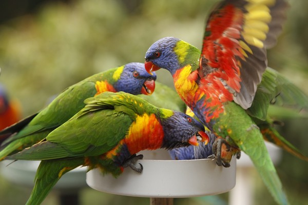
Natural history
Lories and lorikeets live in large flocks in the wild. Depending on the species, lorie sand lorikeets originate from the southeast Asia archipelago or parts of Australia. These birds will fly from island to island in search of food. Lories and lorikeets will eat coconuts andgrapes and they are considered a pest to farmers. The nomadic rainbow lorikeet follows eucalyptus flowers blooming along the Australian coast.
Conservation status
The International Union for Conservation of Nature or IUCN lists the conservation status of most lories and lorikeets as “least concern”, although some species are considered vulnerable or “near threatened”. The red-and-blue lory (Eos histrio), Rimitara lorikeet (Vini kuhlii), and ultramarine lorikeet (Vini ultramarina) are endangered.
Taxonomy
Class: Aves
Order: Psittaciformes
Family: Psittacidae
Subfamily: Loriinae
There are 12 genera of lories and lorikeets with 56 species and numerous subspecies.
The most common pet species is the red or Moluccan lory (Eos bornea).
Rainbow or green-naped lorikeets (Trichoglossus haematodus haematodus) are shown above.
Physical description
- Lorikeets have a longer, more slender tail and are smaller compared to the short, blunt tail of the larger lories.
- Plumage color varies with the species and ranges from red, blue, yellow, green, violet, and olive brown.
- The popular rainbow lory has a yellow collar with blue crown, cheeks and forehead as well as a red chest barred with black and thighs yellow barred with green.
- Most pet lory and lorikeet species are sexually monomorphic, however sexual dimorphism is recognized in some species. For example, the female fairy lorikeet (Charmosyna pulchella) has yellow patches on the sides of her rump, while the underside of the tail can be bright yellow in the male.
Diet
- Lories and lorikeets are nectarivores that feed on pollen, nectar, flowers, soft fruits, and insects.This diet creates watery droppings making lories and lorikeets better suited for aviary life.
- Companion lories and lorikeets must be provided with sufficient water. Change food often to prevent spoilage.
- Grit is not necessary but lories and lorikeets will eat grit when it is provided. In fact, some individuals will overeat grit when ill putting them at risk for impaction.
Dr. Nicole Howard of the Calgary Humane Society writes: My particular dusky lorikeet lived to 20 years of age…on a pelleted diet mixed with water…She was supplemented with organic baby food mixed in with her pelleted diet. I did NOT use corn syrup or any sweeteners. [I also recommend this diet] to new lorikeet owners (email communication Jan 4, 2017).
Husbandry
- Provide daily baths or showers.
- Perch diameter should range from 3/8 to ¾ in (0.95-1.9 cm) depending on the size of the bird. Sand paper perch covers are very abrasive to the feet and are not recommended.
- Great at escaping their cage. Make sure cage doors are locked.
Behavior
- Lories and lorikeets are intelligent and quite playful. They can learn words and tricks quite easily.
- They can be better suited as lively aviary birds, however they are territorial and do not get along with other bird species.
- Rainbow lories may sleep on their backs.
- Foraging is an important part of normal daily parrot activity. Teach and encourage pet birds to play and forage.
Normal physiologic values
| Temperature (average)* | 41.7°C | 107°F |
| Resting heart rate (beats/min) | Variable | 200 g bird ~ 178 |
| 500 g bird ~ 147 | ||
| Resting respiration (breaths/min) | Variable | 30-70 |
| Body weight (g) | 20-280 g | Species dependent |
| Rainbow lorikeet | 130 | |
| Red lory | 170 | |
| Mean life span (years) | 15-20 | Lorikeets |
| 20-30 | Lories | |
| Weaning age (days) | 62-70 | Parent-raised |
| Fledgling age (days) | 42-50 | |
| Puberty (years) | 2 | |
| Mean number of incubation days | 21-25 | |
| Average number of eggs laid | 1-5, usually 2 | Higher numbers with smaller species |
| Target environmental temperature | Mimic natural environment. | Household temperatures of 70-80°F (21-27°C) are generally acceptable, however healthy birds can tolerate hot and cold temperatures. |
| * Routine avian exam does not include measuring bodytemperature | ||
Anatomy and physiology
- Brush-like, papillae-tipped tongues allow lories and lorikeets to eat nectar and pollen.
- These nectarivores possess a weakly muscled crop, proventriculus, and ventriculus or gizzard.
- Anatomic traits of order Psittaciformes include:
- Communication of the right and left nasal sinus
- Simply syrinx
- Craniofacial hinge of beak is a synovial joint
- Ceca absent
- Gall bladder often absent
- Zygodactyl foot:two toes pointed backward and two pointed forward
Restraint
Restrain lories and lorikeets by holding the head between the index and middle fingers. Support the body with the palm of the hand as well as the thumb and little finger.
Venipuncture
Use a 25-gauge needle and 1 to 3-mL syringe to draw blood from the right jugular vein.Collection of up to 1% of body weight is acceptable in healthy patients.
Preventive medicine
- Obtain a complete history and perform a thoroug hannual physical examination.
- Establish baseline data with regular clinical testing (complete blood count, protein electrophoresis, and plasma biochemistries.
- Ensure proper nutrition and husbandry.
- Recommend quarantine of newly acquired birds.
- Perform additional testing for select diseases based on history and physical exam findings:avian polyomavirus, avian chlamydiosis.
- Use of the equine West Nile virus vaccine may be associated with hemolytic anemia in lories.
- Birds housed in large groups or aviaries are at higher risk of Pacheco’s disease virus and use of the vaccine may be indicated.
- The avian polyomavirus vaccine is recommended for breeding populations.
Injections
| Intramuscular (IM) | Reasonably safe, most accurate. Inject middle of muscle mass. |
Ideal location –Pectoral muscle mass |
| Subcutaneous (SQ) | Large volumes can be injected, poor absorption. | Location: Inguinal or precrural fold |
| Intravenous | Effective, narrow safety range. | Right jugular vein or brachial vein is most commonly used. Alternative option: superficial metatarsal vein. |
Important medical conditions
Non-Infectious conditions
|
Infectious Diseases
|
References
Basic Information Sheet: Finch
Finch
Natural history
Finches are found worldwide. The zebra finch, owl finch and Gouldian finch are originally from Australia where large flocks maybe found, mainly in arid grassland areas. Owl finches are also found in woodlands and scrublands. The Bengalese or society finch is a cross between the sharp-tailed munia and striata munia and was never found in nature.
Of family Fringillidae, only the red siskin and yellow siskin are listed in Appendix I and Appendix II of Convention on International Trade in Endangered Species (CITES) listings respectively. Appendix I species are threatened with extinction, and commercial trade is prohibited and importation/exportation for scientific research requires special permits. Appendix II species are not necessarily threatened with extinction, but may become so unless their trade is strictly regulated.
Taxonomy
Class: Aves
Order: Passeriformes
Family: Estrildidae: 140 species
Erythrura gouldiae: Gouldian finch
Lonchura striata: Bengalese or society finch
Taeniopygia bichenovii annulosa: owl finch
Taeniopygia guttata guttata: zebra finch, the most common pet finch sold
Family: Fringillidae: ‘True finches’, more than 125 species
Carduelis cucullata: red siskin
Carduelis xanthogastra: yellow-bellied siskin
Many birds in other families are also called finches.
Physical description
- Gouldian finch – vivid normal coloration with both accepted and “non accepted” color mutations.
- Bengalese/society finch – brown, tan, and white patterns.
- Zebra finch – Black-and-white tail, red beak with color mutations common.
- Nestlings have species-specific luminous mouth parts to attract feeding parents.
Sexual dimorphism
- The Gouldian finch is sexually dimorphic with males much brighter than females.
- The differences between the male and female of other species like the owl finch are extremely subtle.
Diet
Finches are granivores that feed primarily on a variety of seeds in the wild. Captive birds are often fed pellets or seed mixes supplemented with egg food and greens.Only a small amount of grit should be offered.
Husbandry
- Cages housing active birds should have at least two perches.
- Cages should be at least 14 inches long and wide for a pair, a longer cage being important for flight ability. Owl finches should have a cage at least 2×3 feet with access to shade.
- Perches should be 3/8 – ¾ inches.
- Cage bar spacing should not be greater than ½ inch.
Behavior
- High stress levels in Gouldian finches. Barbering (or feather picking of flock members) can be a manifestation of overcrowding and aggression.
- Zebra finches adapt well to captivity.
- Bengalese finches are excellent foster parents to Gouldian finches.
- Need lots of exercise.
Normal physiologic values
| Temperature | 42°C (108°F) | |
| Resting heart rate (beats/min) | Estimated 274 | |
| Respiration (breaths/min) | 90-110 | |
| Body weight (g) | Zebra finch | 10-16 |
| Mean life span (years) | Zebra finch, society finch | 4-7 |
| Fledgling age (days) | Zebra finch, society finch | 18-22 |
| Weaning age (days) | 21-28 | |
| Puberty (months) | Zebra finch, society finch | 9-10 |
| Mean number of Incubation days | About 14 | |
| Average number of eggs laid | 3-6 | Limit clutches with proliferative species – Bengalese and zebra |
| Food intake | Up to 30% of BW/day | Basal metabolic rate 65% higher than non-passerine birds |
| Water intake | 250-300 ml/kg BW/day | Zebra finch and other desert birds are an exception,and some finches can go months without a direct water source. |
| Target environmental temperature | Mimic natural environment. Generally, household temperatures of 21-27°C (70-80°F) are acceptable. | Gouldian finches are less able to withstand extreme temperatures when compared to society finches. |
| *Routine avian exam does not include taking temperature | ||
Anatomy and physiology
Finches share the following anatomic traits with other members of order Passeriformes:
- Very high metabolic rate
- Seven air sacs: the cranial thoracic air sacs are fused to the single median clavicular air sac.
- Neopulmo and paleopulmo divisions of lungs well developed.
- There is no communication between left and right nasal sinuses
- An anisodactylous foot well-developed for perching: one toe (the hallux) is behind and the other three toes are in front
- The spleen is oblong, not spherical.
- The ceca are rudimentary or vestigial.
Restraint
“Lights out/perches out”: To catch up a finch, place the cage on the examination table. Turn the lights off and catch up the bird quickly before it acclimates to the darkness. Restrain the head between the index and middle fingers.Use your thumb and little finger to gently control the body.
Venipuncture
- Using a 27-to 30-gauge needle and 0.5-1.0 mL syringe, draw blood from the right jugular vein.Collection of up to 1% of body weight is acceptable in the healthy patient.
- The predominate white blood cell in passerine birds is the lymphocyte.
Preventive medicine
- Obtain a complete history and perform a thorough annual physical examination.
- Establish baseline data with regular clinical testing (complete blood count, protein electrophoresis, and plasma biochemistries.
- Ensure proper nutrition and husbandry.
- Recommend quarantine of newly acquired birds.
- Perform additional testing for select diseases based on history and physical exam findings:avian polyomavirus, psittacosis.
- Determine the origin and history of newly acquired sick birds to contain and prevent further spread of disease.
Injections
| Intramuscular (IM) | Reasonably safe, most accurate. Inject middle of muscle mass. |
Ideal location–Pectoral muscle mass |
| Subcutaneous (SQ) | Large volumes can be injected, poor absorption. | Location: Inguinal or precrural fold |
| Intravenous | Effective, narrow safety range. | Right jugular vein |
Important medical conditions
|
|
References
Basic Information Sheet: Eclectus Parrot
Eclectus parrot – Eclectus roratus
Natural history
The Eclectus is a native of Australia, Indonesia, Papua New Guinea, and/or the Solomon Islands. This species has also been introduced to Palau. Eclectus parrots have an extremely large range, and the International Union for Conservation of Nature (IUCN) lists their conservation status as “least concern”.
Taxonomy
Class: Aves
Order: Psittaciformes
Family: Psittacidae
Eclectus roratus
E. r. roratus – Grand eclectus parrot
E. r. polychloros – Red-sided eclectus parrot
E. r. solomonensis – Soloman Islands eclectus parrot
E. r. vosmaeri – Vasmaeri electus parrot
Physical description
The Eclectus parrot is a medium-sized parrot measuring 12-14.5 in (31-37 cm) in length. The iris is grayish.
Sexual dimorphism
The Eclectus parrot is the most distinctive example of sexual dimorphism in the bird world. In fact for many years, biologists believed these birds to be two different species. The male is mainly green, while the female is a bright, ruby red.
Diet
- The diet of the free-ranging Eclectus consists mainly of fruits, figs, nuts, flower and leaf buds, as well as some seeds.
- Feed companion birds a varied diet including a formulated diet, whole grains, fruits, and vegetables.
- Since psittacines hull seeds before ingestion, they do not require grit. In fact, some individuals will overeat grit when ill putting them at risk for impaction.
- All-seed diets are deficient in protein, vitamins, and minerals including calcium and vitamin A.
Husbandry
- Many texts suggest a minimum cage size of 24 x 36 x 48 in (61 x 91 x 122 cm), however Eclectus parrots need plenty of room to play and climb so bigger is much better.
- Cage bar spacing should range from 0.75-1.5 in (1.9-3.8 cm).
- Perch diameter should measure approximately 1 in (2.5 cm).
- Unless birds are exposed to natural light, consider offering a full-spectrum light to reduce the risk of vitamin D deficiency.
Behavior
- Highly intelligent, relatively quiet birds.
- Sexual dimorphism may extend to behavior and ease of training. Female Eclectus behavior may range from assertive to aggressive so these birds are only recommended for experienced companion parrot owners. Males can sometimes be more timid.
- Foraging is an important part of normal daily parrot activity. Teach and encourage pet birds to play and forage.
Normal physiologic values
| Heart rate (beats/min) | 340-600 | 107.1 F |
| Respiration (breaths/min) | Approximately 25-45 | |
| Body weight (g) | 375-550 | |
| Life expectancy (years) | 30-40 | Maximum life span reported: 50-60y |
| Sexual maturity (years) | 4-6 | |
| Weaning age | Approximately 3 months in | Other subspecies wean between 4.5-6 months of age.Weaning age varies with the individual, and should never be based on a pre-determined time period. |
| Solomon Island Eclectus | ||
| Fledgling age (days) | 72-80 | |
| Mean number of incubation days | 28-30 | |
| Average number of eggs laid | 2 (1 in rare instances) | |
| Target environmental temperature | Mimic natural environment | Household temperatures of 70-80°F (21-27°C) are generally acceptable, however healthy birds can tolerate hot and cold temperatures. |
| *Routine avian examination generally does not include measuring temperature. | ||
Anatomy and physiology
Eclectus plumage lacks barbules, which gives their feathers a softer appearance.
Anatomic traits of Order Psittaciformes include:
- Communication of the right and left nasal sinus
- The only avian tongue with intrinsic muscles
- Simply syrinx
- Craniofacial hinge of beak is a synovial joint
- Ceca absent
- Gall bladder often absent
- Zygodactyl foot: two toes pointed backward and two pointed forward
Restraint
Eclectus parrots may be restrained by holding the thumb and forefinger under the mandible and securing the outer wing with the remaining three fingers. Use the opposite hand to hold the feet.
Venipuncture
Use a 25-gauge needle and a 1 or 3-mL syringe to draw blood from the right jugular vein. Collection of up to 1% of body weight is acceptable in healthy patients.
Preventive medicine
- Obtain a complete history and perform a thorough annual physical examination.
- Establish baseline data with regular clinical testing (complete blood count, protein electrophoresis, and plasma biochemistries.
- Ensure proper nutrition and husbandry.
- Recommend quarantine of newly acquired birds.
- Perform additional testing for select diseases based on history and physical exam findings: avian polyomavirus, psittacosis.
- Determine the origin and history of newly acquired sick birds to contain and prevent further spread of disease.
- Birds housed in large groups or aviaries are at higher risk of Pacheco’s disease virus and use of the vaccine may be indicated.
- The avian polyomavirus vaccine is recommended for breeding populations.
Injections
| Intramuscular (IM) | Reasonably safe, most accurate.Inject middle of muscle mass. | Ideal location –Pectoral muscle mass |
| Subcutaneous (SQ) | Large volumes can be injected, poor absorption. | Location: Inguinal or precrural fold |
| Intravenous | Effective, narrow safety range. | Right jugular vein or brachial vein is most commonly used.Alternative option: superficial metatarsal vein. |
Important medical conditions
- Avian polyomavirus
- Constricted toe syndrome
- Feather destructive behavior
- Hypovitaminosis A
- Psittacine beak and feather disease
**Login to view references**
References
Basic Information Sheet: Conure
Conure
Natural history
Most conures are found in regions of the Amazon Basin but some species are from the Caribbean islands.
Conservation status
Conures are on Appendix II of the Convention on International Trade in Endangered Species (CITES) list. These species are not necessarily threatened with extinction, but may become so unless their trade is strictly regulated.
Taxonomy
Class: Aves
Order: Psittaciformes
Family: Psittacidae
Genus: Aratinga includes 19 species
Aratinga acuticaudata – Blue-crowned or sharp-tailed conure
Aratinga solstitialis – Sun conure
Aratinga jandaya – Jenday conure
Genus: Pyrrhura includes 18 species
Pyrrhura molinae – Green-cheeked conure
Pyrrhura frontalis –Maroon or red-bellied conure
Genus: Enicognathus contains 2 species
There are also several single-species genera.
Nandayus nenday – Nanday conure
Physical description
- Conures are small to medium-sized birds with strong beaks and long tails.
- Most Aratinga species are green birds with red, brown, or blue markings. The Jenday conure has a yellow head and breast while the sun conure is mostly yellow with green wings and tail as well as orange markings on its head and abdomen.
- Pyrrhura species are mostly green with red-brown tails and some scalloped design to the feathers on the neck. There are also a variety of species-specific markings. The maroon-bellied and green-cheeked are often confused, but the maroon-bellied has a heart-shaped maroon shading compared to the only slightly reddish belly of the green-Cheeked.
- Conures are sexually monomorphic.
Diet
- Conures are granivores and frugivores. Free-ranging birds eat a variety of seeds, fruits, and nuts. Companion birds should be fed a varied diet with sufficient vitamin K.
- Queen of Bavaria conures (Aratinga guarouba) have higher dietary fat requirements. Since psittacine birds hull seeds before ingestion, they do not require grit. In fact, some individuals will overeat grit when ill putting them at risk for impaction.
- All-seed diets are deficient in protein, vitamins, and minerals including calcium and vitamin A.
Husbandry
- Provide frequent water baths or showers to maintain normal skin/feather quality.
- These active birds need enough room for flapping, hopping, and climbing. Minimum cage dimensions should be 20 x 20 x 36 in (51 x 51 x 91 cm). Appropriate cage bar spacing of 0.5-0.75 in (1.3-1.9 cm) is recommended for small conures, while 0.75-1.0 in (1.9- 2.5 cm) is recommended for large conures.
- Perch diameter should be between ½ to 5/8 in (1.3-1.6 cm). Sand paper perch covers are very abrasive to the feet, and are not recommended.
- Toys are extremely important, especially for the blue-crowned conure, which is an avid chewer.
Behavior
- Conures are active, playful birds.
- Pyrrhura species are relatively quiet, while some Aratinga species have very loud, harsh voices.
- Foraging is an important part of normal daily parrot activity. Teach and encourage pet birds to play and forage.
Normal physiologic values
| Temperature (average)* | 41.8°C | 107.1°F |
| Resting heart rate (beats/min) | Variable | 206 (average) |
| Respiration (breaths/min) | Small conure | 40-50 |
| Large conure | 30-45 | |
| Body weight (g) | Green-cheeked | 60-80 |
| Red-bellied | 70 | |
| Sun conure | 120 | |
| Nanday | 140 | |
| Blue-crowned | 130-180 (165) | |
| Lesser Patagonian | 240-310 | |
| Mean life span (years) | 20-25 | 35-40y has been reported |
| Sexual maturity (years) | 1-3 | |
| Weaning age (days) | 45-70 | Parent-raised |
| Fledgling age (days) | 35-40 | |
| Mean number of incubation (days) | 21-30 | Depends on species |
| Average number of eggs laid | 2-4 | Can be up to 7 |
| Water intake | High individual variability | |
| Target environmental temperature | Mimic natural environment. | Household temperatures of 70-80°F (21-27°C) are generally acceptable, however healthy birds can tolerate hot and cold temperatures. |
| * A routine avian exam does not include measuring body temperature. | ||
Anatomy and physiology
- Anatomic traits of Order Psittaciformes include:
- Communication of the right and left nasal sinus
- The only avian tongue with intrinsic muscles
- Simply syrinx
- Craniofacial hinge of beak is a synovial joint
- Ceca absent
- Gall bladder often absent
- Zygodactyl foot: two toes pointed backward and two pointed forward
Restraint
- Conures may be restrained by holding the head between index and middle fingers and supporting the body with the palm of the hand as well as the thumb and little finger.
Venipuncture
Using a 25-gauge needle and a 1 or 3mL syringe, draw blood from the right jugular vein. Up to 1% of body weight is acceptable in healthy patients.
Preventive medicine
- Obtain a complete history and perform a thorough annual physical examination.
- Establish baseline data with regular clinical testing (complete blood count, plasma biochemistry panels, and protein electrophoresis).
- Ensure proper nutrition and husbandry.
- Recommend quarantine of newly acquired birds.
- Perform additional testing for select diseases based on history and physical exam findings: avian polyomavirus, psittacosis.
- Determine the origin and history of newly acquired sick birds to contain and prevent further spread of disease.
- Birds housed in large groups or aviaries are at higher risk of Pacheco’s disease virus and use of the vaccine may be indicated.
- The avian polyomavirus vaccine is recommended for breeding populations.
Injections
| Intramuscular (IM) | Reasonably safe, most accurate.Inject middle of muscle mass. | Ideal location –Pectoral muscle mass |
| Subcutaneous (SQ) | Large volumes can be injected, poor absorption. | Location: Inguinal or precrural fold |
| Intravenous | Effective, narrow safety range. | Right jugular vein or brachial vein is most commonly used.Alternative option: superficial metatarsal vein. |
Important medical conditions
| Infectious conditions | Non-Infectious Diseases
|
| Conures, particularly Nanday and Patagonian conures, may serve as asymptomatic carriers of Pacheco’s disease virus. | |
References
Basic Information Sheet: Cockatoo
Cockatoo – Cacatuidae
Natural history
Cockatoos originate from the Indonesian Islands, Australia, and New Guinea, depending on the species. Habitats range from woodlands and open forest to coastal plains. Cockatoos are listed in Appendix I of the CITES list. Appendix I species are threatened with extinction, and commercial trade is prohibited and importation/exportation for scientific research requires special permits.
Taxonomy
Class: Aves
Order: Psittaciformes
Family: Cacatuidae
Cacatua moluccensis – Moluccan or salmon-crested cockatoo (shown above)
Cacatua galerita – Greater sulfur-crested cockatoo
Cacatua suphurea – Lesser sulfur-crested cockatoo
Physical description
There are 18 species of cockatoos in 6 genera. The most common pet cockatoos are the umbrella, sulphur-crested, lesser sulphur-crested, and Moluccan cockatoo.
- Cockatoos are medium to large-sized parrots with thick, heavy bills that range from 12-28 in (30-70 cm) in length.
- Cockatoos have an erectile crest that rises when the bird is threatened, excited, angry or ready to play.
- White is most the common color. Some species may have orange, pink or yellow, while grey or black coloring is more rare.
- The Umbrella cockatoo has a distinct white crest that rises like an umbrella, which differentiates it from the other white cockatoos.
Sexual dimorphism
The male cockatoo has a black or dark brown iris while the female typically has a light brown or red-brown iris.
Diet
- Dietary strategies vary among species ranging from omnivore to granivore. The diet of free-ranging birds may include fruits, seeds, insects and insect larvae, and flowers. Unlike most parrots, some cockatoos will eat the outer fleshy part of fruit.
- The black cockatoo needs more fat in its diet.
- Since psittacine birds hull seeds before ingestion, they do not require grit. In fact, some individuals will overeat grit when ill putting them at risk for impaction.
- All-seed diets are deficient in protein, vitamins, and minerals including calcium and vitamin A.
Husbandry
- Large cockatoos require tall, roomy cages that are strong and securely welded with adequate door and feeder locks.
- Cage bar spacing should range from 0.5-0.75 in (1.3-1.9 cm) with a perch diameter of 0.75-2 in (1.9-5 cm), depending on species.
- Provide frequent water baths or showers to maintain normal skin/feather quality and to manage the large amounts of powder dust produced by many cockatoo
Behavior
- Cockatoos have an erectile crest they will raise when threatened, excited, angry or ready to play.
- Moluccan cockatoos can be extremely noisy and will scream out seemingly at random.
- Cockatoos are very social and extremely sensitive. These species are at increased risk for feather destructive behavior and self-mutilation.
- Foraging is an important part of normal daily parrot activity. Teach and encourage pet birds to play and forage.
Normal physiologic values
| Temperature (average)* | 41.2 C | 106.2 F |
| 40-50 | 200 g bird = 178 | |
| Heart rate (beats/min) | Variable | 500 g bird = 147 |
| 1000 g bird = 127 | ||
| Respiration (breaths/min) | 15-40 | |
| Body weight (g) | Greater Sulfur-crested | 880g |
| Moluccan | 700-100 (850g) | |
| Umbrella | 400-700 (440g) | |
| Citron-crested (Cacatua sulphurea citrinocristata) | 360-425 (350g) | |
| Goffin’s (Cacatua goffini) | 220-390 (350g) | |
| Mean life span | General | 30-45 years |
| Moluccan | Up to 70 years | |
| Sexual maturity | Medium-sized cockatoos | 3-4 years |
| Large cockatoos | 5-6 years | |
| Mean number of incubation days | General | 23-30 |
| Moluccan | 29.3 | |
| Rose-breasted | 21.9 | |
| Average number of eggs laid | 3-4 | Smaller species may lay up to 7 eggs. |
| Weaning age (days) | Medium-sized cockatoos | 90-120 – Parent-raised |
| Large cockatoos | 120-150 – Parent-raised | |
| Water intake | High individual variability | |
| Target environmental temperature | Mimic natural environment. | Household temperatures of 70-80°F (21-27°C) are generally acceptable, however healthy birds can tolerate hot and cold temperatures. |
| * Routine avian exam does not include measuring body temperature | ||
Anatomy and physiology
- Cockatoos often have more fat in their subcutaneous layer than other parrots.
- The umbrella cockatoo produces a substantial amount of powder down.
- Powder down feathers are obvious in white cockatoos and can be found in patches underneath the wings compared to the scattered powder down feathers of other parrots.
- Unlike most parrots, cockatoos have a gall bladder.
- The male iris is black or dark brown, while females have a light brown or red-brown iris. Immature cockatoos have a pale grey iris.
- Anatomic traits of Order Psittaciformes include:
- Communication of the right and left nasal sinus
- The only avian tongue with intrinsic muscles
- Simply syrinx
- Craniofacial hinge of beak is a synovial joint
- Ceca absent
- Zygodactyl foot: two toes pointed backward and two pointed forward
Restraint
- Large cockatoos can be restrained by holding the thumb and forefinger under the mandible and securing the outer wing with the remaining three fingers. Use the opposite hand to hold the feet.
- Restrain smaller species by holding the head between the index and middle fingers. Support the body with the thumb and little finger.
- Even brief restraint of a cockatoo, should leave powder down on one’s hands and clothes. Absence of powder down can be an early sign of feather dysplasia seen with clinically significant conditions such as Psittacine beak and feather disease.
Using a 26-gauge needle and 3mL syringe, draw blood from right jugular vein. Up to 1% of body weight is acceptable.
Preventive medicine
- Obtain a complete history and perform a thorough annual physical examination.
- Establish baseline data with regular clinical testing (complete blood count, protein electrophoresis, and plasma biochemistries.
- Ensure proper nutrition and husbandry.
- Recommend quarantine of newly acquired birds.
- Perform additional testing for select diseases based on history and physical exam findings: avian polyomavirus, psittacosis.
- Determine the origin and history of newly acquired sick birds to contain and prevent further spread of disease.
- Birds housed in large groups or aviaries are at higher risk of Pacheco’s disease virus and use of the vaccine may be indicated.
- The avian polyoma virus vaccine is recommended for breeding populations.
Injections
| Intramuscular (IM) | Reasonably safe, most accurate.Inject middle of muscle mass. | Ideal location –Pectoral muscle mass |
| Subcutaneous (SQ) | Large volumes can be injected, poor absorption. | Location: Inguinal or precrural fold |
| Intravenous | Effective, narrow safety range. | Right jugular vein or brachial vein is most commonly used.Alternative option: superficial metatarsal vein. |
Important medical conditions
Infectious Diseases
|
Non-Infectious conditions
|
References
Basic Information Sheet: Cockatiel
Cockatiel – Nymphicus hollandicus
Natural history
Cockatiels originate from the non-coastal regions of Australia. The free-ranging population is very large, and the IUCN lists this species’ conservation status of “least concern”. Cockatiels probably represent the smallest of the cockatoos, although there is some controversy surrounding this classification. Cockatiels are common as aviary birds and they make excellent pets.
Taxonomy
Class: Aves
Order: Psittaciformes
Family: Psittacidae
Nymphicus hollandicus
Physical description
The cockatiel is a small parrot (approximately 32 cm or 12.5 in) with an erectile crest and long retrices or tail feathers.
Coloration
The standard or wild type cockatiel is predominately grey with yellow, orange, and white accents. Many color mutations exist including lutino, cinnamon, fallow, whiteface, pied, silver, olive, yellow cheek, and pearl.
Sexual dimorphism
Male cockatiels have yellow foreheads, throats, crests and cheek patches with orange ear coverts. Females have much less yellow and a barred pattern underneath the wings. Males have solid black plumage under their wings.
Sexual dimorphism is most obvious in the standard or wild type cockatiel. Until maturity at 6 months of age, coloration will resemble that of a female including yellow or white bars on the ventral surface of the wings.
Diet
- Cockatiels are granivores and frugivores. Free-ranging birds feed on grass seeds, grains, berries, and other fruits.
- Feed companion birds a varied diet to decrease the likelihood of obesity.
- Since psittacine birds hull seeds before ingestion, they do not require grit. In fact, some individuals will overeat grit when ill putting them at risk for impaction.
- All-seed diets are deficient in protein, vitamins, and minerals including calcium and vitamin A.
Husbandry
- Provide frequent water baths or showers to maintain normal skin/feather quality and to help control powder down dust.
- Cages should be at least 20-24 in (50-60 cm) long and wide.
- Perch diameter should be approximately 5/8 and 1.5 in (1.6-3.8 cm). Provide at least two perches placed far enough apart that the cockatiel can fly or glide. Sand paper perch covers are very abrasive to the feet, and are not recommended.
- Cage bar spacing of 0.5-0.75 in (1.3-1.9 cm) is recommended.
Behavior
- Cockatiels are sociable birds.
- Cockatiels can have traumatic “night frights” which usually involve the bird thrashing around its cage in extreme fright.
- Foraging is an important part of normal daily parrot activity. Teach and encourage pet birds to play and forage.
Normal physiologic values
| Resting heart rate (beats/min) | Approximately 206 | |
| Respiration (breaths/min) | 40-50 | |
| Temperature (average)* | Approximately 41.8°C | 107.1°F |
| Body weight (g) | 80-125 | Average 90 |
| Mean life span (years) | 12-15 | Up to 25y has been reported. |
| Sexual maturity (months) | 6-12 | |
| Weaning age (days) | 47-52 | Parent-raised chicks |
| Fledgling (days) | 32-38 | |
| Mean Incubation (days) | 19-21 | Both the male and the female incubate the eggs. |
| Number of eggs laid per clutch | 3-8 | Average 5 |
| Weaning age (days) | 42-52 | |
| Water intake (average) | 13.6 ml/day | Considerable individual variation exists |
| Target environmental temperature | Mimic natural environment. | Household temperatures of 70-80°F (21-27°C) are generally acceptable, however healthy birds can tolerate hot and cold temperatures. |
| *A routine avian exam does not include measuring body temperature. | ||
Anatomy and physiology
Anatomic traits of Order Psittaciformes include:
- Communication of the right and left nasal sinus
- The only avian tongue with intrinsic muscles
- Simply syrinx
- Craniofacial hinge of beak is a synovial joint
- Ceca absent
- Gall bladder often absent
- Zygodactyl foot: two toes pointed backward and two pointed forward
Restraint
Cockatiels may be restrained by holding the head between index and middle fingers. Support the body with the palm of the hand as well as the thumb and little finger.
Venipuncture
Use a 27-gauge needle and 1 to 3-mL syringe to draw blood from the right jugular vein. Collection of up to 1% of body weight is acceptable in healthy patients.
Preventive medicine
- Obtain a complete history and perform a thorough annual physical examination.
- Establish baseline data with regular clinical testing (complete blood count, protein electrophoresis, and plasma biochemistries.
- Ensure proper nutrition and husbandry.
- Recommend quarantine of newly acquired birds.
- Perform additional testing for select diseases based on history and physical exam findings: avian polyoma virus, psittacosis.
- Determine the origin and history of newly acquired sick birds to contain and prevent further spread of disease.
Injections
| Intramuscular (IM) | Reasonably safe, most accurate. Inject middle of muscle mass. |
Ideal location –Pectoral muscle mass |
| Subcutaneous (SQ) | Large volumes can be injected, poor absorption. | Location: Inguinal or precrural fold |
| Intravenous | Effective, narrow safety range. | Right jugular vein or brachial vein is most commonly used. Alternative option: superficial metatarsal vein. |
Important medical conditions
Infectious Diseases
|
Non-Infectious conditions
|
| Lutino cockatiels are at increased risk for health problems. | |
**Login to view references**
References
Basic Information Sheet: Canary
Canary – Serinus spp.
Natural history
Originating from the Canary Islands, the canary’s song captured the attention of Europeans, who started importing these birds in the late 1500’s. Although breeding for desirable traits has produced many variations, the wild canary is a small, green bird. Free-ranging populations are strong and are found in a wide variety of habitats, which is why the canary was placed in the International Union for Conservation of Nature (IUCN) Red List category of “Least Concern”.
Taxonomy
Class: Aves
Order: Passeriformes
Family: Fringillidae
Serinus canaria
S. c. domestica – Domestic or song canary
Breeds
There are over 200 breeds of canary. Canaries are bred for color (e.g., red factor canary), song (e.g., the Waterslager, German roller, American singer), and form (e.g., frills, crested, or shape groupings). Most canaries sold in pet stores are not of any specific breed, and are often called “common canaries”.
Physical description
- Full plumage is seen in fall and winter when mature feathers are in and birds are ready to mate.
- Color canaries are divided into a melanin group (brown, black, etc.) and lipochrome group (white, red, and yellow).
Sexual dimorphism
- Canaries normally sing during the fall, winter, and spring. Most canaries stop singing while they molt during the summer months.
- Males have a more melodious song.
Diet
- Canaries are granivores. The diet should be supplemented with fresh fruits, vegetables, and a protein source like egg food.
- Color additives from commercial products or a diet high in pigmented foods added to influence feather color, especially for red factors.
- White canaries have a higher dietary vitamin A requirement.
- All-seed diets are deficient in vitamins, mineral, and protein including calcium and vitamin A.
- Canaries do not require grit, however they should be offered a cuttlebone.
Husbandry
- Canaries are mostly a “hands-off” pet, although they can recognize their owner.
- Provide a large cage measuring at least 18 in (46 cm) long and wide.
- Cage bar spacing should be approximately 3/8 in (1 cm).
- Perch diameter should range from 53/8 to 3/4 in (1-1.9 cm) in diameter.
- Provide a bird bath at least twice a week.
Behavior
- Canaries are lively birds.
- During summer molting, singing may decrease or stop in canaries.
Normal physiologic values
| Temperature | 108°F | 42°C |
| Resting heart rate (beats/min) | Approximately 274 | |
| Respiration (breaths/min) | 60-80 | |
| Body weight (g) | 12-30 | |
| Mean life span (years) | 6-12, up to 15 | |
| Mean number of incubation days | 12-14 | |
| Average number of eggs laid (clutch size) | Type canaries (most common canary that can be identified by their shape or appearance) | 4 eggs |
| Color canaries | 4-7 eggs | |
| Fledgling Age (days) | 14 | |
| Weaning Age (days) | 21 | |
| Puberty (years) | < 1 | |
| Food intake | Up to 30% of body weight (BW)/day | Basal metabolic rate 65% higher than non-passerines. |
| Water intake | 250-300 ml/kg BW/day | May die if water withheld for >48 hours. |
| Target environmental temperature | About 75°F (24°C) | Canaries can be acclimated to colder temperatures. |
| *Routine avian exam does not include measuring body temperature | ||
Anatomy and physiology
Song canaries utilize both bronchial ends of the syrinx to produce two sounds at once. Canaries share the following anatomic traits with other members of Order Passeriformes:
- Very high metabolic rate.
- 7 air sacs: the cranial thoracic air sacs are fused to the single median clavicular air sac.
- Neopulmo and paleopulmo divisions of lungs well developed.
- There is no communication between left and right nasal sinuses.
- An anisodactylous foot well-developed for perching: one toe (the hallux) is behind and the other three toes are in front.
- The spleen is oblong, not spherical.
- The ceca are rudimentary or vestigial.
Restraint
- “Lights out/perches out”: To catch up a canary, place the cage on the examination table. Turn the lights off and catch up the bird quickly before it acclimates to the darkness.
- Restrain the canary’s head between the index and middle fingers. Use your thumb and little finger to gently control the body.
Venipuncture
- Using a 27-to 30-gauge needle and a 0.5 or 1.0 mL syringe, draw blood from the right jugular vein. Collection of up to 1% of body weight is acceptable in the healthy patient.
- The predominate white blood cell in passerines is the lymphocyte.
Preventive medicine
- Obtain a complete history and perform a thorough annual physical examination.
- Ensure proper nutrition and husbandry.
- Recommend quarantine of newly acquired birds.
- Perform additional testing for select diseases based on history and physical exam findings
- Determine origin and history of newly acquired sick birds to contain and prevent further spread of disease.
- Poxvirus immunization is indicated for canaries housed outdoors.
Injections
| Intramuscular (IM) | Reasonably safe, most accurate. Inject middle of muscle mass. |
Ideal location –Pectoral muscle mass |
| Subcutaneous (SQ) | Large volumes can be injected, poor absorption. | Location: Inguinal or precrural fold |
| Intravenous | Effective, narrow safety range. | Right jugular vein |
Important medical conditions
Infectious Diseases
|
Non-Infectious conditions
|
**Login to view references**
References
Basic Information Sheet: Amazon Parrot
Amazon parrot-Amazona spp.
Natural History
Amazon parrots originate from a large portion of the Amazon Basin in South America although species-specific ranges vary. Habitats range from savannah, palm grove, scrub forest to rainforest. Wild Amazon parrots are incredibly active, foraging and flying in flocks.
Amazon parrots are listed on Appendix I of the Convention on International Trade in Endangered Species (CITES) list. Appendix I species are threatened with extinction, and commercial trade is prohibited and importation/exportation for scientific research requires special permits.
Taxonomy
Class: Aves
Order: Psittaciformes
Genus: Amazona 27 species and many subspecies
Amazona finschi: Lilac-crowned Amazon parrot
Amazona auropalliata: Yellow-naped Amazon
Amazona amazonica: Orange-winged Amazon
A. ochrocephala oratrix: Double yellow-headed Amazon
A. aestiva: Blue-fronted Amazon
Physical description
- Amazon parrots are stocky birds with rounded, short tails. Members of genus Amazona typically range from 12-15 in (30-38 cm) in length.
- The plumage is mostly green but other colorful markings are seen in various species. For example, orange-winged Amazons have a large orange patch on the underside of their wings.
- Amazon parrots are sexually monomorphic except for the rare yellow-lored Amazon (Amazona xantholora).
Diet
- Amazons may be frugivores, florivores, and/or omnivores so free ranging birds feed on fruits, flowers, seeds, and sometimes insects.
- Feed companion birds a varied diet to decrease the likelihood of obesity.
- Since psittacine birds hull seeds before ingestion, they do not require grit. In fact, some individuals will overeat grit when ill putting them at risk for impaction.
- All-seed diets are deficient in protein, vitamins, and minerals including calcium and vitamin A.
- Amazon parrots are likely to gain weight with inactivity because of their hearty eating habits.
Husbandry
- Provide frequent water baths or showers to maintain normal skin/feather quality.
- Perch diameter should be should be approximately 1 in (2.5 cm).
- Cage bar spacing should range from 0.75-1.0 in (1.9-2.5 cm) for most species. Larger Amazons may require 0.75-1.5 in (1.9-3.8 cm).
- Activity is important for Amazon parrots to prevent obesity and secondary health problems.
Behavior
- Amazon parrots can be noisy.
- Amazons are generally good family pets with a reasonable temperament, however behavior that may be perceived as aggression can be exhibited, particularly during the breeding season.
- Amazons are typically extroverted birds that do not fixate on one person in the household. Social interaction is important for their well-being.
- Foraging is an important part of normal daily parrot activity. Teach and encourage pet birds to play and forage.
Normal physiologic values
| Temperature (average)* | 41.8°C | 107.1°F |
| Heart rate (beats/min) | 340-600 | |
| Respiration (breaths/min) | 15-45 | |
| Body weight (g) | Orange-winged Lilac-crowned Blue-fronted Yellow-naped Double yellow-head | 300-360 (up to 490 recorded) 325 375-400 (up to 500 recorded) 480 (550-680 recorded) 450-545 (up to 650 recorded) |
| Mean life span (years) | 40-60 | |
| Sexual maturity | 4-6 years | |
| Weaning age (days) | 90-120 – Parent- raised 75-90 – Hand-reared | Weaning age varies with the individual, and should never be based on a pre-determined time period. |
| Fledgling age (days) | 45-60 | |
| Mean incubation (days) | 28-29 | Incubation ranges from 24-29 depending on the species. |
| Average number of eggs laid | 3-4 | |
| Target environmental temperature | Mimic natural environment. | Household temperatures of 70-80°F (21-27°C) are generally acceptable, however healthy birds can tolerate hot and cold temperatures. |
* Routine avian exam does not include measuring body temperature
Anatomy and physiology
- Amazon parrots lack a preen or uropygial gland.
- Amazon parrot plumage has a distinctive, musky odor.
- Anatomic traits of Order Psittaciformes include:
- Communication of the right and left nasal sinus
- The only avian tongue with intrinsic muscles
- Simply syrinx
- Craniofacial hinge of beak is a synovial joint
- Ceca absent
- Gall bladder often absent
- Zygodactyl foot: two toes pointed backward and two pointed forward
Restraint
Amazons may be restrained by holding the head between index and middle fingers. Support the body with palm of the hand as well as the thumb and little finger.
Venipuncture
Use a 25-gauge needle and 1 to 3-mL syringe to draw blood from the right jugular vein. Collection of up to 1% of body weight is acceptable in healthy patients.
Preventive medicine
- Obtain a complete history and perform a thorough annual physical examination.
- Establish baseline data with regular clinical testing (complete blood count, protein electrophoresis, and plasma biochemistries.
- Ensure proper husbandry.
- Recommend quarantine of newly acquired birds.
- Perform additional testing for select diseases based on history and physical exam findings: avian polyomavirus and chlamydiosis.
- Determine the origin and history of newly acquired sick birds to contain and prevent further spread of disease.
- Birds housed in large groups or aviaries are at higher risk of Pacheco’s disease virus and use of the vaccine may be indicated.
- The avian polyomavirus vaccine is recommended for breeding populations.
Injections
| Intramuscular (IM) | Reasonably safe, most accurate. Inject middle of muscle mass. | Ideal location –Pectoral muscle mass |
| Subcutaneous (SC) | Large volumes can be injected, poor absorption. | Location: Inguinal or precrural fold |
| Intravenous (IV) | Effective, narrow safety range. | The right jugular vein or brachial vein are most commonly used. Alternative option: superficial metatarsal vein. |
Important medical conditions
| Non-infectious diseases | Infectious disease | Species-specific concern |
| Obesity, hepatic lipidosis | Chlamydiosis | Epilepsy has been reported in red-lored Amazon parrots (Amazona autumnalis autumnalis) |
| Aspergillosis | ||
| Atherosclerosis | ||
| Egg binding | ||
| Hypovitaminosis and secondary sinusitis | ||
| Hemoglobinuria is seen in Amazons with lead toxicity | ||
| Internal papillomatosis |
References
Basic Information Sheet: African Grey Parrot
African grey parrot – Psittacus erithacus
Natural history
African grey parrots are among the most familiar of all parrots. Originating from central Africa, many African cities now have feral populations. The Timneh grey parrot is localized to the Ivory Coast and Sierra Leone. Habitats for grey parrots include savannahs, coastal mangroves, woodland and edges of forest clearings. African greys are listed under Convention on International Trade in Endangered Species (CITES) Appendix II, which means these species are not necessarily threatened with extinction, but may become so unless their trade is strictly regulated.
Taxonomy
Class: Aves
Order: Psittaciformes
Family: Psittacidae
Psittacus erithacus
P. e. erithacus – Congo African grey parrot/p>
P. e. timneh – Timneh African grey parrot
Two subspecies of the grey parrot are the larger, more popular Congo grey parrot and the smaller Timneh.
Physical description
- The African grey is a medium-sized parrot with a bare facial patch.
- Iris color is black for the first 4 months of age, but changes fully to yellow by 4 years old.
- The Timneh grey parrot has a horn-colored beak, dark grey body, maroon tail, and white around the eyes.
- The Congo grey has a black beak, silver-grey body that is lighter around eyes and rump, red tail, and white around the eyes.
- African grey parrots are sexually monomorphic.
Diet
- Greys are granivores and frugivores. Free-ranging birds feed on seeds, figs, and fruits.
- Known to be fussy feeders, African greys prefer seeds, which can lead to nutritional deficiencies, including calcium deficiencies.
- Since psittacine birds hull seeds before ingestion, they do not require grit. In fact, some individuals will overeat grit when ill putting them at risk for impaction.
- All-seed diets are deficient in protein, vitamins, and minerals including calcium and vitamin A.
Husbandry
- Grey parrots produce a lot of powder down. Offer daily baths or showers.
- Offer a full-spectrum light to birds not exposed to natural lighting.
- Cage bar spacing should be 0.75-1.0 in (1.9-2.5cm) with a perch diameter of approximately 1 in (2.5 cm).
- Introduce new objects slowly to reduce stress.
Behavior
- Grey parrots are very intelligent birds. They are often able to mimic many sounds and talk.
- These intelligent birds require plenty of physical and mental stimulation.
- If special efforts are not taken, greys can easily become “one person” birds.
- Feather damaging behavior is a common problem in grey parrots, particularly in individuals that are not given the attention they need or with abrupt changes in routine.
- Foraging is an important part of normal daily parrot activity. Teach and encourage pet birds to play and forage.
Normal physiologic values
| Temperature (average)* | 41°C | 105.8°F |
| Heart rate (beats/min) | 340-600 | |
| Respiration (breaths/min) | Approximately 25-45 | |
| Body weight (g) | 300-400 | |
| Mean life span (years) | 30-40 | Congo grey parrot |
| Sexual maturity (years) | 4-6 years | |
| Weaning age (days) | 100-120 75-90 |
Parent-reared Hand-raised Weaning age varies with the individual, and should never be based on a pre-determined time period. |
| Fledgling age (days) | 50-65 | |
| Mean number of incubation days | 26-28 | |
| Average number of eggs laid | 2-4 | |
| Weaning age | 100-120 days | |
| Target environmental temperature | Mimic natural environment. | Household temperatures of 70-80°F (21-27°C) are generally acceptable, however healthy birds can tolerate hot and cold temperatures. |
| * Routine avian exam does not include measuring body temperature | ||
Anatomy and physiology
- African grey parrots produce lots of powder down.
- Iris color is black for the first 4 months of age, but changes fully to yellow by 4 years.
- Anatomic traits of Order Psittaciformes include:
- Communication of the right and left nasal sinus
- The only avian tongue with intrinsic muscles
- Simply syrinx
- Craniofacial hinge of beak is a synovial joint
- Ceca absent
- Gall bladder often absent
- Zygodactyl foot: two toes pointed backward and two pointed forward
Restraint
- Restrain grey parrots by holding the thumb and forefinger under the mandible and securing the outer wing with the remaining three fingers. Use the opposite hand to hold the feet.
- Facial bruising can be seen if fingers are placed over the bare facial patch.
Venipuncture
Use a 26-gauge needle and 1 to 3-mL syringe to draw blood from the right jugular vein. Collection of up to 1% of body weight is acceptable in healthy patients.
Preventive medicine
- Obtain a complete history and perform a thorough annual physical examination.
- Establish baseline data with regular clinical testing (complete blood count, protein electrophoresis, and plasma biochemistries.
- Ensure proper nutrition and husbandry.
- Recommend quarantine of newly acquired birds.
- Perform additional testing for select diseases based on history and physical exam findings: avian polyoma virus and avian chlamydiosis.
- Determine origin and history of newly acquired sick birds to contain and prevent further spread of disease.
- Birds housed in large groups or aviaries are at higher risk of Pacheco’s disease virus and use of the vaccine may be indicated.
- The avian polyomavirus vaccine is recommended for breeding populations.
Injections
| Intramuscular (IM) | Reasonably safe, most accurate. |
Inject middle of muscle mass.Ideal location –Pectoral muscle massSubcutaneous (SQ)Large volumes can be injected, poor absorption.Location: Inguinal or precrural foldIntravenousEffective, narrow safety range.Right jugular vein or brachial vein is most commonly used.
Alternative option: superficial metatarsal vein.
Important medical conditions
| Non-Infectious Diseases |
- Aspergillosis
- Feather destructive behavior
- Hypocalcemia syndrome
- Hypovitaminosis A
- Oral abscesses
- Rhinoliths
Infectious Diseases
- Psittacine beak and feather disease (PBFD)
- Bornavirus infection (proventricular dilitation disease or PDD)
References
Basic Information Sheet: Cottontail Rabbit
Cottontail Rabbit (Sylvilagus spp.)
Photo credit: Donald Hines via Flickr Creative Commons
Natural history
The cottontail is found throughout the eastern and Midwestern United States, and is common throughout much of its range.
The high reproductive capacity and adaptability of this species has made the cottontail one of the most important game animals in North America.
Taxonomy
Class: Mammalia
Order: Lagamorpha
Family: Leporidae – hares, rabbits
Genus: Sylvilagus – There are approximately ten cottontail species including:
Sylvilagus floridanus – Eastern cottontail (widest distribution)
S. audubonii – Audobon’s cottontail
S. bachmani – Brush rabbit
S. palustris – Marsh rabbit
S. nuttallii – Mountain cottontail
Although distinct species, all are commonly referred to as “cottontails”.
Diet
Cottontail rabbits eat a wide variety of plant foods including grasses, sedges, sprouts, leaves, fruits, buds, and bark. During the summer months, cottontails primarily eat grasses, legumes, succulent annuals, weeds, as well as the occasional garden vegetable. The winter diet includes small grains, as well as twigs, bark, and buds of shrubs and trees.
Favorite food items often include white and crimson clover, Bahia grass, and green succulent vegetation like alfalfa, wheat, barley, ryegrass, and winter peas.
A captive diet frequently consists of a variety of greens, grasses, clover, and hay.
Husbandry
Cottontail rabbits serve as the “poster child” for prey species, as they serve as fair game for nearly all predators. This species tends to be high strung and easily stressed. Cottontails do not bear the rigors of captivity well and should be maintained in captivity for as short a period as possible. Provide adequate visual security like a hide box and house them as far away as possible from the sight, sound, and smell of dogs and cats.
Cottontails are solitary animals that are relatively intolerant of conspecifics.
Cottontails are also crepuscular and nocturnal. Feeding activity peaks 2 to 3 hours after dawn and during the hour after sunset.
Normal physiologic values
| Temperature | 101.3-103 F | 38.0-39.6 C |
| Pulse | not available (n/a) | |
| Respiration | n/a | |
| Mean life span | <3 years | |
| Sexual maturity | 2-3 months | smaller species reach puberty earlier than larger species |
| Gestation | 25-31 days | |
| Litter size | 1-7 (average 3-4) | |
| Birth weight | 23-33 g | |
| Eyes open | Day 4-5 | |
| Gestation | 25-31 days | |
| Litter size | 1-6 (average 2) | |
| Weaning age | 6-8 weeks | |
| Emerge from nest | ˜ Day 14 | |
| Weaning age | Day 16-22 | |
| Independent | Week 7-8 | Eyes open, ears erect, ˜ 4 inches (10 cm) long |
| Body weight, 6 months | 0.5 kg (1.1-1.2 kg) | |
| Adult weight | 1.76-3.37 lb (0.8-1.53 kg) | Eyes open, ears erect, ˜ 4 inches (10 cm) long |
| Target environmental temperature: | 65-70°F | 15.6-21.0°C |
| Target environmental humidity | 30% to 50% | |
| Daily h20 intake | 120 ml/kg/day |
The doe creates a shallow nest made of grass and lined with fur. Here the young remain hidden by litter or leaves.
The doe cottontail may breed the same day the litter is born and may repeat the young rearing process three or four times during the breeding season. A new nest is generally constructed for each litter.
The doe’s milk is extremely rich, and the young are nursed once or twice daily for 3-5 minutes at a time.
Restraint
The principles and precautions for cottontail rabbit restraint are the same as for the European rabbit, however the high-strung nature of the cottontail requires particularly swift and sure technique.
Rabbits possess a relatively lightweight, delicate skeleton paired with extremely strong, well-developed back and leg muscles. With improper restraint, rabbits that struggle or kick run the risk of a broken back or leg. Always restrain rabbits on a non-slip surface such as a large, heavy towel or pad.
Gloves and protective clothing may also be indicated due to potential zoonotic risk (see ‘Important medical conditions’ below).
Venipuncture
Venipuncture: Collect larger volumes from the jugular vein or lateral saphenous vein. Smaller samples may be taken from the cephalic vein.
Antibiotics to Avoid
Avoid antimicrobials that attack only gram-positive bacteria such as beta-lactams.
P.L.A.C.E.:
- Penicillin
- Lincosamide, lincomycin
- Amoxicillin, ampicillin
- Cephalosporins, clindamycin
- Erythromycin
Important medical conditions
Most rabbits presented for medical care are young, less than 1 year of age. Trauma is a common reason for presentation. Other health conditions reported in cottontail rabbits include:
- Ectoparasites like ticks, lice, mites, and especially fleas are frequently observed on cottontails rabbits (see Ectoparasite Control in Small Mammals).
- Cuterebra cutaneous myiasis (also known as “warbles” or “bots”): Large fly larvae are commonly found in the subcutaneous space of the neck and chest during warm weather months. Adult flies lay eggs on the rabbit fur, which later hatch into larvae that bore through the skin. Cuterebra larvae grow until they are approximately 1.5 inches (3.8 cm) long, at which time they emerge from the rabbit’s skin and fall off the rabbit. The larva then burrows into the ground where it pupates and later emerges as an adult fly. Mortality is rare in infected rabbits.
- Coccidiosis is found in most cottontails. Eimeria media and E. perforans can infect the intestines, potentially causing fatal enteritis in stressed juveniles. Hepatic coccidiosis is caused by E. steidae.
- Fibroma virus, a member of the poxvirus, causes Shope’s fibroma or “rabbit horn”. Disease is spread by arthropod vector. Skin lesions are occasionally seen on the legs, feet, and ears of cottontail rabbits. Lesions begin as a slight thickening of subcutaneous tissues, and progress over several months before suddenly regressing.
- Papillomavirus causes horny warts on the neck, shoulders, ears, or abdomen of cottontail rabbits.There is no treatment for either type of skin lesion, and both conditions usually resolve spontaneously over several months. Interestingly these viruses are a possible explanation for myths about the “jackalope”, a mythical rabbit-like creature with the antlers of an antelope.
- Myxomatosis causes fibrotic skin nodules and is also transmitted by arthropod vectors. Wild rabbits are generally considered quite resistant to myxoma virus, however the brush rabbit (S. bachmani) is the natural host of the North American (Californian) strains. Some cottontail species (S. nuttallii, S. audubonii) are also susceptible to South American (Brazilian) strains of myxoma virus. Unlike the disease seen in European rabbits, myxoma virus does not appear to cause disseminated disease in healthy cottontails.
- Staphylococcus aureus infection is transmitted by skin abrasion or insect bites, Infection can manifest as abscessation of lymph nodes, and can even become systemic. Diagnosed is confirmed by bacterial culture.
- Baylisascaris: Aberrant larval migration of Baylisascaris procyonis, B. columnaris has been associated with central nervous system disease (circling, loss of balance, blindness) in cottontail rabbits. Diagnosis is typically made by histopathology of the spinal cord.
- Emerging disease: rabbit hemorrhagic disease virus
Cottontail rabbits can also serve as asymptomatic carriers or intermediate hosts of a number of infectious organisms including Sarcocystis falcatula, Taenia pisiformis, Francisella tularensis (tularemia or “rabbit fever”), Listeria monocytogenes, Pasteurella spp., Yersinia spp., Leptospira spp., and Rickettsia rickettsia (Rocky Mountain spotted fever).
These conditions are very unlikely to be responsible for admission for medical care, however gloves and protective clothing should ideally be worn when handling injured rabbits since they can serve as potential reservoirs of disease.
References
Basic Aviculture Vocabulary & Concepts: Images and Facts to Help You ‘Talk the Talk’
Are you confident in your medical approach to pediatric health problems ranging from constricted toes to omphalitis, but hazy on the details of incubation and hatch? Many avian veterinarians deal with aviculturists only sporadically, which can diminish your ability to extract relevant patient history. Use Aviculture Vocabulary & Concepts to quickly review common breeder concepts and terms, so that you are better able to focus on your patient’s medical care . . .
Pour les vétérinaires. Par les vétérinaires.
Le site Lafervet.com est conçu pour une utilisation par les vétérinaires. Il est ouvert aux vétérinaires diplômés, aux techniciens vétérinaires diplômés, aux animaliers et aux étudiants dans ces domaines.
Créer un compte pour accéder à des articles et des ressources du site.
L'inscription est gratuite.
Para Profesionales Veterinarios. Por Profesionales Veterinarios.
El sitio Lafervet.com es para uso de los profesionales veterinarios. Está abierto a los veterinarios licenciados, técnicos veterinarios licenciados, rehabilitadores licenciados y estudiantes en estos campos.
Cree una cuenta para acceder a los artículos y recursos del sitio.
La registro es gratis.
Already a LafeberVet Member?
Please Login
Reptile History Form
Donated by Dr. Tom Tully of Louisiana State University School of Veterinary Medicine, download the PDF or DOCX version of this reptile history form to begin a productive conversation in the examination room with a reptile owner . . .
Pour les vétérinaires. Par les vétérinaires.
Le site Lafervet.com est conçu pour une utilisation par les vétérinaires. Il est ouvert aux vétérinaires diplômés, aux techniciens vétérinaires diplômés, aux animaliers et aux étudiants dans ces domaines.
Créer un compte pour accéder à des articles et des ressources du site.
L'inscription est gratuite.
Para Profesionales Veterinarios. Por Profesionales Veterinarios.
El sitio Lafervet.com es para uso de los profesionales veterinarios. Está abierto a los veterinarios licenciados, técnicos veterinarios licenciados, rehabilitadores licenciados y estudiantes en estos campos.
Cree una cuenta para acceder a los artículos y recursos del sitio.
La registro es gratis.
Already a LafeberVet Member?
Please Login
Avian Behavioral History Form 3 – Parrot
Provided by Liz Wilson, RVT and Certified Parrot Behavior Consultant
Patrick Redig, DVM, PhD
 Dr. Patrick Redig is a Professor at the University of Minnesota College of Veterinary Medicine as well as the Founder and Director Emeritus of the world-renowned Raptor Center. Dr. Redig has been responsible for many advances in avian orthopedic surgery and raptor medicine; he is also the author of numerous publications.
In 2015, Dr. Redig was named the T.J. Lafeber Avian Practitioner of the Year.
Lynne Seibert, DVM, MS, PhD, DACVB
 Dr. Lynne Seibert received her DVM degree from the University of Tennessee. After 3 years in private practice, she completed Master’s and PhD degrees in psychology at the University of Georgia, with a focus on animal behavior and neuroscience. Lynne completed a residency in behavior medicine at the University of Georgia, achieving board certification with the American College of Veterinary Behaviorists in 2001. She has taught veterinary behavior courses at six veterinary colleges and currently teaches behavior to veterinary students in Alabama. She is a Past-President of the American Veterinary Society of Animal Behavior, and served two terms as secretary-treasurer on the ACVB Board of Regents. She has presented over 2,000 hours of behavior education in 40 states and four countries. Dr. Seibert owns a behavior referral practice in Lawrenceville, Georgia, north of Atlanta, and she is mentoring her second approved non-conforming behavior resident.
Avian Physical Examination Form 1
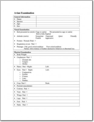 Avian Physical Examination Forms Created by LafeberVet
Egg Necropsy Form
An egg necropsy is an important part of the diagnostic workup to evaluate the cause of dead-in-shell. First, candle the egg and obtain basic measurements. Next enter the egg through the air cell. Carefully examine the shell membranes, then expose the interior of the egg. Collect culture samples as needed. Proceed using this form to gather standardized data and appropriate measurements.

Download the Egg Necropsy Form, created by LafeberVet
Avian Necropsy Form 2
Download this avian necropsy form, adapted from a University of Wisconsin-Madison Research Animal Resources Center form. Additional resources are available at rarc.wisc.edu/pathology/forms.html
Download Avian Necropsy Form 2
Avian Necropsy Form 1
Download this avian necropsy form, adapted from a form by the University of Wisconsin-Madison’s Research Animal Resources Center. Additional resources are available at rarc.wisc.edu/pathology/forms.html.
Download Avian Necropsy Form 1
Avian History Form 5
 Provided by Dr. Heather Barron
Avian History Form 4
Avian History Form 3
This two-page history form was shared by Thomas Tully, DVM, DABVP (Avian Practice) of Louisiana State University
Download Avian History Form 3 PDF
Avian History Form 2

This one-page avian history form was provided by Dr. Tom Tully of Louisiana State University School of Veterinary Medicine.
Download Avian History Form 2 PDF
Thomas Tully, MS, DVM, DABVP (Avian Practice), DECZM
 Thomas Tully, DVM is chief of the Zoological Medicine Service at Louisiana State University and the 2011 recipient of the T.J. Lafeber Avian Practitioner of the Year Award. He is a member of the American Board of Veterinary Practitioners in avian practice as well as a Diplomate of the European College of Avian Medicine and Surgery. Dr. Tully’s research interests include aviculture preventive medicine, feather problems, avian nutrition, infectious disease, and ratites. Dr. Tully has numerous presentations and publications to his credit, including the Handbook of Avian Medicine and the Manual of Exotic Pet Practice.
Avian History Form 1
Provided by Dr. Jörg Mayer from the University of Georgia at Athens
Jörg Mayer, MS, DVM, DABVP (Exotic Companion Mammal Practice), DECZM, DACZM

Dr. Jörg Mayer is an Associate Professor of Zoological Medicine in the Department of Small Animal Medicine and Surgery at University of Georgia College of Veterinary Medicine. Dr. Mayer received his doctorate of veterinary medicine from the University of Budapest in Hungary and his Master’s from the Royal Veterinary College in London. Dr. Mayer is particularly interested in reptiles, amphibians, and fish as well as small mammals such as rabbits and ferrets. He served as the 2010-2012 President of the Association of Exotic Mammal Veterinarians as well as the co-author and co-editor of Clinical Veterinary Advisor: Birds and Exotic Pets and Exotic Pet Behavior.
Reptile Awareness Day Quiz
October 21st is Reptile Awareness Day. Although Reptile Awareness Day was founded with the worthy goal of exploring the ecological challenges that reptiles face, celebrate with LafeberVet by taking our quiz that focuses on clinical information. Are you aware of these reptile-centric facts that will benefit you, your staff, and of course your reptile patients . . .
Pour les vétérinaires. Par les vétérinaires.
Le site Lafervet.com est conçu pour une utilisation par les vétérinaires. Il est ouvert aux vétérinaires diplômés, aux techniciens vétérinaires diplômés, aux animaliers et aux étudiants dans ces domaines.
Créer un compte pour accéder à des articles et des ressources du site.
L'inscription est gratuite.
Para Profesionales Veterinarios. Por Profesionales Veterinarios.
El sitio Lafervet.com es para uso de los profesionales veterinarios. Está abierto a los veterinarios licenciados, técnicos veterinarios licenciados, rehabilitadores licenciados y estudiantes en estos campos.
Cree una cuenta para acceder a los artículos y recursos del sitio.
La registro es gratis.
Already a LafeberVet Member?
Please Login
Gregory A. Rich, DVM
 Dr. Gregory A. Rich (left) is the owner of the Avian & Exotic Animal Hospital of Louisiana in Metairie, Louisiana. Dr. Rich has over 30 years of experience in avian and exotic animal medicine and surgery, and he has been a member of the American Veterinary Medical Association and the Association of Avian Veterinarians (AAV) for 25+ years. Dr. Rich is a past Board Member of the AAV. He has lectured nationally at a wide variety of veterinary and avicultural conferences and has been featured in several veterinary texts and periodicals.
In 2016, Dr. Rich was named the T.J. Lafeber Avian Practitioner of the Year.
Why Does My Rabbit? House Rabbit Behavior
Why Does My Rabbit? Common house rabbit behavior questions answered.
In many instances, rabbits kept in hutches, pet stores, or laboratories do not receive enough stimulation or physical space to demonstrate their full behavioral repertoire. Owners of house rabbits know these are intelligent creatures with distinct personalities and a range of behaviors. In this educational handout, clients can review normal husbandry, age, and sex-related behavior as well as normal sounds and actions like chewing and digging. The challenges of multi-rabbit households are also discussed.
Download the Why Does My Rabbit? PDF Handout
Vitamin A
Vitamin A plays an important role in maintaining a healthy….
Unsafe Foods For Birds
What are unsafe foods for birds? Veterinary health professionals rely on a wide range of information because it is generally considered best to err on the side of caution. The following list divides potential danger foods into two categories…

Download the PDF version of the Unsafe Foods for Birds client education handout.
Training Basic Skills
Many parrots instinctively strive for a position of dominance within their flock (your household). Many of the behavioral problems that can arise from such a situation, may be prevented when…