
View the RACE-approved webinar recording presented by Javier Nevarez, DVM, PhD, DACZM, DECZM (Herpetology). The live event was an updated version of his 2016 presentation. Topics covered include recognizing signs of pain and discomfort, analgesics commonly used in reptiles, as well as balanced anesthetic protocols. Since a key component of successful anesthesia and analgesia is the ability to properly monitor the patient, essential monitoring parameters and equipment are also discussed. This presentation concludes with a review of respiratory stimulus in the reptile and anesthetic recovery . . .
Pour les vétérinaires. Par les vétérinaires.
Le site Lafervet.com est conçu pour une utilisation par les vétérinaires. Il est ouvert aux vétérinaires diplômés, aux techniciens vétérinaires diplômés, aux animaliers et aux étudiants dans ces domaines.
Créer un compte pour accéder à des articles et des ressources du site.
L'inscription est gratuite.
Para Profesionales Veterinarios. Por Profesionales Veterinarios.
El sitio Lafervet.com es para uso de los profesionales veterinarios. Está abierto a los veterinarios licenciados, técnicos veterinarios licenciados, rehabilitadores licenciados y estudiantes en estos campos.
Cree una cuenta para acceder a los artículos y recursos del sitio.
La registro es gratis.
Already a LafeberVet Member?
Please Login
Amphibian Handling and Restraint
Part of the Herptile Handling Teaching Module, this review article addresses handling and restraint of amphibians, a diverse group that consists of over 8,000 extant species belonging to three orders: Anura (frogs and toads), Caudata or Urodela (salamanders, newts, and sirens), and Gymnophiona or Apoda (caecilians). Transport, defense mechanisms that may prove hazardous to handlers, potential dangers for the patient, and specific capture, handling, and restraint techniques are reviewed. Equipment needed, including personal protective equipment, is also discussed as well as measures to reduce the risk of zoonotic disease transmission . . .
Pour les vétérinaires. Par les vétérinaires.
Le site Lafervet.com est conçu pour une utilisation par les vétérinaires. Il est ouvert aux vétérinaires diplômés, aux techniciens vétérinaires diplômés, aux animaliers et aux étudiants dans ces domaines.
Créer un compte pour accéder à des articles et des ressources du site.
L'inscription est gratuite.
Para Profesionales Veterinarios. Por Profesionales Veterinarios.
El sitio Lafervet.com es para uso de los profesionales veterinarios. Está abierto a los veterinarios licenciados, técnicos veterinarios licenciados, rehabilitadores licenciados y estudiantes en estos campos.
Cree una cuenta para acceder a los artículos y recursos del sitio.
La registro es gratis.
Already a LafeberVet Member?
Please Login
Test Your Knowledge: HPAI
The Highly Pathogenic Avian Influenza webinar course was reviewed and approved by the American Association of Veterinary State Boards (AAVSB) Registry of Approved Continuing Education (RACE) program for 1 hour of continuing education credit, in jurisdictions that recognize AAVSB RACE approval. Complete the brief quiz and download your continuing education certificate . . .
Pour les vétérinaires. Par les vétérinaires.
Le site Lafervet.com est conçu pour une utilisation par les vétérinaires. Il est ouvert aux vétérinaires diplômés, aux techniciens vétérinaires diplômés, aux animaliers et aux étudiants dans ces domaines.
Créer un compte pour accéder à des articles et des ressources du site.
L'inscription est gratuite.
Para Profesionales Veterinarios. Por Profesionales Veterinarios.
El sitio Lafervet.com es para uso de los profesionales veterinarios. Está abierto a los veterinarios licenciados, técnicos veterinarios licenciados, rehabilitadores licenciados y estudiantes en estos campos.
Cree una cuenta para acceder a los artículos y recursos del sitio.
La registro es gratis.
Already a LafeberVet Member?
Please Login
Colin McDermott, VMD, DABVP (Reptile & Amphibian Practice), CertAqV
 Colin McDermott lives and works in Hong Kong, where he serves as a veterinary surgeon at Zodiac Pet and Exotic Animal Hospital and a clinical assistant professor in the Department of Veterinary Clinical Sciences of City University. Dr. McDermott earned his doctorate from the University of Pennsylvania School of Veterinary Medicine in 2011. He completed a 2-year specialty internship at the University of Pennsylvania followed by a fellowship in aquarium science and veterinary medicine at the National Aquarium in Washington, D.C. and Baltimore, Maryland. Dr. McDermott worked in private practice for 3 years at an emergency and specialty hospital in New Jersey, establishing their exotic animal medicine and surgery service. He moved to Hong Kong in 2019. where he works at an exotics-focused private practice hospital. Dr. McDermott is board certified in reptile and amphibian practice through the American Board of Veterinary Practitioners. He is also a certified aquatic veterinarian through the World Aquatic Veterinary Medical Association.
Test Your Knowledge: Chelonian Shell Repair
The Chelonian Shell Repair webinar course was reviewed and approved by the American Association of Veterinary State Boards (AAVSB) Registry of Approved Continuing Education (RACE) program for 1 hour of continuing education credit, in jurisdictions that recognize AAVSB RACE approval . . .
Pour les vétérinaires. Par les vétérinaires.
Le site Lafervet.com est conçu pour une utilisation par les vétérinaires. Il est ouvert aux vétérinaires diplômés, aux techniciens vétérinaires diplômés, aux animaliers et aux étudiants dans ces domaines.
Créer un compte pour accéder à des articles et des ressources du site.
L'inscription est gratuite.
Para Profesionales Veterinarios. Por Profesionales Veterinarios.
El sitio Lafervet.com es para uso de los profesionales veterinarios. Está abierto a los veterinarios licenciados, técnicos veterinarios licenciados, rehabilitadores licenciados y estudiantes en estos campos.
Cree una cuenta para acceder a los artículos y recursos del sitio.
La registro es gratis.
Already a LafeberVet Member?
Please Login
Highly Pathogenic Avian Influenza

Photo credit: J. Kelly, USFWS NE region
Abstract
Highly Pathogenic Avian Influenza: What’s different about this strain?
Considerations for wild and captive birds
Since 2021, North America has been responding to an outbreak of highly pathogenic avian influenza (HPAI) (H5N1) clade 2.3.4.4b. This strain has had global spread and implications on both captive and wild bird populations, as well as in a variety of mammalian species. With a large spectrum of clinical presentations, depending on species, HPAI can be particularly deadly to some animals including raptor species. This outbreak has been longer lasting and more geographically widespread than previous outbreaks of other HPAI strains, and thus has created different considerations for management and response.
Since 2022, The Raptor Center (TRC), located at the University of Minnesota, has been in active response for the outbreak. Regularly admitting over 1,000 wild raptors a year for rehabilitation, TRC detected over 200 HPAI positive testing birds in 2022 with active surveillance efforts. All birds were identified through testing immediately upon admission by cloacal and oropharyngeal influenza A matrix RT-PCR testing. Positive samples were referred to the National Veterinary Services Laboratories for further viral characterization. All birds who presented alive on admission received full physical examinations to better understand clinical presentations in different species. Substantial biosecurity and biosafety measures were utilized and validated during the response.
As this strain continues to circulate, it will continue to have important implications to both wild and captive birds. Active surveillance through a variety of routes, including wildlife rehabilitation, will give vital insight into the characteristics of this outbreak and how best to continue to respond, both now and to future strains.
Outline
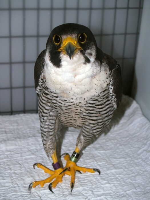
Photo credit: Beka Weiss
|
|
|
About the presenter
Victoria Hall is the Executive Director of The Raptor Center and the Redig Endowed Faculty Chair in Raptor and Ecosystem Health at the University of Minnesota. Dr. Hall earned her Doctorate in 2014 and a Masters of Science (Veterinary Preventive Medicine) in 2015 from Mississippi State University. She is also a Diplomate of the American College of Veterinary Preventive Medicine. Dr. Hall has a strong background in One Health, serving as the first veterinary epidemiologist for the National Zoo. [Learn more].
Webinar recording
Post-test
Complete the brief quiz. With a passing grade of 70% or higher, you can download your continuing education (CE) certificate for 1 hour of CE credit in jurisdictions that recognize American Association of Veterinary State Boards (AAVSB) Registry of Approved Continuing Education (RACE) approval.
Expert Q&A
Although some questions were addressed during the live event, Dr. Hall generously answered the remaining questions. A lightly edited transcript of these questions and answers are posted below:
SIGNALMENT
In mammals is it only carnivore/omnivore or could deer be infected?
There have not been reported positive deer at this time, but broad surveillance is not being conducted. Keep an eye on the USDA HPAI mammal website for the latest information on what has been confirmed in the United States.
I have pet African grey parrots in Texas. What is the relative danger of them being outside?
Everything is a risk assessment, so I would consider multiple factors. First, what is the transmission of AI in your area? This can be hard to determine, but using the USDA website can at least give you some idea. Second would be would the parrot have direct contact with the ground or surfaces wild birds could land on. Third would be if the parrots could have direct contact with wild birds themselves. If you have low transmission in the area and you are taking the birds outside in a manner where they are not having contact with any surfaces that wild birds could have interacted with- then risk would be extremely low. If transmission in your area is high and you are placing them directly on the ground for example, then you would introduce much more risk to the parrots.
CLINICAL PICTURE
What were the primary pathologic lesions noted in the raptors that died of disease?
For full descriptions of lesions, please check out: Wünschmann A, Franzen-Klein D, Torchetti M, Confeld M, Carstensen M, Hall V. Lesions and viral antigen distribution in bald eagles, red-tailed hawks, and great horned owls naturally infected with H5N1 clade 2.3.4.4b highly pathogenic avian influenza virus. Vet Pathol. 2024 Jan 10:3009858231222227. doi: 10.1177/03009858231222227. Epub ahead of print. PMID: 38197395.
Did you see any relationship between viral load, clinical signs, or transmissibility?
We are still diving into that analysis as we match videos of clinical signs to other data including viral load- so stay tuned to some future publications!
DIAGNOSTICS
Did you say how long PCR results took to come back? In that time, did the 16% of asymptomatic birds develop neuro signs?
PCR results can technically come back in a matter of hours if the test is run immediately. Most often though, people need to submit them to a lab and then each lab will have a different window of time that it takes them to return results. At The Raptor Center, we set up our own lab so are fortunately to have very timely results to make decisions based off of. Of the 16% of asymptomatic birds, the majority died overnight, developed clinical signs, or were euthanized for other injury reasons.
Do you think the higher numbers in Minnesota are mainly due to a higher amount of testing?
There is definitely an increase in numbers in Minnesota due to testing. Unfortunately, we do not have robust testing of wildlife in the United States and in the times of a mass die-off of wild birds, if only one bird was tested then only one bird would be reflected on the USDA map (even though many might have died). The Raptor Center itself contributed over 70% of the positive testing raptors to the state of Minnesota Surveillance in 2022, as we felt that having data would help ourselves and others in our state make better risk-based decisions.
Have you identified any different genotypes this year?
We continue to see genotypes that match what USDA is detecting in wild bird surveillance in the flyway.
PREVENTION & CONTROL
How long do you recommend keeping a raptor or other species in quarantine if PCR testing is not available?
We find, for species that are likely to show signs of illness if they get infected (such as raptors), that within 48 hours you are likely to see signs in the majority of infected animals- they tend to get much sicker and/or die. This is based off our experience in 2022/2023, so it is important to note this virus changes over time and that is for the majority (not all) infected raptors seen. Species that are not likely to show signs of illness if infected (i.e. ducks) are much more difficult and might shed for weeks if infected.
Moving out of migration season and into baby season this spring, how do you think your quarantine/isolation procedures may change? What do your quarantine practices look like now?
Our practices change based on risk…The higher the transmission (for example waterfowl are migrating through right now) the more we increase procedures. Right now, we are admitting into a dedicated room in our hospital (versus a separate building) and it is working pretty well.
With babies coming, we actually manage the babies in a separate space and keep biosecurity between babies. That way if they come in just for a day or so, and then we send them back out, we are confident they were not exposed to anything in our care. If we need to foster, we do multiple rounds of HPAI testing on the baby we would like to foster to confirm that we do not accidentally introduce HPAI into a different nest.
Do you have a flow chart/checklist for steps for donning/doffing PPE? Currently seeing both WNV positive and HPAI positive in California.
Check out https://www.cdc.gov/hai/pdfs/ppe/ppe-sequence.pdf and let us know if we can help adjust it for your facility! Having it on the wall is so important to help everyone remember.
Where did you get the blue footbath pads? I’ve been trying to find them online!
We use puppy pads to be honest! Tend to be the cheapest that we find.
Our state [New Mexico] has just sent rehabilitators the first (!) communication about HPAI. They included a chart from USDA which showed the testing process. The conclusion of the chart states that if a bird tested positive, the rehab center would be shuttered- no birds in, no birds out. No mention was made of the biosecurity precautions that might have been in place. Since we are the ones responsible for alerting about a possible case, and for paying for the original testing, that policy seems to be a huge disincentive to reporting. Also, closing rehab centers encourages the public to keep possession of a wild bird that is possibly infected. Both seem to me to be horrible public policy. Also to you?
That is a real problem. When responses are so severe, it makes no sense why anyone would test. And we agree, if rehab centers close, then the public keeps the bird creating other risk. We have been able to advocate in the state of Minnesota that we could safely and responsibly handle potentially positive birds without create risks of it spilling out of our center into other birds, humans, or agricultural systems. We did this with strong biosecurity protocols. [The Raptor Center is] always happy to help join conversations about rehab and biosecurity, so let us know if we can support you!
Do you know if for poultry flock that had a positive HPAI raptor found inside the enclosure, were the owners allowed to test/quarantine flock or were they culled?
I unfortunately cannot speak to the state or federal response for facilities that had positive wildlife found. This would likely vary based by state and situation. Wild birds are considered a different category by the World Organization of Animal Health and can be handled differently based on state/federal response.
Is F10 a good disinfectant…and also can we used as a nebulizer at low concentrations?
Unfortunately, I don’t have experience with that disinfectant. [You could] work …potentially [with] the manufacturer to determine safety and use.
Editor’s note: The U.S. National Science Foundation does briefly mention the use of F10 in Updated Guidance on the Highly Pathogenic Avian Influenza – Environmental Update for USAP Personnel. Also visit the EPA’s Registered Antimicrobial Products Effective Against Avian Influenza.
ZOONOTIC POTENTIAL
Is there any information on the zoonotic potential in beaches and human used bodies of water?
I have not read any studies on the potential of transmission to humans in bodies of water–there is definitely great work with HPAI and water out there and this would be a great topic to explore further!
The following questions were answered by the moderator, Christal Pollock, DVM.
SIGNALMENT
Have any of the penguin colonies in Antarctica been affected by HPAI?
Surveys have confirmed the presence of a low pathogenic H11 avian influenza virus in Antarctica.
Ogrzewalska M, Couto Motta F, Resende PC, Fumian T, Fonseca da Mendonça AC, Appolinario Reis L, Lima Brandao M, Chame M, Arantes Gomes IL, Mendonca Siqueira M. Influenza A(H11N2) Virus Detection in Fecal Samples from Adélie (Pygoscelis adeliae) and Chinstrap (Pygoscelis antarcticus) Penguins, Penguin Island, Antarctica. Microbiol Spectr. 2022 Oct 26;10(5):e0142722. doi: 10.1128/spectrum.01427-22. Epub 2022 Sep 19. PMID: 36121294; PMCID: PMC9603087.
https://www.theguardian.com/environment/2023/dec/08/mass-deaths-elephant-seals-penguins-bird-flu-antarctic-ecological-disaster-aoe. Disease has been seen in skuas in Antarctica and there is fear that disease will reach the penguin colonies.
Addendum: In February 2024, highly pathogenic avian influenza was found on mainland Antarctica.
PATHOGENESIS & TRANSMISSION
Is shedding via respiratory system, fecal also, other eg dander? any info on if vertical as well as horizontal?
The fecal-oral route most commonly transmits the orthomyxovirus causing avian influenza, although virus is also shed in respiratory and ocular secretions. Dander does not play a role in transmission, but water contaminated with virus-laden feces does. Vertical transmission has also been documented, but this mode of transmission is probably limited. Here are the most recent references that I came across in in a quick search:
Yu G, Wang A, Tang Y, Diao Y. Vertical Transmission of H9N2 Avian Influenza Virus in Goose. Front Microbiol. 2017 Aug 15;8:1559. doi: 10.3389/fmicb.2017.01559. PMID: 28861069; PMCID: PMC5559544.
Uchida Y, Takemae N, Tanikawa T, Kanehira K, Saito T. Transmission of an H5N8-Subtype Highly Pathogenic Avian Influenza Virus from Infected Hens to Laid Eggs. Avian Dis. 2016 Jun;60(2):450-3. doi: 10.1637/11312-110315-Reg. PMID: 27309286.
Is it possible that all raptors are infected by eating infected birds/animals?
If this question were part of a quiz, the “ALL” would make it a trick question, as there are rarely absolutes. Carnivorous mammals are at risk of becoming directly infected by feeding on sick or dead wild birds, so it is reasonable to assume the same might be true in raptors. The presence of viral proteins in the GI tract as opposed to the respiratory tract also supports this supposition.
Reperant LA, van Amerongen G, van de Bildt MW, Rimmelzwaan GF, Dobson AP, Osterhaus AD, Kuiken T. Highly pathogenic avian influenza virus (H5N1) infection in red foxes fed infected bird carcasses. Emerg Infect Dis. 2008 Dec;14(12):1835-41. doi: 10.3201/eid1412.080470. PMID: 19046504; PMCID: PMC2634621.
Any information on factors that might affect viral shedding?
Dr. Hall discussed some of these factors: season, geographic location, and host species.
Age at infection (At least in waterfowl, younger birds maintain infection within the population), virulence of the virus strain, and condition of the host would also be important factors. There has also been researching evaluating gene expression in individual birds, as some birds are apparently “super shedders”:
Dolinski AC, Homola JJ, Jankowski MD, Robinson JD, Owen JC. Host gene expression is associated with viral shedding magnitude in blue-winged teals (Spatula discors) infected with low-path avian influenza virus. Comp Immunol Microbiol Infect Dis. 2022 Nov-Dec;90-91:101909. doi: 10.1016/j.cimid.2022.101909. Epub 2022 Nov 7. PMID: 36410069; PMCID: PMC10500253.
Are there any cases where we know that an infected raptor has transmitted the virus to another raptor.
Although transmission was not confirmed, a group of hunting falcons were believed to be infected with HPAI through exposure to free-ranging houbara bustards. I am not aware of any other confirmed cases, but I will ask.
Naguib MM, Kinne J, Chen H, Chan KH, Joseph S, Wong PC, Woo PCY, Wernery R, Beer M, Wernery U, Harder TC. Outbreaks of highly pathogenic avian influenza H5N1 clade 2.3.2.1c in hunting falcons and kept wild birds in Dubai implicate intercontinental virus spread. J Gen Virol. 2015 Nov;96(11):3212-3222. doi: 10.1099/jgv.0.000274. PMID: 26350163.
CLINICAL PICTURE
So clinical signs in raptors look more like poisoning?
Lead toxicity is an important differential diagnosis for birds that present with clinical signs consistent with avian influenza. Other important differentials include West Nile virus and traumatic brain injury.
PREVENTION & CONTROL
What advice should we provide hunters (waterfowl) about the probability and effect of harvesting waterfowl for food?
There are some good resources provided by state and federal sources on this topic. Ducks Unlimited also summarizes these points.
- https://www.aphis.usda.gov/publications/animal_health/fsc_hpai_hunters.pdf
- https://www.ncwildlife.org/Connect-With-Us/waterfowl-hunters-should-take-avian-influenza-precautions
- https://www.ducks.org/conservation/waterfowl-research-science/waterfowl-hunters-and-the-avian-flu-what-you-need-to-know
RACE approval
This program is approved for 1 hour of continuing credit for veterinarians and veterinary technicians in jurisdictions that recognize American Association of Veterinary State Boards (AAVSB) Registry of Approved Continuing Education (RACE) approval.
Victoria Hall, DVM, MS, DACVPM
 Victoria Hall is the Executive Director of The Raptor Center and the Redig Endowed Faculty Chair in Raptor and Ecosystem Health at the University of Minnesota. Dr. Hall earned her Doctorate in 2014 and a Master of Science (Veterinary Preventive Medicine) in 2015 from Mississippi State University. She is also a Diplomate of the American College of Veterinary Preventive Medicine. Dr. Hall has a strong background in One Health, serving as the first veterinary epidemiologist for the National Zoo and a public health officer for the Smithsonian Institution’s COVID-19 response. Dr. Hall formerly served as an Epidemic Intelligence Service Officer for the Centers for Disease Control and Prevention, responding to infectious disease outbreaks, including multidrug resistant tuberculosis, measles virus, and Zika virus.
Understanding the Chelonian Shell
The shell is a bony structure unique to order Chelonia. No other animal, living or extinct, has its body enclosed within a bony shell similarly constructed in its entirety. This review article describes shell structure, pertinent shell vocabulary terms, as well as shell function, morphology, growth, and pathology . . .
Pour les vétérinaires. Par les vétérinaires.
Le site Lafervet.com est conçu pour une utilisation par les vétérinaires. Il est ouvert aux vétérinaires diplômés, aux techniciens vétérinaires diplômés, aux animaliers et aux étudiants dans ces domaines.
Créer un compte pour accéder à des articles et des ressources du site.
L'inscription est gratuite.
Para Profesionales Veterinarios. Por Profesionales Veterinarios.
El sitio Lafervet.com es para uso de los profesionales veterinarios. Está abierto a los veterinarios licenciados, técnicos veterinarios licenciados, rehabilitadores licenciados y estudiantes en estos campos.
Cree una cuenta para acceder a los artículos y recursos del sitio.
La registro es gratis.
Already a LafeberVet Member?
Please Login
Test Your Knowledge: Avian Anesthesia – Practical Applications
. . .
Pour les vétérinaires. Par les vétérinaires.
Le site Lafervet.com est conçu pour une utilisation par les vétérinaires. Il est ouvert aux vétérinaires diplômés, aux techniciens vétérinaires diplômés, aux animaliers et aux étudiants dans ces domaines.
Créer un compte pour accéder à des articles et des ressources du site.
L'inscription est gratuite.
Para Profesionales Veterinarios. Por Profesionales Veterinarios.
El sitio Lafervet.com es para uso de los profesionales veterinarios. Está abierto a los veterinarios licenciados, técnicos veterinarios licenciados, rehabilitadores licenciados y estudiantes en estos campos.
Cree una cuenta para acceder a los artículos y recursos del sitio.
La registro es gratis.
Already a LafeberVet Member?
Please Login
Chelonian Shell Repair
J. Jill Heatley, MS, DVM, DABVP (Avian Practice, Reptile & Amphibian Practice), DACZM presented a live, interactive webinar, RACE-approved for 1 credit hour and hosted by LafeberVet. This distance-learning event compared and contrasted multiple shell repair techniques. Additional topics reviewed included wound management, shell preparation, and fixator removal . . .
Pour les vétérinaires. Par les vétérinaires.
Le site Lafervet.com est conçu pour une utilisation par les vétérinaires. Il est ouvert aux vétérinaires diplômés, aux techniciens vétérinaires diplômés, aux animaliers et aux étudiants dans ces domaines.
Créer un compte pour accéder à des articles et des ressources du site.
L'inscription est gratuite.
Para Profesionales Veterinarios. Por Profesionales Veterinarios.
El sitio Lafervet.com es para uso de los profesionales veterinarios. Está abierto a los veterinarios licenciados, técnicos veterinarios licenciados, rehabilitadores licenciados y estudiantes en estos campos.
Cree una cuenta para acceder a los artículos y recursos del sitio.
La registro es gratis.
Already a LafeberVet Member?
Please Login
Avian Anesthesia: Practical Applications
Jody Nugent-Deal, RVT, VTS (Anesthesia), VTS (Exotic Companion Animal) presented this RACE-approved webinar recording. This distance-learning event first reviews avian anatomy and physiology, emphasizing clinically relevant ways that birds differ from mammals. This course also goes over anesthesia monitoring equipment and what actually works on birds, as well as machine and basic equipment set-up. She concludes with a discussion of common anesthetic protocols used in avian patients. View the recording, then complete (and pass) the brief post-test to earn 1 hour of continuing education credit . . .
Pour les vétérinaires. Par les vétérinaires.
Le site Lafervet.com est conçu pour une utilisation par les vétérinaires. Il est ouvert aux vétérinaires diplômés, aux techniciens vétérinaires diplômés, aux animaliers et aux étudiants dans ces domaines.
Créer un compte pour accéder à des articles et des ressources du site.
L'inscription est gratuite.
Para Profesionales Veterinarios. Por Profesionales Veterinarios.
El sitio Lafervet.com es para uso de los profesionales veterinarios. Está abierto a los veterinarios licenciados, técnicos veterinarios licenciados, rehabilitadores licenciados y estudiantes en estos campos.
Cree una cuenta para acceder a los artículos y recursos del sitio.
La registro es gratis.
Already a LafeberVet Member?
Please Login
Kay Backues, DVM, DACZM
 Kay Backues is the Director of Animal Health at Tulsa Zoo & Living Museum in Tulsa, Oklahoma. Dr. Backues has practiced general zoo medicine at Tulsa Zoo since 1998. Dr. Backues is also an adjunct assistant professor at Oklahoma State University College of Veterinary Medicine Veterinary Clinical Sciences. Kay is a 1991 graduate of Kansas State University College of Veterinary Medicine. She is a Diplomate of the American College of Zoological Medicine (ACZM) and a Past President of ACZM (2018) and the American Association of Zoo Veterinarians (2011). Dr. Backues is also a 2007 recipient of the ACZM Presidential Service Award. As a general zoo practitioner, Dr. Backues has co-authored publications on a variety of topics, from elephant care and vaccination in llamas to pharmacokinetics in flamingos and gastritis in lemurs. Dr. Backues also authored the 2015 chapter on Anseriformes in Fowler’s Zoo and Wild Animal Medicine, Volume 8.
Dr. Backues is shown here with her 37 year old umbrella cockatoo, Cobber, whom she has owned since she was 5 months old.
Raptor Anatomy Fundamentals
What is a raptor?
Raptors, or birds of prey, consume foods almost exclusively of animal origin and are characterized by a strong beak, curved sharp talons, and keen vision. Raptors are an extremely diverse and wide-ranging group that fall within four different taxonomic orders (Table 1). Nocturnal raptors or owls belong to order Strigiformes (Fig 1). For example, the typical owls (order Strigiformes, family Strigidae) are a large group that are found worldwide except for Antarctica and some very remote islands.9,37,49 Diurnal raptors are represented by orders Accipitriformes, Falconiformes, and Cathartiformes (Fig 2) with members of order Accipitriformes also found worldwide in almost every terrestrial habitat, from the arctic tundra to the tropical rainforest.34,37
Table 1. Taxonomic classification in birds of prey 9,10,27,37,38
Class Aves
Order Strigiformes
Family Tyonidae: barn owls
Family Strigidae: typical owls
Order Accipitriformes
Family Accipitridae: hawks, eagles, kites, harriers, Old World vultures, buzzards
Family Pandionidae: osprey
Family Sagittariidae: secretary bird
Family Cathartidae: New World vultures, condors
Order Falconiformes
Family Falconidae: falcons, kestrels, merlins, hobbies, caracaras
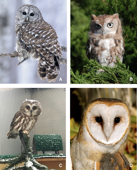
Figure 1. Owls. According to the International Ornithological Committee (IOC), there are over 200 species of “typical owls” worldwide belonging to the family Strigidae. (A) barred owl (Strix varia) (B) Eastern screech owl (Megascops asio) (C) Northern saw-whet owl (Aegolius acadicus). (D) The IOC also reports approximately 18 species in the genus Tyto of family Tytonidae. Shown here, a common barn owl (Tyto alba). Photo credit: Jeff Fischer/The Raptor Center, UMN. Click image to enlarge.
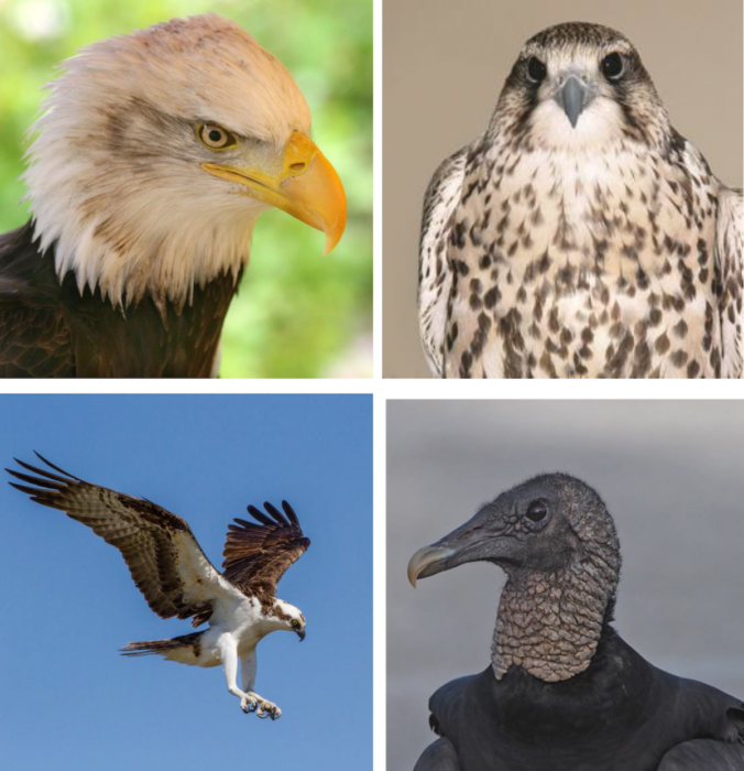
Figure 2. Examples of diurnal raptors include: (A) American bald eagle (Haliaeetus leucocephalus) Photo credit: Linda Cronin. (B) saker falcon (Falco cherrug) Photo credit: Dr. Jaime Samour. (C) osprey (Pandion haliaetus) Photo credit: Andy Morffew/Flickr Creative Commons. (D) black vulture (Coragyps atratus) Photo credit: Judy Gallagher/Flickr Creative Commons. Click image to enlarge.
Veterinary health professionals may be presented with raptors from the wild or those held for education, research, captive breeding, or falconry. If you are comfortable with the basics of avian anatomy and physiology, then you are well on your way to understanding raptors. However, this taxonomic group has many unique anatomic and physiologic adaptations that allow these birds to pursue and catch prey, including flight, beak, and talon modifications. The following collection of raptor anatomy and physiology facts should serve the reader well during physical examination, clinical care, and/or necropsy.
Integument
There are several anatomical structures derived from bird skin, such as the hard protective covering of talons and beaks, papilla on feet, feathers, and glands.
Talon sheaths
The feet of most raptors are armed with strong, needle-sharp, highly curved claws or talons. Each talon consists of a bone that is covered with a sheath made of sheets of keratin, a hard fibrous protein derived from skin (Fig. 2). Talons are used to catch, hold, and often kill prey.16,32,34,52 They are blunted in vultures since they do not need to capture live prey.34,74
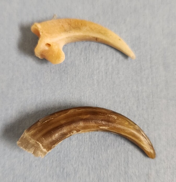
Figure 3. The talon bone (top) and protective sheath (bottom) of a bald eagle (Haliaeetus leucocephalus), Photo credit: The Raptor Center, UMN. Click image to enlarge.
Clinical Tip: In many species, the talon of the third digit has a specialized sharp edge on its medial surface that is used for feather grooming. In some owls, like the common barn owl (Tyto alba), this edge is pectinate and sometimes referred to as a “feather comb” (Fig 4). In all species, this edge should be preserved during talon maintenance in captivity.16,30,34,37
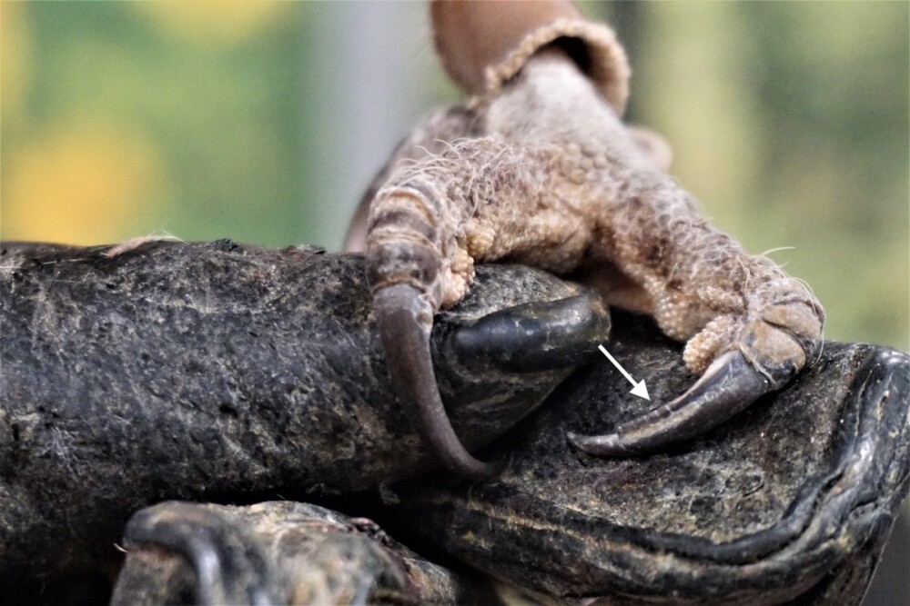
Figure 4. Feather comb on the edge of talon number 3 (arrow) in a common barn owl (Tyto alba). Photo credit: Gail Buhl/The Raptor Center, UMN
Rhamphotheca
Similar to talons, the raptor beak has a hard protective keratin covering called the rhamphotheca. As in other types of birds, the rhamphotheca in raptors continually grows and needs to be worn down to maintain a normal length and shape. In the wild, this happens naturally; in captivity, filing often has to be performed manually by caregivers in a process referred to as coping.
Clinical Tip: In captivity, raptors require regular maintenance of the keratin covering their beak and talons. If talon sheaths become too long, they can result in puncture wounds to the metatarsal pad or toes and/or uneven weight bearing that will also affect the health of the plantar foot surface. An overgrown rhamphotheca may develop chips or cracks that can extend toward the growth plates. Beak coping is often done with a rotary tool, but only experienced individuals should perform this technique in a live bird. Beak coping can be done without general anesthesia for some individuals, but is required for birds that become highly stressed.
Foot papillae
While thick scales protect the dorsal surface of the feet from injury, roughened papillae on the plantar surface assist in grasping.16,34 In some raptors that hunt birds or fish, the ventral surface of the toe pads have wart-like projections or spicules that create a sandpaper-like surface that allows them to grasp slippery prey (Fig 5).23,34
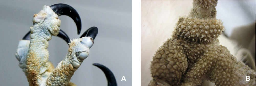
Figure 5. Spicule-like papillae on the plantar surface of an osprey (Pandion haliaetus) foot. Photo credit: (A) Illinois Raptor Center (B) The Raptor Center, UMN. Click image to enlarge.
Flight feathers
The integrity of the flight feathers is of the utmost importance for raptors destined for release.34 The tail feathers or rectrices of hospitalized raptors may be protected with a tail guard made from materials such as light but sturdy cardboard or file folders (Fig 6). The tail guard is placed over the rectrices and secured to the covert feathers with adhesive tape.
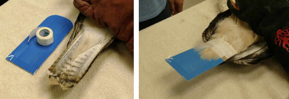
Figure 6. (A). Properly sized and (B) secured tail guard. Photo credit: The Raptor Center, UMN
Nocturnal owls have several modifications that allow them to be remarkable, silent hunters.16,33 The leading edge of the tenth primary feather, and in some species the ninth or more, is combed or serrated rather than smooth on each wing (Fig 7).16,33,37 This modified edge has the effect of reducing air turbulence and the noise normally created during wing flapping.16,33
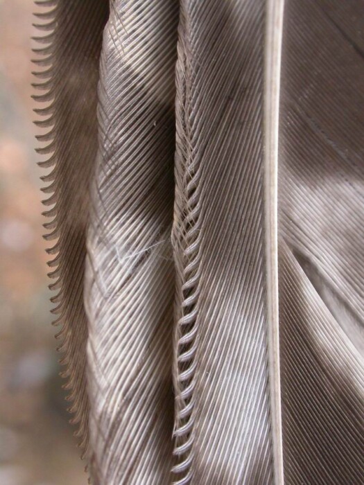
Figure 7. Close-up of flight feathers from a long-eared owl (Asio otus). Photo credit: “snowy owls”/ Wikimedia Commons. Click image to enlarge.
Quiet flight is further enhanced by the velvety pile on the dorsal surface of the vanes of most flight feathers. This soft, downy surface is composed of long, distal barbules that stick out from the otherwise flat surface of the feather vanes and reduce the sound of feathers over one another (Fig 8).37 The visual effect of these elongated barbules covering the pennaceous feather vanes reduces the glossiness of nocturnal owl plumage.37
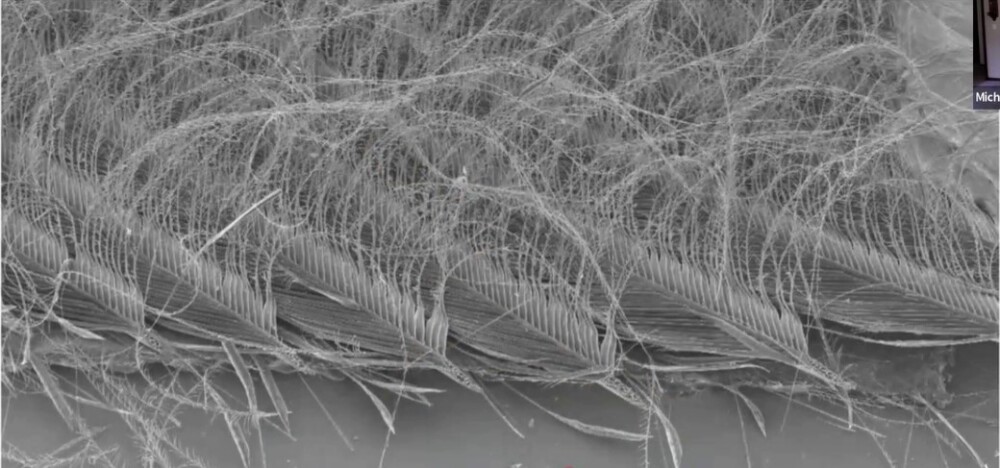
Figure 8. Electron microscopic lateral view of a flight feather of a common barn owl (Tyto alba). Photo credit: The Raptor Center, UMN. Click image to enlarge.
Head feathers
Vultures and condors routinely scavenge large carcasses and have minimal feathering on their head (Fig 9).31 This adaptation minimizes soiling of the head while the birds are consuming entrails.31
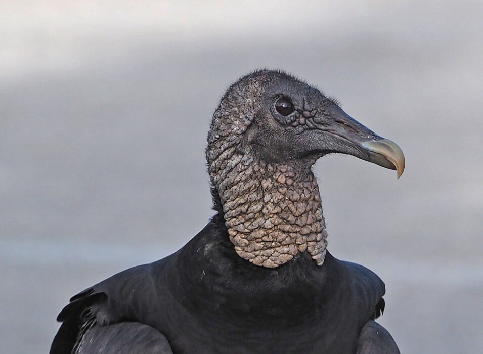
Figure 9. The heads of vultures and condors are bald to sparsely feathered, as shown in this black vulture (Coragyps atratus). Photo credit: Frank Wouters/Flickr Creative Commons. Click image to enlarge.
Molting
Molting, the process of natural feather replacement, is a gradual process that takes approximately 6 months in most raptors, although molt may last up to 2 to 3 years in Old World vultures.4,34 Most raptors molt feathers once per year in symmetrical pairs, usually after breeding.34 However, some of the smaller owls, such as the Northern pygmy owl (Glaucidium gnoma) and the burrowing owl (Athene cunicularia), molt all their rectrices at once. Some diurnal raptors molt only partially each year.34 These birds have one or more immature or subadult plumages, which allows these years to be distinguished from adult plumage (Fig 10).34,46 Once adult plumage is obtained, age cannot be determined using plumage character.
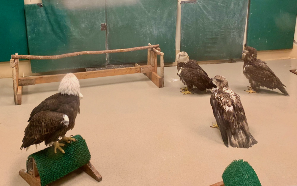
Figure 10. A few plumages in the bald eagle (Haliaeetus leucocephalus). From left to right: Adult (at least 5 years old), end of 4th year, end of second year, and first year. Photo credit: The Raptor Center, UMN. Click image to enlarge.
Uropygial gland
Like other birds, raptors have few glands; however, most birds of prey possess a uropygial or preen gland. The uropygial gland is located on the dorsal surface at the upper base of the tail, and varies in size with the species. When stimulated by the bird’s beak, this gland secretes an oily, fatty substance that is spread during preening to clean, waterproof, and condition the feathers (Fig 11).1
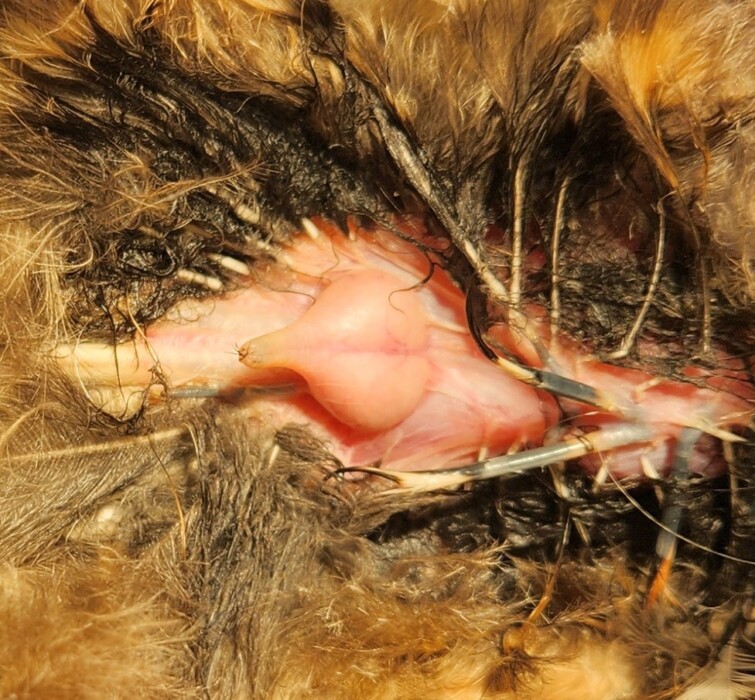
Figure 11. Uropygial gland of a barred owl (Strix varia). Photo credit: The Raptor Center, UMN. Click image to enlarge.
Musculoskeletal
Feet
Many raptors use their feet to capture and sometimes kill prey. The classic raptorial foot is armed with sharp, strongly curved talons that form the tips of the distal phalanges (Fig 3, Fig 12).32,34,37,52 Many raptors, including owls, eagles, and hawks, capture and kill prey by stabbing and crushing with their powerful feet.32,34,74
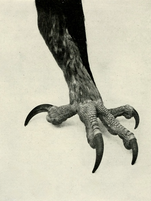
Figure 12. The anisodactyl foot of a golden eagle (Aquila chrysaetos). Photo credit: William Beebe/Wikimedia Commons. Click image to enlarge.
As in most birds, the raptor foot conforms to one of two basic arrangements. The anisodactyl foot of hawks, eagles, falcons, Old World vultures, and kites is adapted for perching and firmly grasping prey.23,30 The first digit, or hallux, faces backward, while digits II, III, and IV are directed forward (Fig 12, Fig 13).23,30,34 The zygodactyl foot, seen in owls and osprey allows an even stronger hold.23,30,49,52 When perched, digits II and III of the zygodactyl foot face forward and digits I and IV face backward (Fig 14). Digit IV is opposable and can be swiveled into an anterior or posterior position that allows a firm grasp on slippery or squirming prey.23,34,49
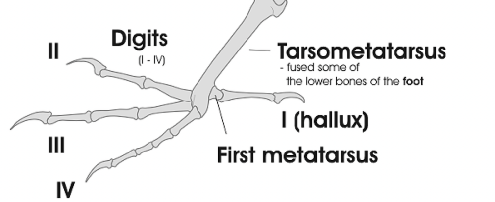
Figure 13. The hallux and digit II in many raptorial species are used together to immobilize prey and are sometimes referred to as the “killing talons” or “power toes”. The remaining digits are used for grasping. Photo credit: Darekk2 via Wikimedia Commons. Click image to enlarge.
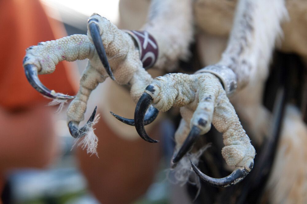
Figure 14. Zygodactyl toe placement in an osprey (Pandion haliaetus). Digits II and III face forward and digits I and IV face backward. Digit IV is opposable and can be swiveled into an anterior or posterior position to adjust the grip on squirmy prey. Photo credit: Getty Images. Click image to enlarge.
Falcons have flat feet and relatively long, thin toes (Fig 15). Unlike most raptors, whose feet are used to kill, the feet of falcons are primarily used to catch and immobilize prey. To actually kill their prey, falcons have a specialized feature on each side of their rhinotheca, a tomial tooth.32
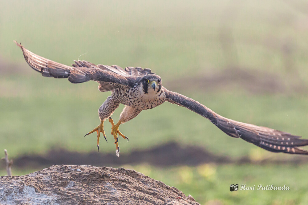
Figure 15. A peregrine falcon (Falco peregrinus) takes off in flight. Note the long, thin toes. Photo credit: Hari K. Patibanda via Flickr Creative Commons. Click image to enlarge.
Clinical Tip: When housed in captivity, falcons require a broad, flat perch, referred to as a “block”, that is covered with padding like artificial turf. Diurnal raptors that prefer to grasp should be provided with curved, rounded perches, such as “bow” perches, wrapped with turf or a suitable type of rope (Fig 16).32,34
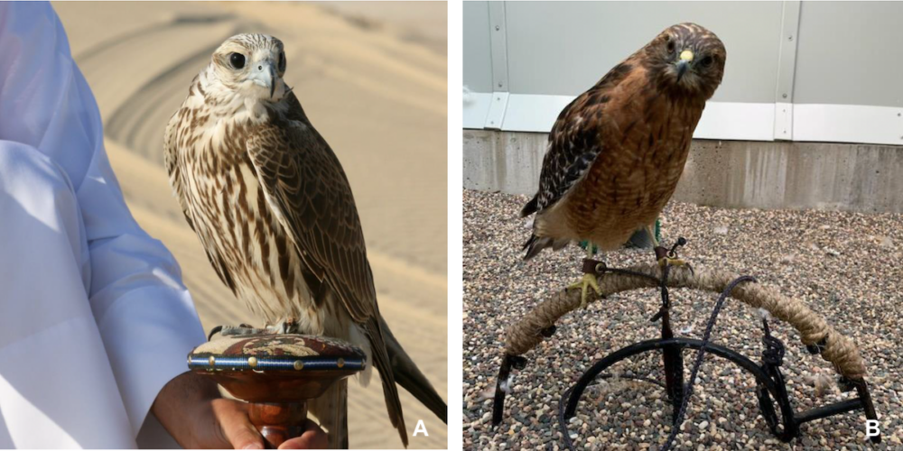
Figure 16. (A) A saker falcon (Falco cherrug) on a block perch. Photo credit: Qatar Pharoah Hound. (B) A red-shouldered hawk (Buteo lineatus) on a bow perch. Photo credit: The Raptor Center, UMN. Click image to enlarge.
New World Vultures have comparatively weak feet similar to those of chickens.59 Their feet are not useful for grasping prey but can be used to brace a cadaver while it is rended with the beak.59
Clinical Tip: Raptors possess two potentially dangerous weapons, the talons and beak. Therefore proper restraint requires training and should not be attempted without observation and supervision by others skilled in handling techniques.
Clinical Tip; It is also incredibly difficult (and hazardous) to pry open the feet of a restrained raptor.34 This is because the digital flexor tendons have unidirectional, interlocking ratcheting mechanisms that resist digital extension when the toes are clenched.34 Straightening the leg at the ankle (hock) joint can loosen this mechanism making extending the toes easier and it is recommended to pull back on digits I and III (Fig 17).
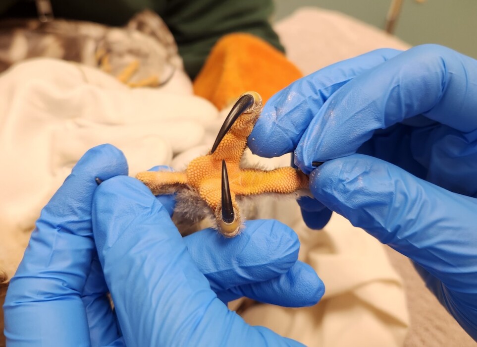
Figure 17. Extending the toes of a barred owl (Strix varia). Note the distinct papillae on the metatarsal pad and plantar surface of the toes. Photo credit: The Raptor Center, UMN. Click image to enlarge.
Pneumatic bones
Raptors share many of the features of the unique avian respiratory system. For instance, much of the avian skeleton is pneumatized, with diverticula of the air sacs. In birds of prey, pneumatization of the skull appears to be maximally developed in large owls.30 The sternum, femur, and humerus are also frequently pneumatic bones in raptors.23,34
Wing shape
Diurnal raptors demonstrate a variety of wing shapes that are related to their preferred prey and hunting technique(s).32,58 Many diurnal raptors are capable of soaring for long distances in search of food at high altitudes up to 4,000 meters.16,32,58,74 The typical soaring wing is relatively long relative to body size (low to medium wing-loading), particularly the antebrachium and manus (Fig 18).30,58,74 Soaring is also enhanced by a broad tail.74
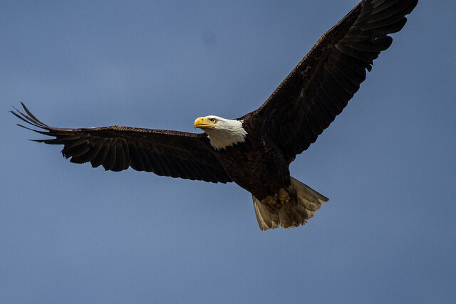
Figure 18. Separation of the tips of the primary remiges, as shown in this bald eagle (Haliaeetus leucocephalus), provides additional lift and propulsion for soaring birds.16,32 Photo credit: David Mitchell via Flickr Creative Commons. Click image to enlarge.
True falcons are characterized by long, pointed wings, narrow tails, and high wing-loading (Fig 19).32,58,74 Falcons are capable of rapid flight that can reach 200 km/h or more, when the birds go into a stoop or dive.32,58,74 The agility of another group of raptors, accipiters, is enhanced by relatively short wings and long tails.74
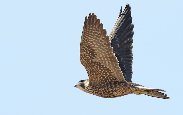
Figure 19. The peregrine falcon (Falco peregrinus) is an example of high wing-loading, with a relatively large body mass and proportionally smaller wings. Photo credit: USFWS Midwest/Flickr Creative Commons. Click image to enlarge.
Tendon ossification
Ossification of tendons may be observed on radiographs in a variety of avian species, including raptors.3,74 The flexor tendons associated with the muscles of the tibiotarsus and toes can become calcified (Fig 20).49,74 It has been theorized that tendon ossification may confer strength and prevent stretching of flexor tendons during catch and transport of heavy prey.23,49 Some antebrachial and tarsal ligaments and cartilaginous structures also become ossified in birds of prey.23
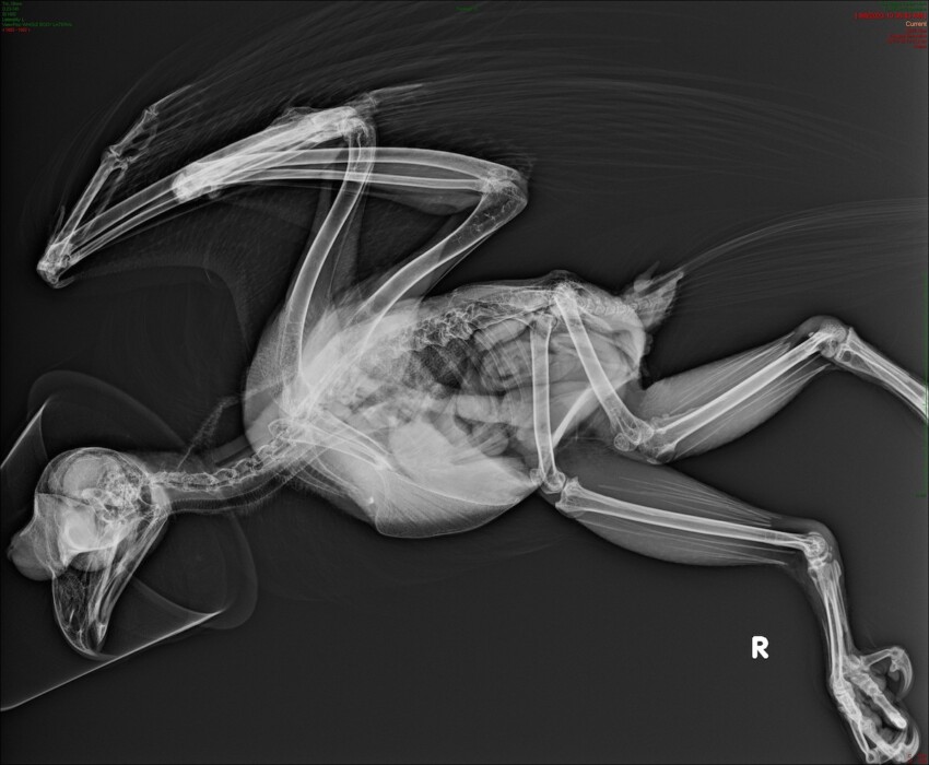
Figure 20. Lateral radiograph showing ossification of tendons associated with tibiotarsus muscles in a great horned owl (Bubo virginianus). Photo credit: The Raptor Center, UMN. Click image to enlarge.
Respiratory
The basic design of the raptor respiratory system is consistent with that seen across class Aves. The raptor cere is often firm, waxy, and varies in color based on species and age (Fig 21, Fig 22). The nares of falcons, Buteo hawks, and eagles have a keratinized flap or operculum (Fig 21).23,30,34 In most birds there are three nasal conchae, however the caudal nasal conchae are absent in some raptors.23,30
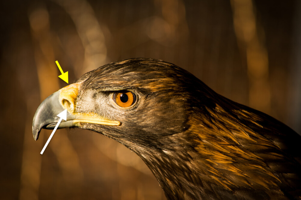
Figure 21. The base of the raptor beak is covered by the cere, a waxy membrane that reaches up to the nares (yellow arrow). Also note the operculum (white arrow) in this golden eagle (Aquila chrysaetos), which serves as a baffle, facilitating air flow in the nostrils during flight. Click image to enlarge.
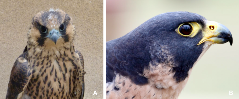
Figure 22. (A) Young peregrine falcons (Falco peregrinus) have a bluish cere which turns (B) bright yellow as they become adults. Photo credit: The Raptor Center, UMN. Click image to enlarge.
Finally, most avian species possess a tracheobronchial syrinx, however, a bronchial syrinx that consists entirely of bronchial elements has been described in owls.30
Gastrointestinal
Most raptors are carnivores that consume other birds, small mammals, reptiles, and occasionally fish.31 Some birds of prey are piscivores that eat primarily fish but may also consume some amphibians, small mammals, and birds. Many smaller species are insectivorous and will consume terrestrial invertebrates, like worms, spiders, and crustacea, as well as small vertebrate prey.11b,23 Vultures are scavengers34, although a few species, like the black vulture (Coragyps atratus) and white-headed vulture (Trigonoceps occipitalis), are known to hunt and kill prey.42 Finally, some raptors are highly specialized feeders, such as the fish-eating osprey, the hook-billed kite (Chondrohierax uncinatus), and snail kite (Rostrhamus sociabilis).16,58
Beak
All raptors possess a sharply hooked bill or beak and sharp cutting edges, or tomia, designed for holding and tearing flesh.31,32,34,52 The “tomial tooth” is a protrusion of the maxillary beak or rhinotheca of falcons that allows them to easily sever the neck of vertebrate prey using plier-like action (Fig 23).34,52Each tomial tooth corresponds with a notch in the lower beak or gnathotheca.23
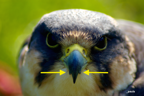
Figure 23. Tomial teeth (arrows) in a peregrine falcon (Falco peregrinus). Photo credit: _pauls. Click image to enlarge.
Clinical Tip: Attempt to preserve the tomial teeth when grooming the falcon beak.13 If there is a crack in the corner of one tomial tooth leading to delamination of the beak, the tooth will need to be ground out and debris removed. A new tomial tooth will reform as the beak grows out.
Tongue
The raptor tongue is adapted for manipulation of food. Distinct highly keratinized papillae on the tip of the tongue create a tough, raspy surface that allows for greater food manipulation and rapid swallowing (Fig 24).31
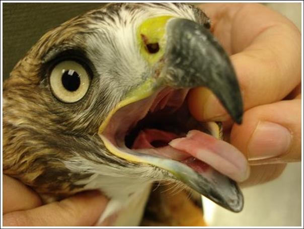
Figure 24. The raptor tongue is relatively long with a sharp-ended apex and a distinct, median groove. Photo credit: Dr. Julia Ponder/The Raptor Center, UMN. Click image to enlarge.
Esophagus
Raptors have a highly elastic esophagus that allows the passage of large food items due to its comparatively wide diameter and numerous longitudinal folds on its inner surface.30,31 Many diurnal birds of prey also have a distinct crop or ingluvies for the storage of food (Fig 25).31,34 The bearded vulture (Gypaetus barbatus) is the only vulture known to lack a crop, presumably due to a diet consisting primarily of bones which would be difficult to store.25,34 Owls do not possess a true crop, but merely a fusiform enlargement of the cervical esophagus.23,49
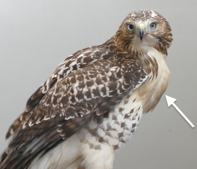
Figure 25. Diurnal raptors store food in the crop. When full, it can be seen as a large bulge over the throat region (arrow) as shown in this juvenile red-tailed hawk (Buteo jamaicensis). Photo credit: The Raptor Center UMN.
Stomach
Raptors that feed on relatively large soft food items, such as meat- or fish-only diets, possess a relatively large sac-like stomach.31,34 The glandular stomach, or proventriculus, expands to accommodate large foodstuffs. The ventriculus functions to mechanically breakdown what cannot be digested chemically (Fig 26).30,34,45 The raptor stomach is relatively thin-walled and poorly muscled with a light cuticle or koilin layer that primarily serves to protect mucosa from the gastric juice produced by the proventriculus. 23,30,31,45,56 Externally, the junction between the proventriculus and ventriculus or isthmus, is often difficult to identify.45
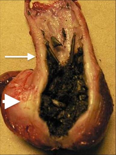
Figure 26. Sac-like stomach in the great horned owl (Bubo virginianus) consisting of the glandular stomach or proventriculus (arrow) and the muscular ventriculus (arrowhead). Photo credit: Dr. Julia Ponder/The Raptor Center, UMN. Click image to enlarge.
The proventriculi of fish and meat-eating birds secrete large quantities of gastric juice. Diurnal raptors are able to more thoroughly digest bones due to an extremely acidic stomach pH that measures approximately 1.7 prior to meals in hawks.14,31,34,74 A gastric pH of 1.0 was recorded in an African white-backed vulture (Gyps africanus) and gastric pH as low as 0.7 has been recorded in other diurnal raptors.24,25 In owls, gastric pH averages 2.2 to 2.5 which does not provide sufficient acidity to break down bone and therefore influences the composition of pellets.14,49,62,74
Intestines
Raptors generally have medium-length to short intestines when compared to other avian species.31 The intestinal tract is comparatively longer in scavenging birds.25,31 The rectum of the American kestrel (Falco sparverius) has been described as unusually long, a feature which may aid in water resorption.31
Ceca
Ceca are paired organs projecting from the proximal colon where it meets the small intestine. Among bird species, there is a great deal of variability in size, shape, and proposed function of the ceca.11 Owls possess large, paired, water-absorbing glandular ceca (Fig 27), while the ceca in diurnal birds of prey are simple, vestigial lymphatic structures or absent.

Figure 27. The distal part of the glandular ceca are enormously expanded in owls. Photo credit: Dr. Julia Ponder/The Raptor Center, UMN. Click image to enlarge
Accessory glandular structures
A well-developed gallbladder is present in most raptors.4,23,34 The pancreas is relatively small in birds of prey when compared to granivorous and herbivorous avian species, presumably due to the high digestibility of animal-origin foods.31 Among birds of prey, the pancreas is relatively large in owls, occupying approximately half of the duodenal loop (Fig 28). The pancreas is smaller in buteos, eagles, and falcons.34,74

Figure 28. The pancreas is found between the ascending and descending limbs of the duodenum. Shown here, the relatively large pancreas of a barred owl (Strix varia). Photo credit: Dr. Julia Ponder/The Raptor Center, UMN. Click image to enlarge.
Pellets
The consumption of whole animals provides complete nutrition, and small prey items are often consumed whole headfirst, especially by owls (Fig 29).31 In fact, some owls will only eat the head if food is abundant. The beak is used to tear large prey items into smaller pieces (Fig 30).31 Poorly digestible components, like the head or skin, may not be consumed.31 The gastrointestinal tract may also be discarded when an adequate food supply is available.31
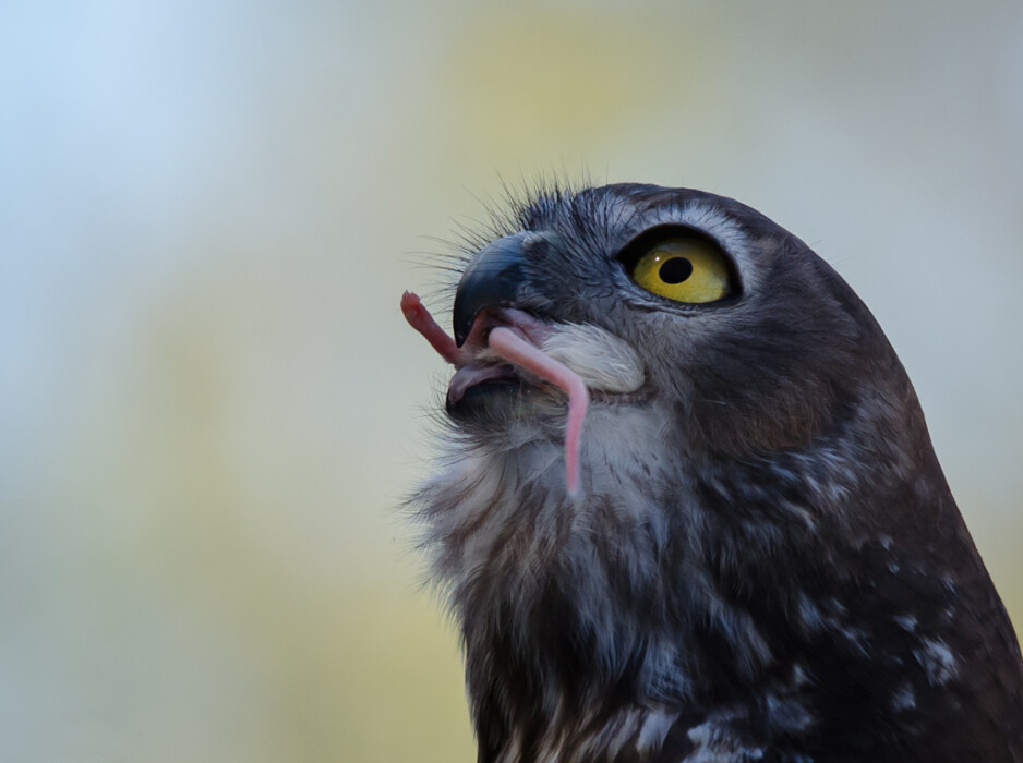
Figure 29. A barking owl (Ninox connivens) consumes a mouse whole. Photo credit: James Niland/Flickr Creative Commons. Click image to enlarge.
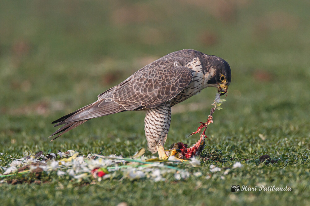
Figure 30. A peregrine falcon (Falco peregrinus) consuming a bird by ripping and tearing the flesh. Photo credit: Hari K. Patibanda/Flickr Creative Commons. Click image to enlarge.
In the final phase of gastric digestion in raptors (and several other types of birds), indigestible material, such as fur, feathers, exoskeletons, fins, scales, shells, teeth, and claws, is compacted into a pellet within the ventriculus (Fig 31).31 Antiperistaltic muscular contractions move the pellet up into the lower esophagus and oropharynx.49 Oral expulsion of the pellet is called egestion or “casting”.23,34 Egestion is a unique physiologic event distinct from vomiting or regurgitation.
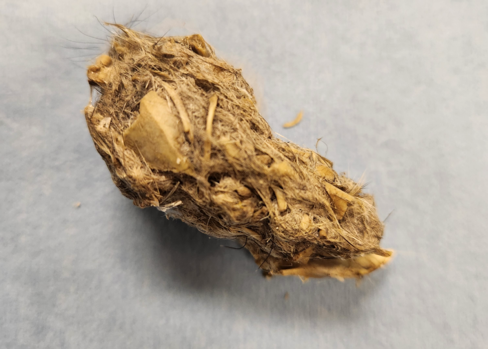
Figure 31. Compacted indigestible material or pellet collected from an owl. Photo credit: Lori Arent. Click image to enlarge.
Pellets are only egested once gastric digestion of a meal is complete. The average interval from feeding to egestion in owls ranges from 10 to 13 hours.49 The interval averages from 19.5 to 23.5 hours in hawks. Owls normally produce a pellet with each meal while hawks can eat more than one meal before casting.34 In the wild, most pellets are egested before midmorning or after killing prey, but before the first feeding of the day.
Clinical Tips: It is important for clinicians to monitor pellet egestion in a raptor patient. Failure to produce a pellet when expected can indicate gastrointestinal tract dysfunction.45
Juvenile birds may have difficulty casting some material, such as fur, and raptors should generally not be provided food with indigestible material until they are over 12 days old (>20 days in some species).23
Feces and fecal flora
Normal raptor feces tend to be soft, viscous, and depending on the bird’s diet can range in color from yellow to dark brown to dark green.32 Fecal samples normally harbor a wide variety of enteric bacteria. Large numbers of Gram-negative coliforms and clostridial organisms are part of normal gastrointestinal flora in birds of prey. Aerobic bacteria isolated from the cloaca of red-tailed hawks (Buteo jamaicensis) and Cooper’s hawks (Accipiter cooperii) include coagulase-negative Staphylococcus, coagulase-positive Staphylococcus, Micrococcus sp., Streptococcus sp., Escherichia sp., and Salmonella spp.34,35 It has also been proposed that raptors can harbor Campylobacter spp., which potentially poses a zoonotic risk.12,47
For additional information: Visit Raptor Gastrointestinal Anatomy and Physiology.
Reproduction
Sex determination
In raptors, visually distinguishing between the sexes can be challenging. However, there are a couple of easily identifiable features that help determine the sex of some species.
SIZE DIMORPHISM
Although there can be considerable overlap in body size between the sexes in some species.33,34, many raptors display reversed sexual dimorphism in which the male is approximately 30% smaller than the female.32,58,72,74 This phenomenon is most pronounced in diurnal species, especially raptors that feed on other birds, such as falcons and accipiters.32,68In fact, the falconry term “tiercel” denotes a male falcon or hawk and is derived from the Latin word for third: “tertius”.32
Male and female New World vultures and condors typically show little differences in size.16,38 If there is a difference, males tend to be larger than females although the size distinction is usually minimal.34,38 Among members of family Cathartidae, the Andean condor (Vultur gryphus) exhibits the most pronounced conventional sexual dimorphism.2
COLOR DIMORPHISM
In a limited number of raptor species, plumage is dimorphic between the sexes. American kestrels, merlins (Falco columbarus), Northern harriers (Circus cyaneus), snowy owls (Bubo scandiacus), osprey, and white-headed vultures (Fig 32-Fig 34) are examples of color dimorphism.33,34,41,58
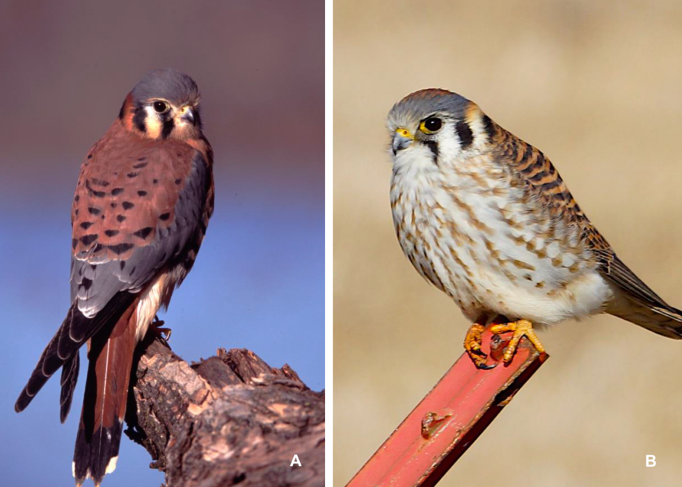
Figure 32. (A) The male American kestrel (Falco sparverius) has a blue/gray cap and wings and solid cinnamon-colored tail feathers with a dark terminal band. Photo credit: Jeff Fischer of The Raptor Center, UMN. (B) The female kestrel is more uniformly cinnamon colored, lacking the blue/gray coloring on her wings and also has a barred tail. Photo credit: John Schmoll/Flickr Creative Commons. Click image to enlarge.
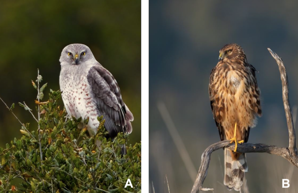
Figure 33. (A) The male Northern harrier (Circus cyaneus) is gray above and whitish below with black wingtips. (B) Females and immature harriers are brown. Photo credit: Channel City Camera Club/Flickr Creative Commons. Click image to enlarge.
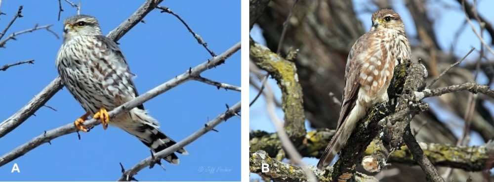
Figure 34. (A) The adult male merlin (Falco columbarius) is blue or gray, while (B) the female is brown. Photo credit: Jeff Fischer/The Raptor Center, UMN. Click image to enlarge.
Gender can be verified in sexually monomorphic species using endoscopic visualization of the gonads, DNA analysis, or intracloacal ultrasonography.22,34,64,72 Fecal steroid hormone immunoassays have also been utilized in research settings.13,34
Ovaries
Although paired ovaries and oviducts develop in the embryo, the right ovary and oviduct rapidly regress after hatch in most avian species.23,30,32,53,55 Although conventional wisdom has held that the right ovary is rarely functional 23,30,34, a study evaluating histological and immunohistochemical data in the goshawk, long-eared owl (Asio otus), common buzzard (Buteo buteo), and sparrow hawk (Accipiter nisus) suggests that folliculogenesis and ovulation may occur in the right ovary.53 In addition, in some Falconiformes, such as peregrine falcons, the right ovary has been reported to be not only present but also may be partially active.18 The right oviduct in several birds of prey has been reported to undergo involution and remain vestigial even when both gonads are functional.23,30,32,53
Breeding age and sex
Small raptors reach sexual maturity at a younger age than large raptors and in many species, male raptors tend to reach sexually maturity before females. Among diurnal species, small accipiters, small falcons, harriers, and Harris’s hawks (Parabuteo unicinctus) usually begin breeding at 1 to 2 years of age.13,46 Buteo hawks, kites, and large falcons breed at 2 to 3 years.46 Ospreys begin breeding at 3 years or more.46 Depending on the species and the individual bird, eagles begin breeding at 4 to 9 years.46 Vultures and condors may begin breeding between 6 to 12 years of age.38,46
Owls also show a similar pattern between size and breeding age. Screech owls (Megascops spp.) have been recorded to breed during their first year.67 The common barn owl (Tyto alba) also reaches sexual maturity very early in life.54 In fact, all members of family Tytonidae can reproduce in their first year, although the rare bird delays reproductive activity until their second year.13,54 The Eurasian eagle owl (Bubo bubo), a member of family Strigidae, reaches sexual maturity between 1-3 years.13
Clutch size
Several factors can influence the number of eggs laid in a clutch. First is the species and its natural history, such as the length of parental care needed post fledging. Large vultures and condors lay only one egg per clutch as their chicks can require care for up to an entire year.26,38,43,46 The smallest raptors typically lay up to 6 eggs per clutch (Table 2). Clutch sizes can also vary dramatically during any breeding season with the latitude, weather, food supply and general health of the female.21,37,60
| Table 2. An overview of eggs per clutch in raptors 21,26,36,38,43,46 | |
|---|---|
| Species | |
| Condors, large vultures* | |
| Eagles, small vultures | |
| Large kites, Buteo hawks, caracaras, ospreys | |
| Small kites, large falcons, large accipiters | |
| Small falcons, small accipiters, harriers | |
| *The Egyptian vulture (Neophron percnopterus) and bearded vulture (Gypaetus barbatus) are the only Old World vultures whose clutch usually has two eggs 21 | |
Most female members of family Strigidae lay an average clutch of two to seven eggs. In times of abundance, some species may lay larger clutches.9 The great horned owl (Bubo virginianus) generally lays two to three eggs and barn owls are quite prolific, laying up to 10 eggs.54,60
Eggs are laid over several days. Although variation occurs among individuals, small raptors lay an egg once every other day, medium-sized species lay eggs every 2 to 3 days, and large species lay eggs at intervals of 2 to 5 days.36,37,46
Incubation
Most raptors begin incubation before they complete laying their clutches. Some birds, like the common barn owl and great horned owl (Bubo virginianus) will begin to incubate as soon as the first egg is laid.16,33,60,71 However, pygmy owls (genus Glaucidium) begin incubating only after the last or penultimate egg is laid.33 Diurnal raptors also begin brooding with the second or third egg (or the first with eagles and vultures).36 Incubation periods in birds of prey tend to be relatively long, ranging from 4 weeks in smaller birds to 7 or 8 weeks in larger species.46
In most raptors, the female protects the nest, eggs and young birds and broods the eggs for almost their entire incubation (Table 3).21,33,36,37,54,60 The male is primarily responsible for hunting and providing food to the brooding female.21,36,37,54 He broods the eggs only occasionally when the female preens or feeds.30,36 Both Old and New World vultures tend to share incubation between the male and female.21
| Table 3. Incubation periods and clutch sizes of selected birds of prey 13,21,36,38,46 | ||||
|---|---|---|---|---|
| Species | ||||
| American kestrel | Falco sparverius | |||
| Bald eagle | Haliaeetus leucocephalus | 34-35 35-38 |
||
| Barn owl (common) | Tyto alba | |||
| Black kite | Milvus migrans | 2-3 | 31-37 |
|
| Black vulture | Coragyps atratus | |||
| California condor | Gymnogyps californianus | 55 |
||
| Cinereous vulture | Aegypius monachus | |||
| Common buzzard | Buteo buteo | 2-4 | 33-35 36 |
|
| Cooper’s hawk | Accipiter cooperii | |||
| Eurasian eagle owl | Bubo bubo | |||
| European sparrowhawk | Accipiter nisus | 4-6 | 35 |
|
| Golden eagle | Aquila chrysaetos | |||
| Griffon vulture | Gyps fulvus | 1 | 52-59 |
|
| Gyrfalcon | Falco rusticolus | 35 |
||
| Harris’ hawk | Parabuteo unicinctus | 2-4 | 33-36 |
|
| Lanner falcon | Falco biarmicus | 3-4 | 32-35 |
|
| Merlin | Falco columbarius | 3-5 | 28-32 |
|
| Mississippi kite | Ictinia mississippiensis | |||
| Northern goshawk | Accipiter gentilis | 3-4 | 35-38 |
|
| Osprey | Pandion haliaetus | |||
| Peregrine falcon | Falco peregrinus | 3-4 | 32-34 31 |
|
| Red kite | Milvus milvus | 1-3 | ||
| Red-shouldered hawk | Buteo lineatus | |||
| Red-tailed hawk | Buteo jamaicensis | |||
| Saker falcon | Falco cherrug | |||
| Turkey vulture | Cathartes aura | |||
| White-tailed sea eagle | Haliaeetus albicilla | 1-3 | 38 |
|
Most female raptors develop an incubation or brood patch during incubation.30,54,69 The brood patch develops in both sexes in species that share incubation duties, like vultures.21,69 In species where the male incubates for only short periods, the brood patch is absent or poorly developed in species.21
Asynchronous hatching
Since most raptors begin incubation before they complete their clutches, eggs hatch over an extended period and clutches can consist of chicks that are several days to a week or more apart in age (Fig 35).46,54,60In the wild, it is not unusual for the youngest nestlings to become progressively weaker from starvation, injury, and/or chilling.46,60 These chicks may die or even be killed and eaten by their older siblings.46,60 As the young grow, they become less aggressive. If the smaller chicks can survive the first few weeks, they have a good chance of fledging.46
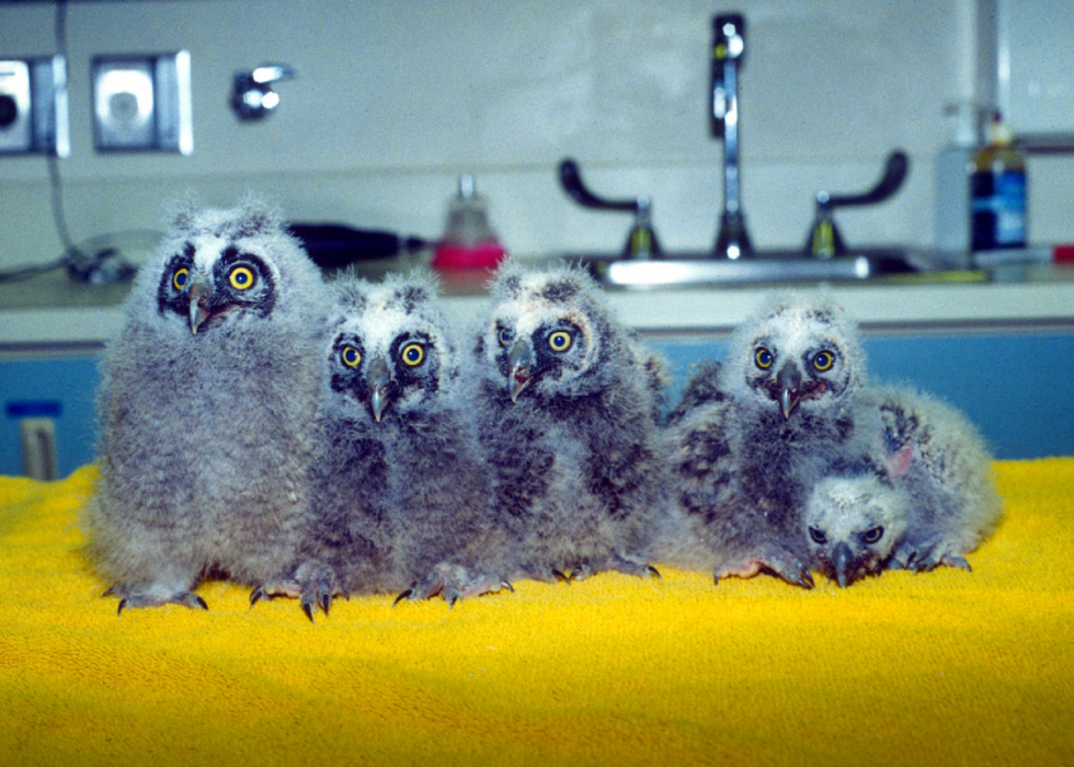
Figure 35. A brood of long-eared owls (Asio otus) demonstrating different ages due to asynchronous hatching. Photo credit: The Raptor Center, UMN
Nearly all raptors outside the tropics raise one brood each year.38,46 However, Harris’s hawks have been known to have more than one brood in resource-rich years; a few species, like the California condor (Gymnogyps californianus), lay one clutch every other year.15,38
Raptors are semi-altricial
Hatchling birds of prey are weak, unable to stand or self-feed.46,60,71 Chicks are covered with an insulating layer of natal down feathers for much of their flightless period which, depending on the species, can last between 4-31 weeks (Fig 36, Fig 37).28,33,37,46 Diurnal raptors may hatch with their eyes open or closed, but usually by day 2 their eyes have begun to open.28 The eyes are fully open by day 6.28 Owls hatch with closed eyes.28,33 Their eyes begin to open at 4–6 days of age and are fully open by day 9 or 10.28,60
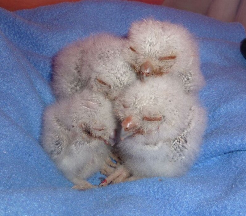
Figure 36. Owls, like these screech owls (Megascops asio), typically hatch with eyes closed and a whitish natal down. Photo credit: The Raptor Center, UMN
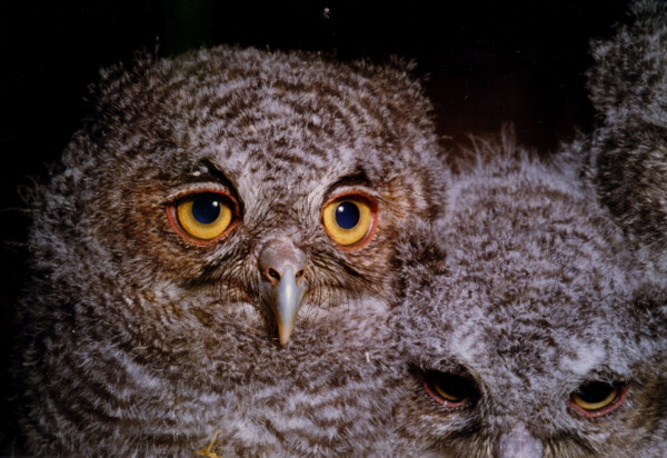
Figure 37. The second set of down feathers or mesoptile of raptor chicks is a fluffy, downy plumage that covers the head and body, while flight and tail feathers resemble those of adults.33 Shown here, screech owlets (Megascops asio). Click image to enlarge.
Life span
In the wild, the life span of birds of prey is often limited by anthropogenic causes, such as obstacles, environmental toxins, and/or human activity that reduces habitat and prey resources.5,11b In captivity, however, raptors often outlive their wild counterparts and may face geriatric conditions, such as arthritis, atherosclerosis, and ocular abnormalities.8,65 It is common that with proper care, small species can live to approximately 15 years of age, medium-sized species to their mid-20s to early 30s, and large species to their mid-30s and 40s.
Special senses
Vision
All birds have high visual acuity and relatively large eyes; however, the forward-facing eyes of raptors provides a large binocular visual field of vision and greater depth perception.29,32,49 Binocular vision is particularly well developed in predators. and this trend is most pronounced in owls. Owls possess the most rostrally-directed eyes with a wide visual field of 150 degrees reported in barn owls.32 How is the raptor eye unique?
GLOBE
The globe is rounded in diurnal raptors and long and tubular in owls (Figure 38).29,30,49,52,70 The tubular shape of the globe gives owls telescopic vision, allowing them to pick out small shapes (like prey) from a distance.52 Extraocular muscles are reduced in all birds; however, the large size of the globe and its tight fit within the orbit, means that eye mobility is particularly limited in owls.10,29,32 Fortunately, this limitation is countered by the highly mobile cervical spine.32
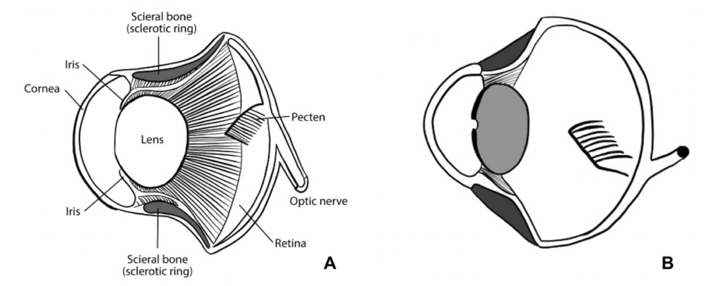
Figure 38. Globe shape in birds of prey. (A) tubular shape of owls (B) spherical shape of Accipitriformes, Cathartiformes, and Falconiformes. Photo credit: The Raptor Center, UMN. Click image to enlarge.
Scleral cartilage serves to provide some internal reinforcement of the eye.19,32 In many falcons and eagles, the scleral cartilage surrounding the optic nerve is ossified to form a U-shaped bone, the os nervi optici.19,29,30 Scleral ossicles are also particularly numerous among owls.5b,19,73
NICTITATING MEMBRANE
The nictitating membrane, or third eyelid, is semi-translucent in some raptors and opaque white in others, such as barred owls (Strix varia) (Fig 39).29,32 The nictitans sweeps across the cornea in a nasal to lateral direction and functions to protect the cornea and provide moisture.
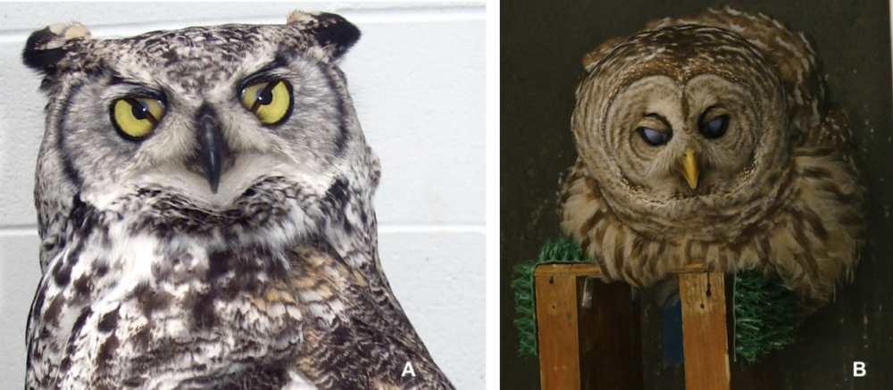
Figure 39. Nictitating membranes in (A) great horned owl (Bubo virginianus) and (B) barred owl (Strix varia). Photo credit: The Raptor Center, UMN. Click image to enlarge.
Many raptors close the nictitating membrane prior to striking their prey.23 Falcons use their nictitans as eye protection during hunting dives, while osprey close their third eyelid under water.23 Movement of the third eyelid over the cornea is typically rapid in diurnal birds, but is relatively slow in owls.29
In contrast to most birds, the upper eyelid of owls is also larger and more mobile.29,32
CORNEA
Raptors possess relatively large corneas, which allow them to gather the maximum amount of light.49 The typical bird cornea is thin; however, several exceptions have been described in diurnal birds of prey.29,32,44 The thickest portion of the golden eagle (Aquila chrysaetos) cornea is found at the limbus (1.2 μm) and the thinnest at the central cornea (0.64 μm).44
The cornea is kept moist by secretions of the gland of the nictitating membrane, as well as the lacrimal and Harderian glands in most species.29 Owls, which produce only a small volume of tears, lack a lacrimal gland.29,32
The anterior sclerocorneal muscle is largest and most developed in hawks and owls.23 This muscle pulls the corneoscleral junction posteriorly, thereby increasing the curvature of the cornea at its center.23,30
IRIS and CILIARY BODY
The avian iris is thin and contains variable amounts of striated dilator and constrictor muscles instead of smooth muscles.23 There is also striated musculature in the ciliary body.23
Clinical Tip: Pupillary light reflexes can be observed in avian patients but are complicated by the fact that birds have voluntary control over constriction and dilation. Complete decussation of the optic nerves also means that there is no true consensual pupillary light response in birds.70
Iris color can vary between the sexes and at different ages within one species.70 For example, the iris of the juvenile red-tailed hawk is yellow and darkens with age becoming chocolate brown in the mature bird.58 The juvenile accipiter has a yellow iris that becomes ruby red in the 5-year old adult (Fig 40).58 The iris of the Northern harrier transitions from brown to yellow.16
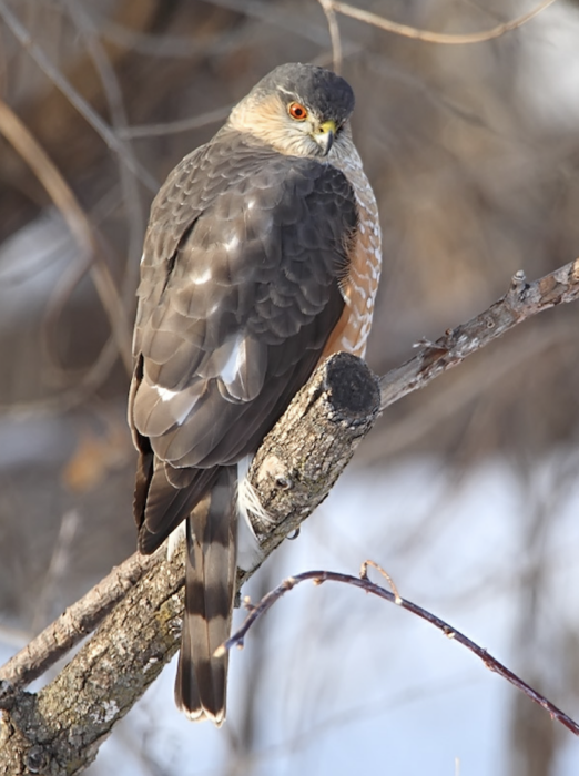
Figure 40. The accipiter iris turns ruby red at 5-years of age, as shown in this adult Cooper’s hawk (Accipiter cooperii). Photo credit: Jeff Fischer/The Raptor Center, UMN. Click image to enlarge.
LENS – ACCOMODATION
Accommodation refers to active changes in the refractive power of the lens, which is achieved by modifying lens shape. The shape of the lens is controlled by its internal elasticity as well as the force exerted by zonule fibers, which flatten the lens when stretched. The anterior surface of the lens is relatively flat in diurnal birds compared with a more spheroid shape in nocturnal species.23,30,32,70 All avian lenses are softer than in mammals, enabling quick accommodation; however, the owl lens is hard when compared to diurnal raptors.23,32 This reflects the fact that owls rely almost exclusively on corneal accommodation instead of lenticular accomodation.32 Lenticular accommodation in diurnal raptors is partially achieved by actions of the ciliary processes. The ciliary body suspends the lens by the zonular fibers and also forms the ciliary processes, which are kept in close contact with the lens by the action of ciliary muscles.29,44
The avian lens has an annular pad around its central core.29 This pad is particularly well developed in fast-flying diurnal raptors.23,32 The function of the annular pad is not completely understood, but it is believed to play an important role in using the lens to focus the eye and increase refractive power (lenticular accommodation).29,32 The annular pad is reduced in owls.23,32
RETINA
In diurnal birds of prey, the retina is heavily dominated by cone photoreceptors; rods are fewer in number and restricted to the margins in the diurnal raptor eye.32 The owl retina, on the other hand, possess a large number of densely packed rods and is thus specialized for dim-light vision.11,12,20,30,32,49 Up to 56,000 rods per millimeter square have been identified in the tawny owl (Strix alluco). Rod photoreceptors also contain high levels of rhodopsin, a light-absorbing pigment.32,49 Although rods make up to 90% of photoreceptors, owls can also see perfectly well in daylight.30,32,49
Most birds, including raptors, possess a pleated or plicated type of pecten oculi (Fig 41).29,30,34 This structure provides nourishment and oxygen to the avascular retina.10 Diurnal raptors usually have a larger pecten with more folds than nocturnal species.29,30,63 For example, the red-tailed hawk has a very large pecten with 17 to 18 folds, while the pecten in the great horned owl (Bubo virginianus) has seven or eight folds.6,7,29,44 The avascular avian retina also receives oxygenation from the choroid layer, which consists primarily of thin-walled blood vessels (Fig 42).29 In diurnal birds of prey, the choroid also contains heavily pigmented connective tissue that gives the fundus its typical gray, brown, or reddish-brown color. Little if any pigment is present in owls.32
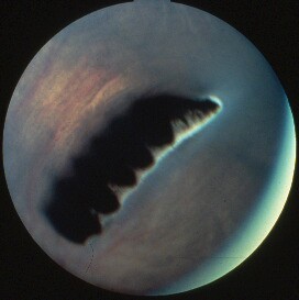
Figure 41. The pecten is a heavily pigmented, highly vascularized, pleated structure that projects into the vitreous from a base situated on the avian optic disk. Image from a great horned owl (Bubo virginianus) . Photo credit: Dennis E. Brooks, DVM PhD Dipl ACVO.
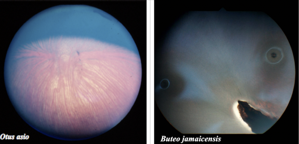
Figure 42. Choroidal vessels in an Eastern screech owl (Otus asio) (left). Choroidal vasculature is clearly visible in most young owls, which have a paucity of pigment in their retinal epithelium. Photo credit: Dr. Christopher Murphy. Choroidal vessels are more difficult to appreciate in most diurnal raptors, such as this red-tailed hawk (Buteo jamaicensis) (right). Photo credit: The Raptor Center, UMN. Click image to enlarge.
Many eagles, hawks, and falcons are bifoveate (Fig. 42).30 A fovea is a small, specialized depression within the retina where visual acuity is highest.29,66,70 The temporal or lateral fovea is dorsal and provides better binocular vision. The central fovea is located medially. Since the eyes of most diurnal raptors are located somewhat laterally in the head, the central fovea participates in lateral monocular vision and fixation of distant objects.23,29,30,32,34,66 Two foveae confer heightened perception of distance and the relative velocity of moving objects (prey) during flight.23,29,34,66,70
Owls only possess a single temporal fovea in each eye, while the Andean condor and black vulture have only a nasal fovea.29,30,32,34
For additional information: Visit the Raptor Ophthalmology series: Anatomy of the Avian Eye, The Ophthalmic Exam, and Ocular Lesions.
Hearing
All birds possess a highly developed sense of hearing, but hearing is the principle sense used for hunting in owls.52 Owls are most accomplished in their ability to localize sound.12,52 Hearing is so finely tuned in the common barn owl that prey can be located in total darkness.48,74
LARGE, ASSYMETRIC EAR OPENINGS
The large ear openings are placed asymmetrically on each side of the head to facilitate vertical location of sound in up to one third of all owl species worldwide.49 The right opening points upward and the left downward (Fig 43). This asymmetrical placement is critical for pinpointing sounds.49 Localization of sound is aided by the wide skull. Differences in intensity and the time that it takes for a sound to reach each ear helps the bird determine whether a sound comes from the right, left, or straight ahead.16,49
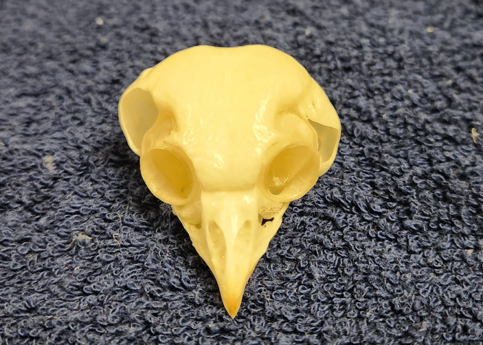
Figure 43. Asymmetrical ear placement in the boreal owl (Aegolius funereus). Photo credit: The Raptor Center, UMN. Click image to enlarge.
FACIAL DISK
The facial disk acts like a funnel to direct sound into the ears (Fig 44). Underlying muscles allow fine positional adjustments of these funnels.32 This tight funnel is particularly prominent in owls and a few diurnal raptors, like osprey and harriers.30,74
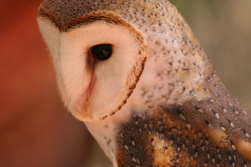
Figure 44. Concentric rings of small, curved, stiff periauricular feathers focus sound into the ear openings in this common barn owl (Tyto alba) similar to a person cupping hands behind their ears. Photo credit: Stephen Barnett. Click image to enlarge.
OPERCULUM
In many owls, the rostral border of the external meatus has a vertical skin flap or operculum that bears a row of feathers along and at right angles to its edge. Striated muscles can be used to erect the operculum, which assists in locating sound.30
EAR COVERTS
In most birds, the external acoustic meatus is covered by specialized contour feathers or ear coverts; however, this region is naked in vultures (Fig 45).
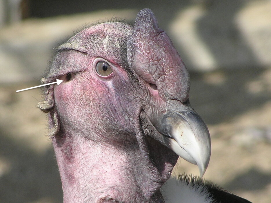
Figure 45. The external acoustic meatus (arrow) is naked in vultures and condors, as seen in this Andean condor (Vultur gryphus). Photo credit: Frank Wouters via Flickr Creative Commons. Click image to enlarge.
Olfaction
Although black vultures (Coragyps atratus) depend more on sight to locate prey, the sense of smell is highly developed in some vultures.32,52 Research has confirmed that the turkey vulture (Cathartes aura) primarily uses its sense of smell to locate carrion.20,61 Scent-guided foraging is associated with an expansion of the olfactory bulbs in vertebrates, and the turkey vulture has the largest olfactory lobes among the New World vultures.20,30,32
What is the secretary bird?
Secretary birds (Sagittarius serpentarius) are large, distinctive birds of sub-Saharan Africa that are characterized by an eagle-like head and long, crane-like legs (Fig 46).50,51 From a phylogenetic perspective, family Sagittariidae represents the deepest divergence of Accipitriformes; however molecular analyses suggests that secretary birds are most closely related to ospreys.39 Adults weigh between 2.3 to 4.27 kg and have an average lifespan of 18.6 years in captivity.57
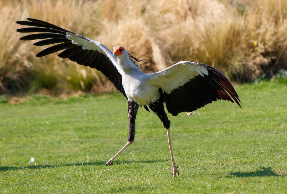
Figure 46. Secretary birds (Sagittarius serpentarius) stand between 0.9 (2.95 ft) to 1.5 m (4.9 ft) tall with a wingspan up to 2.1 m (6.9 ft).50,57 Their exceptionally long legs can be attributed to unusually long tibiotarsus and tarsometarsus bones. Photo credit: Nigel Hoult Flickr Creative Commons. Click image to enlarge.
Secretary birds sometimes kill or stun prey with forceful, lightning-quick kicks or stamps to the head. Visit Cell.com for high-speed camera footage of a secretary bird kicking a rubber snake.51
In addition to their exceptionally long legs, another unique anatomic characteristic of the secretary bird are two pairs of ceca.40 Unlike most raptors, secretary birds are also sexually monomorphic, although females may be slightly smaller than males.51,57
Summary
Raptors are also referred to as birds of prey because they consume foods almost exclusively of animal origin. They possess distinct morphological traits designed for localizing, pursuing, catching, and metabolizing a variety of prey types. These traits begin with the derivatives of the integument – the covering of their beaks and talons, the specialized skin on their foot pads, and modifications to their flight feathers – and are present in many of their major body systems. The esophagus and stomach are specially designed to accommodate large food items and quantities. In the final phase of gastric digestion, the ventriculus compacts indigestible material, such as fur, feathers, and bones (owls), into a pellet which then, through antiperistalsis, is eliminated through the mouth in a process called egestion or “casting”.
As for reproduction, size dimorphism between the sexes, with the males of many species being up to a third smaller than the females, may aid in raising a brood. This difference in size may help each sex to fulfill their primary roles throughout the breeding season.
One additional trait raptors possess is enhanced vision and hearing. The large eye is the most important sensory organ in many birds of prey. Based on the species and its natural history, it has specific features which is critical for survival in the wild. For example, owls have a wider field of binocular vision than diurnal species. The retinas also differ in the number of fovea and type of photoreceptors. Owls only have one fovea and their retinas are specialized for dim-light vision with a large number of densely packed rods. Hawks, on the other hand, are bifoveate and their retinas have a large number of cones. Owls also possess a highly developed sense of hearing due to the presence of large, asymmetric ear openings as well as structures that direct sound into the ear, such as the operculum and facial disk.
Acknowledgement
Thank you to Dr. Patrick Redig for helpful feedback.
References
Avian Anatomy Teaching Module
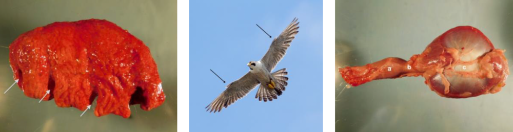
Photo credit: Dr. Nobuko Wakamatsu-Utsuki (left, right), Lori Arent (middle)
Objectives
An understanding of avian anatomy is essential for all facets of medical care. This teaching module is based on completion a reading assignment, which begins with a brief summary of Class Aves before exploring organ systems. Important vocabulary terms and key concepts are emphasized. Upon completion of this review article, the participant will have a basic clinical understanding of avian anatomy.
Syllabus
 |
Complete the reading assignment: Avian Anatomy & Physiology Basics
|
|

RACE approval has expired
This program was approved by the American Association of Veterinary State Boards (AAVSB) Registry of Continuing Education (RACE) to offer a total of 1.00 CE credits to any one veterinarian and/or 1.00 veterinary technician CE credit, however approval has expired.
STAY TUNED: Renewal of this program is pending.
Waterfowl Anatomy & Physiology Basics
Waterfowl belong to Order Anseriformes. Virtually all anseriforms belong to family Anatidae, which consists of ducks, geese, and swans. If you are comfortable with psittacine anatomy and physiology, then many features of waterfowls will be familiar. LafeberVet has listed twelve interesting and clinically significant facts about waterfowl . . .
Pour les vétérinaires. Par les vétérinaires.
Le site Lafervet.com est conçu pour une utilisation par les vétérinaires. Il est ouvert aux vétérinaires diplômés, aux techniciens vétérinaires diplômés, aux animaliers et aux étudiants dans ces domaines.
Créer un compte pour accéder à des articles et des ressources du site.
L'inscription est gratuite.
Para Profesionales Veterinarios. Por Profesionales Veterinarios.
El sitio Lafervet.com es para uso de los profesionales veterinarios. Está abierto a los veterinarios licenciados, técnicos veterinarios licenciados, rehabilitadores licenciados y estudiantes en estos campos.
Cree una cuenta para acceder a los artículos y recursos del sitio.
La registro es gratis.
Already a LafeberVet Member?
Please Login
Avian Anatomy Teaching Module Quiz
. . .
Pour les vétérinaires. Par les vétérinaires.
Le site Lafervet.com est conçu pour une utilisation par les vétérinaires. Il est ouvert aux vétérinaires diplômés, aux techniciens vétérinaires diplômés, aux animaliers et aux étudiants dans ces domaines.
Créer un compte pour accéder à des articles et des ressources du site.
L'inscription est gratuite.
Para Profesionales Veterinarios. Por Profesionales Veterinarios.
El sitio Lafervet.com es para uso de los profesionales veterinarios. Está abierto a los veterinarios licenciados, técnicos veterinarios licenciados, rehabilitadores licenciados y estudiantes en estos campos.
Cree una cuenta para acceder a los artículos y recursos del sitio.
La registro es gratis.
Already a LafeberVet Member?
Please Login
2023 T.J. Lafeber Avian Practitioner of the Year
Dr. Michelle G. Hawkins named
Avian Practitioner of the Year
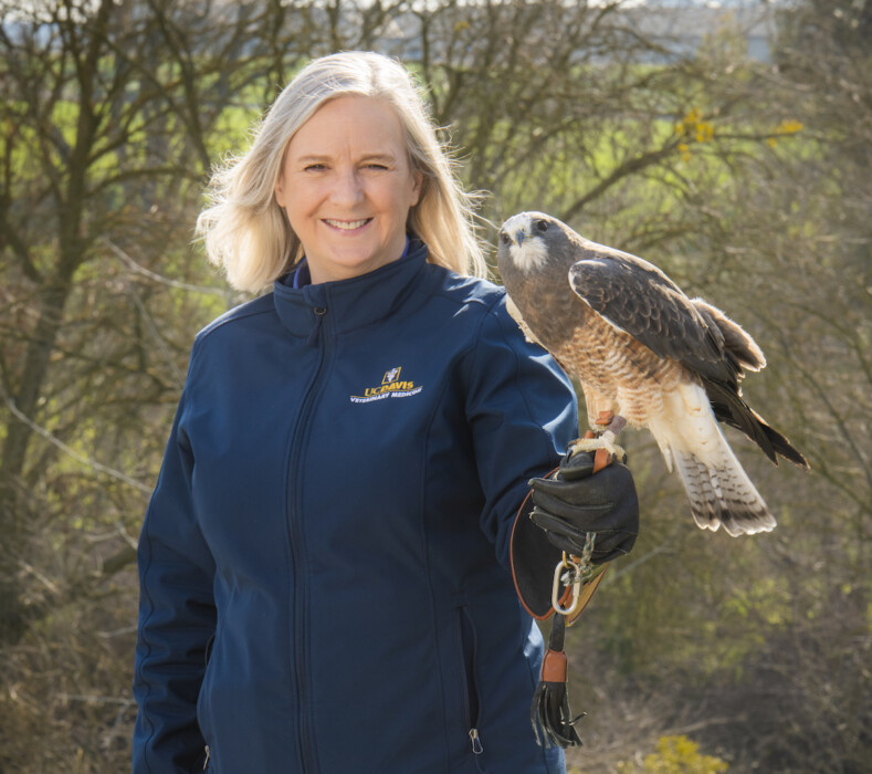
Dr. Michelle Hawkins holds “Grasshopper” a male Swainson’s hawk (Buteo swainsoni) at the California Raptor Center at the University of California School of Veterinary Medicine.Photo credit: Don Preisler/UCDavis
Twenty-one exceptional avian veterinarians from all around the world were nominated for the 2023 T.J. Lafeber Avian Practitioner of the Year. The independent Selection Committee narrowed this list to three finalists and the 2023 T.J. Lafeber Avian Practitioner of the Year, Dr. Michelle Hawkins, was announced during the Plenary Session at the 2023 ExoticsCon.

Michelle Hawkins, DVM, DABVP (Avian Practice) is a Professor of Avian and Exotic Animal Medicine and Surgery at the School of Veterinary Medicine, University of California, Davis and Director of the California Raptor Center.

Share the news
Download and share the press release.

Award recipients
Visit Lafeber.com to learn more about Dr. Hawkins and to see a list of previous Award recipients.

Did you know…?
The T.J. Lafeber Avian Practitioner of the Year is nominated and selected by their peers. The Awardee is not, and has never been, selected by Lafeber Company. The 2023 autonomous Award Selection Committee, consisting of Association of Avian Veterinarians members, was led by Dr. Kenneth Welle.
Raptor Gastrointestinal Anatomy and Physiology
Raptors are a diverse group of birds consisting of three taxonomic orders: Strigiformes (owls), Falconiformes (falcons and caracaras), and Accipitriformes (hawks, eagles, and remaining families). Although the normal diet of free-living raptors varies considerably among species, all raptors hunt and feed on other animals. Meat and fish-eaters possess unique gastrointestinal characteristics that reflect their predatory lifestyle. This review article begins with unique features of the bill and oropharynx, then describes distinctive features of gastrointestinal tract before concluding with pellet formation and egestion and digestive strategies . . .
Pour les vétérinaires. Par les vétérinaires.
Le site Lafervet.com est conçu pour une utilisation par les vétérinaires. Il est ouvert aux vétérinaires diplômés, aux techniciens vétérinaires diplômés, aux animaliers et aux étudiants dans ces domaines.
Créer un compte pour accéder à des articles et des ressources du site.
L'inscription est gratuite.
Para Profesionales Veterinarios. Por Profesionales Veterinarios.
El sitio Lafervet.com es para uso de los profesionales veterinarios. Está abierto a los veterinarios licenciados, técnicos veterinarios licenciados, rehabilitadores licenciados y estudiantes en estos campos.
Cree una cuenta para acceder a los artículos y recursos del sitio.
La registro es gratis.
Already a LafeberVet Member?
Please Login
Passerine Anatomy & Physiology Basics
Perching birds or songbirds belong to order Passeriformes, which makes up the largest taxonomic group of birds. Passeriforms make up nearly 60% of all birds with over 5,000 species belonging to this group. If you are comfortable with psittacine anatomy and physiology, then you are well on your way to understanding passerines. LafeberVet has listed ten interesting, clinically significant facts about passerine anatomy and physiology . . .
Pour les vétérinaires. Par les vétérinaires.
Le site Lafervet.com est conçu pour une utilisation par les vétérinaires. Il est ouvert aux vétérinaires diplômés, aux techniciens vétérinaires diplômés, aux animaliers et aux étudiants dans ces domaines.
Créer un compte pour accéder à des articles et des ressources du site.
L'inscription est gratuite.
Para Profesionales Veterinarios. Por Profesionales Veterinarios.
El sitio Lafervet.com es para uso de los profesionales veterinarios. Está abierto a los veterinarios licenciados, técnicos veterinarios licenciados, rehabilitadores licenciados y estudiantes en estos campos.
Cree una cuenta para acceder a los artículos y recursos del sitio.
La registro es gratis.
Already a LafeberVet Member?
Please Login
Pigeon Anatomy & Physiology: 15 Facts
Although pigeons and doves are a diverse group of birds, they do share some clinically significant anatomy and physiology, including a large, bilobed crop or ingluvies, crop milk production, as well as a vascular plexus found in the subcutis of pigeons. This post also touches on specialized anatomic features unique to fruit pigeons before summarizing some features of the columbid integumentary system, musculoskeletal system, and urogenital tract . . .
Pour les vétérinaires. Par les vétérinaires.
Le site Lafervet.com est conçu pour une utilisation par les vétérinaires. Il est ouvert aux vétérinaires diplômés, aux techniciens vétérinaires diplômés, aux animaliers et aux étudiants dans ces domaines.
Créer un compte pour accéder à des articles et des ressources du site.
L'inscription est gratuite.
Para Profesionales Veterinarios. Por Profesionales Veterinarios.
El sitio Lafervet.com es para uso de los profesionales veterinarios. Está abierto a los veterinarios licenciados, técnicos veterinarios licenciados, rehabilitadores licenciados y estudiantes en estos campos.
Cree una cuenta para acceder a los artículos y recursos del sitio.
La registro es gratis.
Already a LafeberVet Member?
Please Login
Galliform Anatomy Basics
Order Galliformes is a large, diverse taxonomic group with a worldwide distribution. More than 250 species have a chicken-like appearance and short, rounded wings. LafeberVet has listed twelve interesting and clinically significant facts about galliform anatomy and physiology including important vocabulary terms . . .
Pour les vétérinaires. Par les vétérinaires.
Le site Lafervet.com est conçu pour une utilisation par les vétérinaires. Il est ouvert aux vétérinaires diplômés, aux techniciens vétérinaires diplômés, aux animaliers et aux étudiants dans ces domaines.
Créer un compte pour accéder à des articles et des ressources du site.
L'inscription est gratuite.
Para Profesionales Veterinarios. Por Profesionales Veterinarios.
El sitio Lafervet.com es para uso de los profesionales veterinarios. Está abierto a los veterinarios licenciados, técnicos veterinarios licenciados, rehabilitadores licenciados y estudiantes en estos campos.
Cree una cuenta para acceder a los artículos y recursos del sitio.
La registro es gratis.
Already a LafeberVet Member?
Please Login
Avian Anatomy and Physiology Basics
This article is the focus of LafeberVet’s Avian Anatomy Teaching Module, which is RACE-approved for 1 hour of continuing education credit. This review begins with a brief summary of Class Aves before exploring various organ systems. Important vocabulary terms and key concepts are emphasized throughout the text. The reader should have a basic understanding of mammalian anatomy. Birds are quite different both structurally and functionally when compared to mammals, therefore comparisons are frequently drawn between these two taxonomic groups. Since form follows function, descriptions of avian anatomy will also merge into physiology and other life sciences, where appropriate . . .
Pour les vétérinaires. Par les vétérinaires.
Le site Lafervet.com est conçu pour une utilisation par les vétérinaires. Il est ouvert aux vétérinaires diplômés, aux techniciens vétérinaires diplômés, aux animaliers et aux étudiants dans ces domaines.
Créer un compte pour accéder à des articles et des ressources du site.
L'inscription est gratuite.
Para Profesionales Veterinarios. Por Profesionales Veterinarios.
El sitio Lafervet.com es para uso de los profesionales veterinarios. Está abierto a los veterinarios licenciados, técnicos veterinarios licenciados, rehabilitadores licenciados y estudiantes en estos campos.
Cree una cuenta para acceder a los artículos y recursos del sitio.
La registro es gratis.
Already a LafeberVet Member?
Please Login
Post-Test: Clinical Approach to Avian Feather Destructive Behavior
. . .
Pour les vétérinaires. Par les vétérinaires.
Le site Lafervet.com est conçu pour une utilisation par les vétérinaires. Il est ouvert aux vétérinaires diplômés, aux techniciens vétérinaires diplômés, aux animaliers et aux étudiants dans ces domaines.
Créer un compte pour accéder à des articles et des ressources du site.
L'inscription est gratuite.
Para Profesionales Veterinarios. Por Profesionales Veterinarios.
El sitio Lafervet.com es para uso de los profesionales veterinarios. Está abierto a los veterinarios licenciados, técnicos veterinarios licenciados, rehabilitadores licenciados y estudiantes en estos campos.
Cree una cuenta para acceder a los artículos y recursos del sitio.
La registro es gratis.
Already a LafeberVet Member?
Please Login
Patricia Macwhirter BVSc (Hons), PhD, FANZCVS
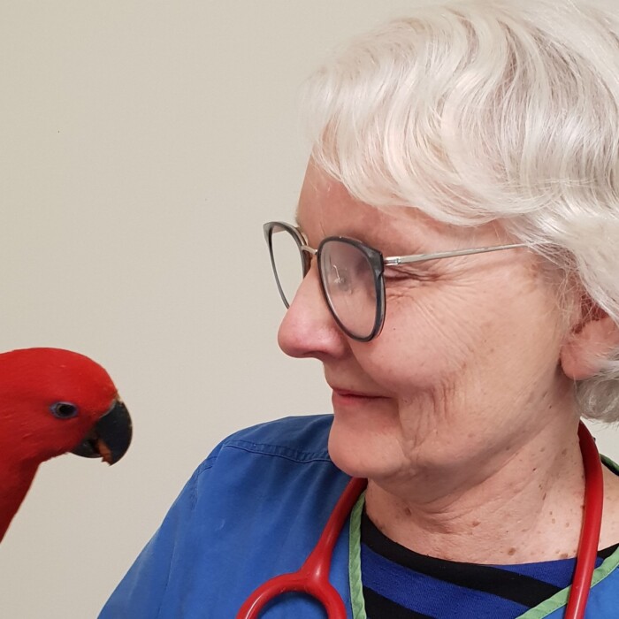 Pat Macwhirter is the co-owner of Melbourne Bird Veterinary Clinic. Dr. Macwhirter earned a Bachelor of Veterinary Science with honors from the University of Sydney University. She then completed an internship and fellowship at the University of Pennsylvania School of Veterinary Medicine, before returning to Australia in 1980 where she opened Burwood Bird and Animal Hospital. Dr. Macwhirter owned or co-owned this practice until 2015, when it was sold, and she enjoyed a brief year’s retirement before returning to clinical practice. In 1993, Dr. Macwhirter became the first person in the Australian state of Victoria to gain board recognition as a Specialist in Avian Medicine. Dr. Macwhirter has always had an interest in birds, bird watching and avian medicine and surgery. Over the years, she has kept and bred budgerigars, Major Mitchell cockatoos, galahs, peacocks, and Quaker parrots. She has authored or co-authored numerous scientific papers, books, and conference proceedings and has been invited to speak at veterinary conferences in Asia, Central America, North America, Europe, and Australia.
Test Your Knowledge: GnRH Agonists in Avian Practice
. . .
Pour les vétérinaires. Par les vétérinaires.
Le site Lafervet.com est conçu pour une utilisation par les vétérinaires. Il est ouvert aux vétérinaires diplômés, aux techniciens vétérinaires diplômés, aux animaliers et aux étudiants dans ces domaines.
Créer un compte pour accéder à des articles et des ressources du site.
L'inscription est gratuite.
Para Profesionales Veterinarios. Por Profesionales Veterinarios.
El sitio Lafervet.com es para uso de los profesionales veterinarios. Está abierto a los veterinarios licenciados, técnicos veterinarios licenciados, rehabilitadores licenciados y estudiantes en estos campos.
Cree una cuenta para acceder a los artículos y recursos del sitio.
La registro es gratis.
Already a LafeberVet Member?
Please Login
Clinical Approach to Feather Destructive Behavior
Feather destructive behavior is one of the most challenging clinical concerns faced by avian veterinarians and owners alike. In this RACE-approved webinar recording, Lauren Thielen, DVM, DABVP (Avian Practice) discusses the systematic hunt for underlying causes as well as case management strategies once feather damaging behavior has been observed. Review of the current literature in feather destructive behavior is discussed as a way to understand why parrots exhibit this behavior and what caretakers can do to reduce the incidence of this problem . . .
Pour les vétérinaires. Par les vétérinaires.
Le site Lafervet.com est conçu pour une utilisation par les vétérinaires. Il est ouvert aux vétérinaires diplômés, aux techniciens vétérinaires diplômés, aux animaliers et aux étudiants dans ces domaines.
Créer un compte pour accéder à des articles et des ressources du site.
L'inscription est gratuite.
Para Profesionales Veterinarios. Por Profesionales Veterinarios.
El sitio Lafervet.com es para uso de los profesionales veterinarios. Está abierto a los veterinarios licenciados, técnicos veterinarios licenciados, rehabilitadores licenciados y estudiantes en estos campos.
Cree una cuenta para acceder a los artículos y recursos del sitio.
La registro es gratis.
Already a LafeberVet Member?
Please Login
The Use of GnRH Agonists in Avian Practice
Avian veterinarian Brian Speer presented this live webinar, RACE-approved for 1.5 credit hours. Reproductive activities, particularly those that are recurrent, pose a significant threat to the health and behavioral well-being of many pet birds. This presentation began by reviewing the basics of female reproductive anatomy and endocrinology before discussing the role of environmental and behavioral stimuli on reproductive endocrinology. Dr. Speer then presented an ethical scale for selecting and prioritizing recommendations for intervention, before discussing medical therapy as well as the environmental and behavioral interventions that can be employed. Dr. Speer concluded with a discussion of behavioral . . .
Pour les vétérinaires. Par les vétérinaires.
Le site Lafervet.com est conçu pour une utilisation par les vétérinaires. Il est ouvert aux vétérinaires diplômés, aux techniciens vétérinaires diplômés, aux animaliers et aux étudiants dans ces domaines.
Créer un compte pour accéder à des articles et des ressources du site.
L'inscription est gratuite.
Para Profesionales Veterinarios. Por Profesionales Veterinarios.
El sitio Lafervet.com es para uso de los profesionales veterinarios. Está abierto a los veterinarios licenciados, técnicos veterinarios licenciados, rehabilitadores licenciados y estudiantes en estos campos.
Cree una cuenta para acceder a los artículos y recursos del sitio.
La registro es gratis.
Already a LafeberVet Member?
Please Login
Jennifer Graham, DVM, DABVP (Avian Practice), DABVP (Exotic Companion Mammal Practice), DACZM
 Jennifer Graham graduated from Auburn University in 1999. She completed an avian/exotic internship at the University of Georgia at Athens followed by a 3-year residency in avian/exotic animal medicine at the University of California at Davis. Dr. Graham worked at Angell Animal Medical Center in Boston from 2006-2012 and she ran the Zoological Companion Animal Medicine Service at the Cummings School of Veterinary Medicine at Tufts University from 2012-2022. Dr. Graham consults on zoological companion animal cases through Moichor, University of Miami, and Zoetis, while also running her own consulting business through Graham Veterinary Consulting. She currently holds adjunct positions with Tufts University, Tuskegee University, and the University of Miami. Jennifer is board certified with the American Board of Veterinary Practitioners in Avian and Exotic Companion Mammal practice categories as well as the American College of Zoological Medicine. Dr. Graham is a member of multiple professional organizations, including the Association of Avian Veterinarians, the Association of Exotic Mammal Veterinarians, Association of Reptilian and Amphibian Veterinarians, and the American Association of Zoo Veterinarians. Jennifer has authored numerous research papers, chapters, and books including the Five-Minute Veterinary Consult: Avian and Exotic Animal Emergency and Critical Care Medicine. You can learn more about Dr. Graham on ResearchGate.
Fawzi Mohamed, BVSC, MS, PhD
 Fawzi Mohamed is a veterinary medical officer and pathologist for the United States Department of Agriculture (USDA). Dr. Mohamed is based out of Plum Island Animal Disease Center. Fawzi earned a PhD in veterinary pathology from the University of Georgia. He has co-authored several papers on rabbit hemorrhagic disease virus and other important infectious diseases, such as African swine fever and foot-and-mouth disease.
Molly Gleeson, DVM, DACZM

Molly Gleeson is currently in private practice in California, where she treats a variety of non-traditional pets. Dr. Gleeson is board certified in zoological companion animal practice through the American College of Zoological Medicine (ACZM). Prior to becoming a Diplomate, she completed a small animal rotating internship and exotics specialty internship at Gulf Coast Veterinary Specialists in Texas, and a residency in zoological companion animal medicine and surgery at the University of California at Davis.
Lymphoma in the Ferret: An Overview of Diagnosis and Treatment
Lymphoma is one of the most commonly diagnosed diseases in the domestic ferret. Hemolymphatic tumors are the third most common neoplasm described in ferrets, with lymphoma representing from 8.6% to 19.3% of ferret neoplasms in retrospective studies. Ferrets of any age or sex can be affected, with lymphoma described in patients as young as 2 months. A recent retrospective found 5 years as the median age of diagnosis in ferrets treated for lymphoma, and was the same for small, intermediate, and large cell lymphomas . . .
Pour les vétérinaires. Par les vétérinaires.
Le site Lafervet.com est conçu pour une utilisation par les vétérinaires. Il est ouvert aux vétérinaires diplômés, aux techniciens vétérinaires diplômés, aux animaliers et aux étudiants dans ces domaines.
Créer un compte pour accéder à des articles et des ressources du site.
L'inscription est gratuite.
Para Profesionales Veterinarios. Por Profesionales Veterinarios.
El sitio Lafervet.com es para uso de los profesionales veterinarios. Está abierto a los veterinarios licenciados, técnicos veterinarios licenciados, rehabilitadores licenciados y estudiantes en estos campos.
Cree una cuenta para acceder a los artículos y recursos del sitio.
La registro es gratis.
Already a LafeberVet Member?
Please Login
Lafeber’s New Diet Conversion Kits
The majority of bird owners need guidance when it comes to avian nutrition, and it can be time consuming!
Our new Diet Conversion Kits help your team save time by providing clients with instructions and tips on how to convert from a seed mix diet to nutritionally balanced diets along with 5 trial size portions of food to get them started.
Featuring:
- 5 convenient trial sizes for parrots or cockatiels: (1) Classic Nutri-Berries, (1) Tropical Fruit Nutri-Berries, (1) Classic Avi-Cakes, (2) Tropical Fruit Gourmet Pellets.
- Pellet conversion instructions.
Reptile Nutrition 101: Veggie & Insectivores Delight
Introduction
La’Toya Latney, DVM, DECZM (ZHM), DABVP (Reptile & Amphibian Practice), CertAqV presented this talk for the University of Minnesota College of Veterinary Medicine Zoo, Exotics, Aquatics & Wildlife Medicine Club as part of the Lafeber Company Student Program.
Abstract
Reptile nutrition is a growing field. Despite the large amount of biological data known about reptiles, there is much less known about how these species process the foods offered in captivity. This non-interactive webinar recording reviews what we know about reptile nutrition, including the nutritional strategies utilized as well as the comparative analysis of commonly fed vegetables and insects. The basic components of reptile nutrition are explored, from metabolizable energy, protein, and fat, to vitamins and minerals. Food preparation is then discussed in detail. Depending on the source, this process can be quite confusing, however, there are measures that can be taken to manipulate the nutritive profile of feeder insects.
Outline
Download a PDF of this detailed outline.
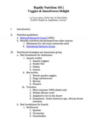
I. Introduction
II. Nutrition guidelines
-
- National Research Council (NRC)
- Herptile nutrition is extrapolated from other species
- Minimums for rats are most commonly used
- Nutritional Advisory Group
III. Nutritional strategies per taxonomic group
- Diet breakdown for chelonians
- Aquatic turtles
- Aquatic veggies
- Feeder fish
- Pellets
- Insects
- Box turtles
- Weeds, garden veggies
- Slugs, earthworms
- Berries
- Flowers
- Aquatic turtles
-
- Tortoises
- Most consume 100% plants only
- Weeds, fibrous roots
- Adapted to live in the desert
- Exceptions: South American spp., African forest tortoises
- Tortoises
- Diet breakdown for lizards
- Herbivores
- Omnivores
-
- Insectivores
- Leopard geckos
- Monitors
- Some chameleons
- Frilled dragons
- Insectivores
-
- Specialized
- Fruit/nectar feeders
- Crested geckos
- Columbian tegus
- Specialized
- Almost all snakes are carnivores
- Temperate
- Examples: corn snake, milk snakes, rat snakes, kingsnakes
- Tropical
- Examples: pythons & boas, snakes from Central & South America
- Temperate
- Specialized diets
- Insects, amphibians, feeder fish
- Examples: garter snakes, rainbow boas, ring-neck snakes, hog nose, egg-eating snake
IV. Nutritional strategies
- Herbivores
- Vegetable dinner wheel
- Dandelion greens
- Romaine lettuce
- Collard greens
- Mustard greens
- Red leaf lettuce
- Escarole
- Endive
- Swiss chard
- Bok choy
- Kale
- Spinach
- Hay science
- Alfalfa
- Botanical hay
- Oat hay
- Orchard grass
- Organic meadow
- Western timothy
- Grasses
- Vegetable dinner wheel
- Insectivore
- Insect composition
- Domestic cricket (Acheta domesticus)
- Mealworm (Tenebrio molitor)
- Mealworm-beetle life cycle
- Earthworm (Lumbricus terrestris)
- Silkworm (Bombyx mori)
- Phoenix worm larvae (Hermetia illucens)
- Madagascar hissing cockroach (Gromphadorhina portentosa)
- Turkistan or red rusty cockroach (Blatta lateralis)
- Butterworm or tebo worm (Chilecomadia moorei)
- Dubia cockroach (Blaptica dubia)
- Hornworm (Maduca quinquemaculata)
- Not readily on the market, but occasionally available
- False katydid (Microcentrum rhombifolium)
- Wood louse (Procellio scaber)
- Carnivores
- Omnivores
- Super-specialized
V. Basic components of nutrition
- Metabolizable energy (ME)
- Definition: Net energy gained from food after digestion and absorption
- Standard metabolic rates for daily energy needs at optimal temps= 32 x BW kg (0.75)
- 500-gram reptile requires 9.5-57 kcal/day at 86°F (30°C)
- Measurement
- Invertebrates: 7-2.7 kcal ME/g
- Butterworms: 977 ME/g
- Cricket: 402
- Earthworm: 708
- House fly: 918
- Mealworm karvaeL 378
- Phoenix worms: 994
- Red Turkish roach: 244
- Superworm larvae: 423
- Wax worms: 747
- Invertebrates: 7-2.7 kcal ME/g
- Factors that impact ME
- Species
- Age
- Activity
- Environmental temperature
- In reptiles, temperature is not maintained by ME
- Fewer calories needed than mammals
- Digestive efficiency is the same as mammals
- Reptiles – Metabolic rate based on metabolic body size
- Protein
- Definition: Amino acid composition and availability
- Nitrogen availability
- Measurement: g/kg or % DM
- Insect protein sources
- Exoskeletons contain amino acids
- Bioavailability may be limited
- Phoenix worms and mountain chicken frogs (Dierenfeld 2008)
- Phoenix worms and leopard geckos (Boykin 2021)
- Definition: Amino acid composition and availability
- Fat
- NRC min for rats
- 5% (50 g/kg) of diet fed (growing)
- Necessary for fat-soluble vitamins
- Lipid content in feeder inverts: 3-6X higher than rodent diets
- Is fat bad for herps?
- Species dependent: strict herbivores rely on fat more than omnivores
- Activity level
- NRC min for rats
| Invertebrates | Crude fat (g/kg) | % fat |
| Butterworms | 294 | |
| Crickets | 68 | As high as 22.8% |
| Earthworms | 16 | 12.6% |
| False katydids | 9% | |
| Fruit flies | 19 | |
| Hissing cockroach | 20.3% | |
| Mealworm larvae | 54 | As high as 31.1% |
| Phoenix worms | 140 | |
| Red rusty cockroach | 14.5% | |
| Silkworms | 14 | |
| Superworm larvae | 177 | As high as 40.1% |
| Tenebrio beetles | 17.7% | |
| Waxworms | 249 | |
| Wood louse | 11.3% | |
| Zoophobas beetles | 14.3% |
- Vitamins
- Vitamin A
- Definition: retinol, beta-carotene, retinyl esters
- Fat-soluble, light sensitive
- Measurement (IU/kg)
- Clinical relevance
- Poor levels in most insects
- Hypovitaminosis A
- A significant and common clinical problem in herptiles
- Squamous metaplasia
- Palpebral edema
- Aural abscess
- Respiratory epithelium compromise
- Lingual squamous metaplasia (“short tongue syndrome” in amphibians)
- Ulcerative chelitis
- Vision loss
- Growth deficits
- Gastrointestinal bloat
- Dermal ulcerations
- Septicemia
- Acute death
- Supplementation with beta carotene is not as effective, must use retinol or retinyl esters
- NRC min retinol for rats: 2300 IU/kg
- Do we know minimum requirements for herps?
- Box turtles: 3-6 IU/g diet DM
- Aquatic turtles: 2-8 IU/g diet DM
- Chameleons: 5-9 IU/ cricket DM
- Foam nesting frogs: 230,000 IU vitamin A/kg of supplement
- Vitamin A
| Invertebrates | IU/kg | ug/kg |
| Butterworms | <300 | |
| Crickets | <1000 | |
| Earthworms | <1000 | |
| False katydids | 2953 | |
| Hissing cockroach | 182 | |
| House flies | <300 | |
| Mealworm larvae | <1000 | |
| Phoenix worms | <300 | |
| Red rusty cockroach | 120 | |
| Silkworms | 1580 | |
| Superworm larvae | <1000 | |
| Tenebrio beetles | 12 | |
| Waxworms | <1000 | |
| Wood louse | 170 | |
| Zoophobas beetles | 41 |
- Vitamin E
- Definition: antioxidant vitamins, alpha-tocopherol
- Measurement
- mg/kg
- % fat in diet
- Clinical relevancy
- Invertebrates have adequate levels, ranging from 5-166 mg/kg
- Steatitis in deficient patients
- NRC rat min 18 mg/kg
- Vitamin D3
- Definition
- 25-hydroxycholecalciferol or calcidiol
- Activated by photosynthetic mechanisms (UVB driven) in diurnal and crepuscular herps and people
- Sun >
- UVB radiation >
- Vitamin D precursor in the skin >
- 25-hydroxycholecalciferol
- 1, 25- hydroxycholecalciferol
- Absorbed via intestine
- Calcium absorption and homeostasis
- Serum levels are dependent on musculoskeletal, renal, integumentary, and GI health
- Measurement: ug/kg or IU/kg
- Production and calcium homeostasis
- Clinical relevance
- Calcium deficiencies across many orders of herptile spp.
- Well studied in diurnal herbivores
- Insectivore supplementation?
- Diurnal or crepuscular
- House gecko (Carmen 2000)
- Jamaican anole (Ferguson 2005)
- Nocturnal
- Leopard geckos (Mitchell, ARAV 2013)
- Homemade vs oral supplements
- UVB first
- Level comparable to natural history
- Even nocturnal spp. can use UVB
- Supplementation
- Caution: Inadvertent toxicity can occur
- Insects “groom” powders off exoskeleton
- If utilized, do so for 1 week or less
- UVB first
- Diurnal or crepuscular
- Definition
- Minerals
- Calcium & phosphorus
- NRC min for calcium in rats: 5-5 g/kg
- NRC min for phosphorus in rats: 3 g/kg
- Insects
- Ca: 089-9.3 g/kg
- P: 5-3.7 g/kg
- Phoenix worms
- 9.3 g/kg
- Wood louse
- 14% calcium DM
- 11.79 Ca:P ratio
- Digestibility?
- Calcium & phosphorus
VI. Food prep
- Commercial diets
- Canned/dried prey items
- Pellets
- Powder/gel food
- Advantages
- Shelf life of products
- Don’t have to handle or care for live prey
- Consistent nutrition profile
- Challenges
- Acceptance by reptiles
- Variable quality/nutritional content
- Care and feeding of prey
- General concepts
- Do not feed directly from pet store of post-shipment
- Need appropriate planning
- Housing examples
- Containers and containment
- Stocking density/surface area
- Substrate/hides
- Crickets
- The development and evaluation of a gut-loading diet for feeder crickets formulated to provide a balanced nutrient source for insectivorous amphibians and reptiles (Attard 2013)
- Ingredients
- Soybean flour
- Sweet potato flour
- Red lentil flour
- Spirulina
- Crushed rabbit alfalfa pellets
- Gut load x 24h
- Ingredients
- The development and evaluation of a gut-loading diet for feeder crickets formulated to provide a balanced nutrient source for insectivorous amphibians and reptiles (Attard 2013)
- Beetle larvae
- Similar setups for mealworms and superworms
- Hydration is important
- Breeding is easy
- Can be gut loaded
- Roaches
- Popular
- Death head
- Lobster
- Dubias
- Dubia blaptica
- Cannot climb glass
- Adults live 1-2 years
- Breed constantly
- Quiet, no odor
- Dubia blaptica
- Turkish
- Hissing
- Madagascar hissing roaches (Gromphadorhina portentosa)
- Large, easy to care for
- Tropical
- Can climb glass
- Adults: 2-3 years
- Juveniles take 3 months to mature
- Madagascar hissing roaches (Gromphadorhina portentosa)
- Popular
- Butterworms or trevoworms (Chilecomadia moorei)
- Chilean carpenter moth – invasive species
- Irradiated prior to import to prevent molting
- Store in fridge for weeks
- Gut load 24 hours prior?
- General concepts
-
-
- Reptiworms, calciworms, and Phoenix worms
- Phoenix worms are the larvae of the black solder fly (Hermetia illucens)
- Well studied, used in aquaculture, poultry, vermiculite
- Stored in cups at room temp
- Phoenix worms are the larvae of the black solder fly (Hermetia illucens)
- Hornworms
- Grow rapidly and eat non-stop
- Feed to insectivores within 7-10 days depending on desired size
- Large specimens
- May require cuticle laceration
- Silkworms
- Picky eaters: mulberry leaves (vitamin A)
- Most vendors sell with a food substrate
- Store at room temperature and feed them within 7-10 days
- Moisture of feed can promote fungal growth
- Earthworms
- Dirt substrate
- Feed composting scraps
- Can gut load 24h prior
- Moth and fly larvae
- Drosophila melanogaster
- Reptiworms, calciworms, and Phoenix worms
-
-
- Prey health is important for insectivore health
- Well hydrated
- Well nourished, high-quality diets
- Infectious disease
- Prey health is important for insectivore health
-
- Augmenting nutrition profile (vitamin/mineral)
- Dusting
- Shake invert in small particle dust prior to feeding
- Imprecise way to guarantee supplementation
- Insects can groom off dust or dust falls off
- Gut loading
- Short-term feeding nutrient dense diet to prey
- Many studies confirm this method works for calcium
- Do both!
- Dusting
- Augmenting nutrition profile (vitamin/mineral)
VII. “Happy Meal”® menu
- Larval insects
- High in fat
- Deficient in vitamins and minerals
- Variable protein bioavailability
- Adult beetles, earthworms, roaches
- Lower fat
- Good protein levels (especially earthworms)
- Most insects have poor calcium levels
- Increase dietary calcium with supplementation
- Exception: Phoenix worms have high calcium content
- Macerate to increase bioavailability
- Insect care
- Well hydrated
- Well nourished
- Feed calcium-rich diet at least 24h prior (not >9% total calcium)
- Can be kept or bred at home
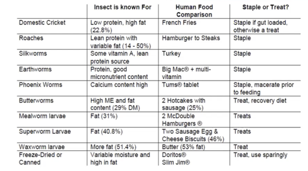
Recording
Client education
Download this list of husbandry resources that Dr. Latney shares with her herptile and piscine clients.
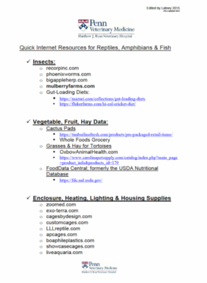
Garden resources
Explore these resources shared by Dr. Latney for building economical, effective gardens for tortoises.
- Starting a Back to Eden Garden From Scratch
- The Best DIY Cinder Block Raised Garden Bed – The Owner-Builder Network
- How to Build a Cinder Block Raised Garden Bed
RACE approval
This program was reviewed and approved by the American Association of Veterinary State Boards (AAVSB) Registry of Approved Continuing Education (RACE) program for 1 hour of continuing education credit for veterinarians and veterinary technicians in jurisdictions that recognize AAVSB RACE approval.
References
Adrenocortical Disease in Ferrets
Hyperadrenocorticism is a common and complex clinical condition in the pet ferret. This disease occurs most frequently in ferrets three years or older but has been reported in animals as young as one year of age. Presumptive diagnosis of adrenal disease in the ferret is based on history, clinical signs, imaging diagnostics, and steroid hormone analysis. Medical therapy using deslorelin implants, though not curative, is recommended. Ferrets may remain asymptomatic for a median of 1-1.5 years. Adrenalectomy may be indicated in case non-responsiveness to medical treatment, although adrenalectomy of the right adrenal gland is difficult . . .
Pour les vétérinaires. Par les vétérinaires.
Le site Lafervet.com est conçu pour une utilisation par les vétérinaires. Il est ouvert aux vétérinaires diplômés, aux techniciens vétérinaires diplômés, aux animaliers et aux étudiants dans ces domaines.
Créer un compte pour accéder à des articles et des ressources du site.
L'inscription est gratuite.
Para Profesionales Veterinarios. Por Profesionales Veterinarios.
El sitio Lafervet.com es para uso de los profesionales veterinarios. Está abierto a los veterinarios licenciados, técnicos veterinarios licenciados, rehabilitadores licenciados y estudiantes en estos campos.
Cree una cuenta para acceder a los artículos y recursos del sitio.
La registro es gratis.
Already a LafeberVet Member?
Please Login
Test Your Knowledge: Avian Ganglioneuritis and Avian Bornavirus
. . .
Pour les vétérinaires. Par les vétérinaires.
Le site Lafervet.com est conçu pour une utilisation par les vétérinaires. Il est ouvert aux vétérinaires diplômés, aux techniciens vétérinaires diplômés, aux animaliers et aux étudiants dans ces domaines.
Créer un compte pour accéder à des articles et des ressources du site.
L'inscription est gratuite.
Para Profesionales Veterinarios. Por Profesionales Veterinarios.
El sitio Lafervet.com es para uso de los profesionales veterinarios. Está abierto a los veterinarios licenciados, técnicos veterinarios licenciados, rehabilitadores licenciados y estudiantes en estos campos.
Cree una cuenta para acceder a los artículos y recursos del sitio.
La registro es gratis.
Already a LafeberVet Member?
Please Login
A Clinician’s Perspective on Avian Ganglioneuritis and Avian Bornavirus
This non-interactive, RACE-approved webinar recording by Dr. Brenna Fitzgerald reviews avian ganglioneuritis (formerly proventricular dilatation disease or PDD) and avian bornavirus, which are extremely complex and potentially confusing topics. This lecture begins with what is currently known (including varying viewpoints among the scientific community) and emphasizes what clinicians need to know about this disease, including diagnostic and treatment options. The latest discoveries reported in the literature are discussed to help guide best practices . . .
Pour les vétérinaires. Par les vétérinaires.
Le site Lafervet.com est conçu pour une utilisation par les vétérinaires. Il est ouvert aux vétérinaires diplômés, aux techniciens vétérinaires diplômés, aux animaliers et aux étudiants dans ces domaines.
Créer un compte pour accéder à des articles et des ressources du site.
L'inscription est gratuite.
Para Profesionales Veterinarios. Por Profesionales Veterinarios.
El sitio Lafervet.com es para uso de los profesionales veterinarios. Está abierto a los veterinarios licenciados, técnicos veterinarios licenciados, rehabilitadores licenciados y estudiantes en estos campos.
Cree una cuenta para acceder a los artículos y recursos del sitio.
La registro es gratis.
Already a LafeberVet Member?
Please Login
Parrot Anatomy Basics
Parrots are primarily arboreal, diurnal birds found in tropical and subtropical regions of the world. Parrots belong to Order Psittaciformes and are divided into three families. There are over 350 species of psittacine birds or parrots. If you are comfortable with the basic principles of avian anatomy and physiology, then you are well on your way to understanding psittacine birds. LafeberVet has listed 16 interesting facts about parrot anatomy and physiology that may serve you well during physical examination, clinical care, and/or necropsy. This post also brief describes the Quaker or monk parrot as well as unique features of . . .
Pour les vétérinaires. Par les vétérinaires.
Le site Lafervet.com est conçu pour une utilisation par les vétérinaires. Il est ouvert aux vétérinaires diplômés, aux techniciens vétérinaires diplômés, aux animaliers et aux étudiants dans ces domaines.
Créer un compte pour accéder à des articles et des ressources du site.
L'inscription est gratuite.
Para Profesionales Veterinarios. Por Profesionales Veterinarios.
El sitio Lafervet.com es para uso de los profesionales veterinarios. Está abierto a los veterinarios licenciados, técnicos veterinarios licenciados, rehabilitadores licenciados y estudiantes en estos campos.
Cree una cuenta para acceder a los artículos y recursos del sitio.
La registro es gratis.
Already a LafeberVet Member?
Please Login
Walter Rosskopf, DVM
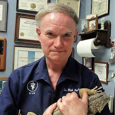 Walter Rosskopf is the owner of the Avian & Exotic Animal Hospital in Hawthorne, California. Dr. Rosskopf has been in avian and exotic animal practice since he graduated from the University of California at Davis in 1969, and he has pioneered numerous avian and exotic medical and surgical procedures. Dr. Rosskopf was one of the first clinicians to establish pet bird normal blood values and he has authored hundreds of scientific articles on avian medicine. Dr. Rosskopf was a Diplomate of the American Board of Avian Practitioners in avian practice from 1994-2014. He was also the President of the Association of Avian Veterinarians (AAV) from 1987-1988. In the 1980s and 1990s, Walter presented at annual AAV conferences over 65 times. He has also lectured internationally at over 100 veterinary conferences and veterinary medical schools. Dr. Rosskopf also taught avian and exotic animal medicine at Oregon State College of Veterinary for 37 years and at Mississippi State for 5 years. Dr. Rosskopf was the co-author of the seminal textbook Diseases Of Caged And Aviary Birds, an author and contributor to Veterinary Clinics of North America, and numerous other textbooks.
Fast Facts on Rabbit Hemorrhagic Disease
INTRODUCTION
Rabbit hemorrhagic disease (RHD) is a highly infectious, fatal viral hepatitis affecting lagomorphs (rabbits, hares, and pikas), including the European rabbit (Oryctolagus cuniculus).10,24 RHD is considered a reportable disease of international concern by the World Organization for Animal Health (formerly OIE).10,20
Rabbit hemorrhagic disease virus (RHDV) is a non-enveloped, single-stranded, positive-sense RNA virus belonging to family Caliciviridae and genus Lagovirus.2,10,22 Historically, RHDV split into six highly pathogenic genotypes (G1-G6), however in 2010, a new, antigenically distinct genotype emerged: rabbit hemorrhagic disease virus type 2 (RHDV2) (Box 1).9,24,25
| *RHDV1 and RHDVa are closely related |
|
The European brown hare syndrome virus is another lagovirus that causes RHD-like disease in hares (Lepus spp.).10,24
RHDV1 | RHDV2 |
|
|---|---|---|
GEOGRAPHIC RANGE | Rabbit hemorrhagic disease virus type 1 (RHDV1) was first reported in China in 1984, resulting in the loss of millions of rabbits in less than 1 year. 6,8,11,22 Two years later, RHDV1 was first reported in Europe. | In 2010, a new form of RHD, rabbit hemorrhagic disease virus type 2, emerged in France in wild and farmed rabbits. 6,7,8,18 |
| Since that time, RHD has been reported in over 40 countries, occurring regularly across parts of Europe, Asia, Africa, the Americas, and Oceania.21 Outbreaks have also occurred in the Middle East.21 | Within 5 to 6 years, RHDV2 had spread across Europe and reached Africa, Israel, Australia, New Zealand, and North America.3,6,7,18 RHDV-2 has now become the dominant subtype of RHD in endemic countries. | |
| Isolated cases have appeared intermittently in the US and Canada. | The first confirmed case of RHDV2 reported in North America was seen in Quebec in 2016.18 The first case in the US was seen in 2018. Since then, RHVD2 outbreaks have been seen sporadically throughout the US and Canada.7,9,18 | |
SIGNALMENT |
||
| Species | Wild and domestic European rabbits (O. cuniculus)1,7,18 | Wider host range, including many species of wild rabbits, such as hares (Lepus spp.) and cottontails (Sylvilagus spp.), pikas, and the domesticated European rabbit3,7,9,14,18 |
Since the emergence of RHDV2, viral RNA has been identified in a few rodent species, presumably through the ingestion of rabbit-infected tissues or feces during scavenging.1, 5, 15 Although virus was found in wild mice living near RHDV2 outbreaks, the mice did not show clinical signs of disease.15 |
||
| Age | Adult rabbits 7,21 | Adults as well as kits as young as 7 to 15 days old 20 |
| Zoonotic potential | RHD is not considered a food safety concern and is not associated with infection or disease in humans.12,21 |
|
TRANSMISSION | RHDV is extremely contagious. RHDV is easily transmitted through direct rabbit-to-rabbit contact with bodily fluids (urine, feces, respiratory secretions) or hair from an infected animal.7,21 Transmission mainly occurs by the oral route but nasal or conjunctival exposure is also possible.2,21 RHDV is extremely stable in the environment.2,7 Therefore exposure to fomites or mechanical vectors can transmit disease.7,21 Virus can also persist for months in decomposing carcasses in the environment as well as frozen infected rabbit meat.2,7,21 Importation of rabbit meat and byproducts may play an important role in the introduction of RHDV into new geographic regions.7,21 |
|
INCUBATION | ||
CLINICAL PICTURE |
||
| Mortality rate | 70-90% in adult rabbits 6,7,18,20 | RHDV2 strains have become progressively more virulent over time, ranging from 70-100%6,7,20 |
| Duration | Death usually occurs 12–36 hours after the onset of fever 7,18,20,21 | More prolonged period of illness before death (up to 5 days)16 |
| The clinical syndromes caused by RHDV and RHDV2 are similar.6,7,18 RHD often presents as peracute to acute disease. In peracute cases, the only clinical signs may be sudden death without premonitory clinical signs except for vocalizations followed rapidly by collapse and death.7,18 The acute form of disease often begins as fever (>40°C or 104°F) followed by a wide range of progressive clinical signs related to circulatory shock, liver failure, and disseminated intravascular coagulation or DIC: 2,7,8,18,20
Approximately 5 to 10% of rabbits display a subacute to chronic clinical course.1,21 The clinical picture is often similar to that seen with the acute form, but with less severe clinical signs:4,7,18,21
|
||
DIAGNOSIS | A presumptive diagnosis of RHD is often based on history, risk assessment, and clinical findings. American veterinarians that suspect RHD should contact their state veterinarian to receive instructions on testing and reporting. |
|
Minimum database 7,18 | Complete blood count |
|
Coagulation tests |
|
|
Biochemistry panel |
*Liver enzymes can be decreased 6 or elevated (M. Gleeson, written communication, Mar 18, 2023). |
|
Urinalysis |
|
|
| Gross necropsy lesions | Common necropsy lesions include pallor and a friable liver due to necrosis, splenomegaly, and epistaxis. There may also be evidence of disseminated intravascular coagulation, such as petechiation and hyperemia or hemorrhage of the trachea, lungs, heart, liver, spleen, and kidneys.2,7,21 It is also possible for there to be no gross necropsy findings in rabbits that die peracutely.14 |
|
| Histopathology | The most common histopathologic findings include extensive, multifocal to massive hepatocellular necrosis and multifocal to diffuse splenic necrosis. 7,18 Bronchopneumonia, pulmonary hemorrhage or pulmonary edema, and cardiomyocyte necrosis, may be seen in some subacute to chronic cases.7 |
|
| Laboratory testing | Reverse transcription (RT) PCR and antigen ELISA are the most commonly used tests to identify the presence of virus.2,6,7,20,21 Test results typically take only 1-5 business days, but unfortunately, many animals have already succumbed to the virus. Most animals will also die before an antibody response can be detected by Ab ELISA. Therefore testing is most helpful in caring for other animals at risk of exposure and preventing viral spread. PCR testing is best performed on fresh or frozen liver samples, but virus may also be detected in the spleen.2,7,20 Formalin fixed tissue can also be submitted for histopathology.7,21 For antemortem testing, PCR can be performed on urine, feces, or serum collected from convalescent rabbits up to 15 weeks post-infection but results can be inconsistent.7,20 Laboratories currently offering RHD testing include (but are not limited to):
Additional test methods that are used less commonly include electron microscopy, immunohistochemistry, Western blotting, in-situ hybridization, next-generation sequencing, and hemagglutination inhibition.2,7,20 **Plum Island currently does not offer antemortem testing because of inconsistent test results; Ab ELISA is performed for export purposes. |
|
| Definitive diagnosis | Like most caliciviruses, RHDV cannot be grown in cell culture. Therefore rabbit inoculation is necessary to isolate virus. This method is not used for routine diagnosis due to animal welfare concerns. 2,7,20,21 When sudden deaths of multiple rabbits occur within a household or collection, the most useful diagnostic test is necropsy and testing of samples from deceased rabbits. |
|
THERAPY | There is no specific treatment for RHDV, however supportive care can be rewarding, particularly in adult rabbits suffering from the subacute to chronic form of disease.7 |
|
PREVENTION AND CONTROL |
||
| Where RHD is absent... | Prevent the introduction of virus through restrictions on importation of animals or products from endemic areas.7,19 |
|
| When an outbreak occurs... | Eradicate disease through strict quarantine, depopulation, disinfection, and serosurveillance.7 Vaccination of all animals is also recommended as successful post-exposure prophylaxis has been seen.7 |
|
When disease is endemic... |
Cross protection between RHDV2 vaccines and RHDV/RHDVa vaccines is poor.6,7,20,21 Current recommendations in endemic countries is to vaccinate against both variants RHDVa and RHDV-2. |
|
SUMMARY
Rabbit hemorrhagic disease is a highly infectious, fatal viral hepatitis of lagomorphs caused by a calicivirus. There are three major pathogenic rabbit hemorrhagic disease virus genotypes: classical RHDV, RHDVa, and RHDV2. Rabbit hemorrhagic disease virus type 2 has now become the dominant subtype in endemic countries, and outbreaks affecting both domestic and wild rabbits have occurred in North America. Virus is easily transmitted through direct rabbit-to-rabbit contact with bodily fluids or hair through the oral, nasal, or conjunctival routes. Disease can also be transmitted through indirect contact with fomites or mechanical vectors. The incubation period often ranges from 1 to 6 days. While disease caused by RHDV1/RHDVa is restricted to domestic European rabbits, the host range is wider for RHDV2 and includes not only the European rabbit but several species of wild lagomorphs. Also, disease caused by RHDV1/RHDVa occurs primarily in adult rabbits, while disease caused by RHDV2 is seen in rabbits of all ages as young as 7-15 days old.
Rabbit hemorrhagic disease is characterized by high morbidity and high mortality. Clinical signs associated with acute disease can include neurologic and respiratory signs, non-specific signs of illness, jaundice, and bleeding. Death usually occurs 12–36 hours after the onset of fever. A presumptive diagnosis of RHD is often based on history, risk assessment, and clinical findings. When sudden deaths of multiple rabbits occur within a household or collection, the most useful diagnostic test is necropsy and testing of samples from deceased rabbits. Viral loads are particularly high in the liver and spleen. PCR is most commonly used to identify the presence of virus. Prevention and control of disease relies upon strict biosecurity protocols, stringent disinfection, and vaccination.
REFERENCES
Rabbit Hemorrhagic Disease Virus
The objective of this resource is to educate pet rabbit owners about rabbit hemorrhagic disease. The reader will review the distinction between rabbit hemorrhagic disease virus type 1 and type 2, as well as the basics of disease transmission, signs of disease, diagnosis, prevention and control.

Download the PDF version of this client education handout, or modify the DOCX version for your veterinary hospital.
Exotic Small Mammal Anesthesia
Exotic small mammals can be challenging to safely induce, maintain and recover from general anesthesia. View the recording of this RACE-approved webinar, which explores clinical anesthesia in exotic companion mammals from patient assessment and anesthetic induction to monitoring and recovery. This recording discusses the importance of proper physical examinations, the pharmacology of common anesthetic drugs or fluids, including the use of constant rate infusions and locoregional anesthesia techniques, intravenous and intraosseous catheterization, intubation techniques, and how to choose monitoring equipment in common companion exotic small mammal species . . .
Pour les vétérinaires. Par les vétérinaires.
Le site Lafervet.com est conçu pour une utilisation par les vétérinaires. Il est ouvert aux vétérinaires diplômés, aux techniciens vétérinaires diplômés, aux animaliers et aux étudiants dans ces domaines.
Créer un compte pour accéder à des articles et des ressources du site.
L'inscription est gratuite.
Para Profesionales Veterinarios. Por Profesionales Veterinarios.
El sitio Lafervet.com es para uso de los profesionales veterinarios. Está abierto a los veterinarios licenciados, técnicos veterinarios licenciados, rehabilitadores licenciados y estudiantes en estos campos.
Cree una cuenta para acceder a los artículos y recursos del sitio.
La registro es gratis.
Already a LafeberVet Member?
Please Login
Test Your Knowledge: Exotic Small Mammal Anesthesia
. . .
Pour les vétérinaires. Par les vétérinaires.
Le site Lafervet.com est conçu pour une utilisation par les vétérinaires. Il est ouvert aux vétérinaires diplômés, aux techniciens vétérinaires diplômés, aux animaliers et aux étudiants dans ces domaines.
Créer un compte pour accéder à des articles et des ressources du site.
L'inscription est gratuite.
Para Profesionales Veterinarios. Por Profesionales Veterinarios.
El sitio Lafervet.com es para uso de los profesionales veterinarios. Está abierto a los veterinarios licenciados, técnicos veterinarios licenciados, rehabilitadores licenciados y estudiantes en estos campos.
Cree una cuenta para acceder a los artículos y recursos del sitio.
La registro es gratis.
Already a LafeberVet Member?
Please Login
Reptile Case Challenge Teaching Module
Husbandry-related conditions are very common in reptiles. This case-based teaching module explores a condition frequently encountered by reptile veterinary health professionals. This teaching module is approved for 1 hour of continuing credit for veterinarians and veterinary technicians by the American Association of Veterinary State Boards (AAVSB) Registry of Approved Continuing Education (RACE . . .
Pour les vétérinaires. Par les vétérinaires.
Le site Lafervet.com est conçu pour une utilisation par les vétérinaires. Il est ouvert aux vétérinaires diplômés, aux techniciens vétérinaires diplômés, aux animaliers et aux étudiants dans ces domaines.
Créer un compte pour accéder à des articles et des ressources du site.
L'inscription est gratuite.
Para Profesionales Veterinarios. Por Profesionales Veterinarios.
El sitio Lafervet.com es para uso de los profesionales veterinarios. Está abierto a los veterinarios licenciados, técnicos veterinarios licenciados, rehabilitadores licenciados y estudiantes en estos campos.
Cree una cuenta para acceder a los artículos y recursos del sitio.
La registro es gratis.
Already a LafeberVet Member?
Please Login
Rabbit Hemorrhagic Disease and Vaccination

Photo credit: David-O via Flickr Creative Commons
Abstract
Rabbit hemorrhagic disease (RHD) is a usually fatal disease affecting almost exclusively lagomorphs caused by the rabbit hemorrhagic disease viruses (RHDV). While initial outbreaks of RHDV-1 occurred in Europe and Asia, RHDV-2 has emerged and spread across the globe within the span of the last 10 years. RHDV-2 has resulted in major outbreaks of RHD in wild and domestic rabbits in Washington State and the North American Southwest (USA and Mexico) since 2020.
RHDV-2 causes a severe necrotizing hepatitis in lagomorphs that results in secondary disseminated intravascular coagulation and death. Death is often peracute and other signs may not be recognized prior to death. RHDV-2 is a calicivirus that is environmentally stable and transmitted primarily by fomites via the oral-fecal route. Due to the ease in inadvertent exposure via fomites and the seriousness of the disease, vaccination is recommended as the primary means to prevent infection. Initial importation of unapproved European killed vaccines was allowed for affected states only and were difficult and expensive to acquire.
Medgene Labs has recently received United States Department of Agriculture Center for Veterinary Biologics (USDA-CVB) emergency use approval for their inactivated recombinant subunit protein vaccine, increasing availability and ease of acquiring vaccines across the country. Due to vaccine developments, vaccination against RHDV-2 for domestic rabbits is now widely available and strongly encouraged.
Outline
|
 Photo: Alex via Flickr Creative Commons
|
About the presenter
Dr. Amanda Jones is a consultant for Medgene Labs, and a supervising veterinarian for the Medgene Labs Texas Field Safety Site. Dr. Jones is also the owner of Central Texas Rabbit Herd Management in Killeen, Texas and an associate veterinarian for Animal Emergency Services of Killeen. Dr. Jones is a 2014 Cum Laude graduate of the Texas A&M College of Veterinary Medicine and… [MORE]
Webinar recording
PLEASE NOTE: RACE approval has expired for this course. STAY TUNED: Renewal of this program is pending.
Expert Q&A
Dr. Jones generously answered virtually every question entered during the live event. A few remaining questions were answered by email and are now posted below:
I have heard of some practices opening the vaccine, drawing it up into individually dosed syringes, and they will then use these syringes up to at least several days after they have been drawn into the syringes. Generally, with other vaccines, they are supposed to be discarded if they have been in the syringe over 24 hours, so I was curious if this is a safe way to try to use the vaccine over a longer period of time or if this is not recommended. If this is a possible solution to make the vaccine last longer, how long can they be kept in the syringes?
I would not recommend storing the vaccine in individual syringes over keeping it in the glass vial it comes in. We know that the vaccine is stable in the glass vial and that glass is unreactive. Plastic can occasionally bind proteins and no research has been done on the vaccine stability stored in a syringe. Without research to verify there is no impact on the vaccine, I would not recommend it as a general practice.
How do you handle a case where you have suspicion for this disease but unconfirmed and owner declines any treatment other than supportive and takes pet home to die later and was buried by owner.
Unfortunately, there isn’t much that can be done. If you are very suspicious, you could call the state vet for guidance and if they have additional resources to ensure proper testing.
At the USAHA meeting, Medgene gave a presentation that discussed some mortality cases (multiple but details were not provided on exact numbers) shortly after vaccination that was attributed to transport stress. Was curious how that information ties into the information provided today?
As I did not attend that presentation, I am not sure of exactly what was said. Medgene strived to have necropsies performed for any rabbit that died in temporal association with the vaccine. All necropsies I reviewed showed either an underlying condition that was responsible for death or were inconclusive due to advanced decomposition. There have been some cases where rabbits had episodes of GI stasis in close timing with vaccination that may have been associated with the stress of traveling, but those rabbits recovered with supportive care.
RACE approval has expired
This webinar recording was approved for 1 hour of continuing education credit for veterinarians and veterinary technicians in jurisdictions that recognize American Association of Veterinary State Boards (AAVSB) Registry of Approved Continuing Education (RACE) approval, however RACE approval has expired for this course.
References
Amanda Jones, DVM
 Dr. Amanda Jones is a consultant for Medgene Labs, and a supervising veterinarian for Medgene Labs Texas Field Safety Site. Dr. Jones is also the owner of Central Texas Rabbit Herd Management in Killeen, Texas and an associate veterinarian for Animal Emergency Services of Killeen. Dr. Jones is a 2014 Cum Laude graduate of the Texas A&M College of Veterinary Medicine and a 2010 Summa Cum Laude graduate from Eastern Kentucky University. Amanda’s professional affiliations include the American Veterinary Medical Association, Texas Veterinary Medical Association, and the Association of Exotic Mammal Veterinarians.
Lori Arent, MS
 Lori Arent is the Assistant Director at The Raptor Center (TRC), College of Veterinary Medicine at the University of Minnesota (MN). She has a Master’s of Science in Veterinary Physiology from the University of MN. Prior to her current role at TRC, she managed the rehabilitation of 800-1000 raptor patients annually, with a special focus on reconditioning and pre-release preparations. She has authored multiple resources, such as Raptors in Captivity: Guidelines for Care and Management, the resource used by the United States Fish and Wildlife Service for managing a permanent collection of education raptors, as well as the chapter on Avian Anatomy and Physiology in Clinical Anatomy and Physiology for Veterinary Technicians. Lori has also developed a suite of online training courses for raptor rehabilitation professionals and helps teach undergraduate courses and in-person workshops.
Amphibian Sedation and Anesthesia
RACE approval has expired for this webinar recording by Douglas Whiteside,DVM, DVSc, DACZM, DECZM (ZHM), however, renewal of this course is pending. Similar to other vertebrate species, sedation or anesthesia may be required in amphibians for various diagnostic, clinical, and surgical interventions. An understanding of clinically relevant anatomy and physiology, a pre-anesthetic protocol, the selection of appropriate anesthetic drugs to safely conduct a desired procedure, appropriate anesthetic monitoring, and post-anesthetic planning all are key to successfully managing the amphibian patient through an anesthetic event . . .
Pour les vétérinaires. Par les vétérinaires.
Le site Lafervet.com est conçu pour une utilisation par les vétérinaires. Il est ouvert aux vétérinaires diplômés, aux techniciens vétérinaires diplômés, aux animaliers et aux étudiants dans ces domaines.
Créer un compte pour accéder à des articles et des ressources du site.
L'inscription est gratuite.
Para Profesionales Veterinarios. Por Profesionales Veterinarios.
El sitio Lafervet.com es para uso de los profesionales veterinarios. Está abierto a los veterinarios licenciados, técnicos veterinarios licenciados, rehabilitadores licenciados y estudiantes en estos campos.
Cree una cuenta para acceder a los artículos y recursos del sitio.
La registro es gratis.
Already a LafeberVet Member?
Please Login
Care of Senior or Geriatric Domestic Rabbits
The objective of this educational resource is to empower owners on how to best care for senior or geriatric pet rabbits. The reader will learn how to ensure common geriatric conditions do not become a welfare concern for their pet. The goal is not to “cure” but rather manage geriatric diseases, such as cataracts, arthritis, and even cognitive decline.
Download the PDF version (American English) or PDF (British English version) of this client education handout, or modify the DOCX version (American English) or DOCX (British English) for your veterinary hospital.
Senior Pet Questionnaire
Quality-of-life questionnaires are an excellent tool for the veterinary team. This questionnaire can be completed in the waiting room or during consultation with a veterinary nurse. Use of a comprehensive history form paired with this quality-of-life questionnaire can greatly assist in beginning difficult conversations with owners of senior pets and managing geriatric patients. Many age-related diseases are manageable with correct and appropriate husbandry changes.
Download the PDF version of this client education handout, or modify the DOCX version for your veterinary hospital.
Note: This form was adapted from a Geriatric Questionnaire by Today’s Veterinary Practice.
Rabbit History Form
An in-depth clinical history can assist in providing the information needed for a successful treatment plan. Use this questionnaire to obtain a complete and thorough clinical history of your rabbit patient. If the pet rabbit is older, pair this form with the senior rabbit questionnaire, which is intended to evaluate quality of life. These forms can be completed in the waiting room or during consultation with a veterinary nurse.

Download the PDF version of this client education handout, or modify the DOCX version for your veterinary hospital.
Chloe McMenamin RVN, BSc VN, CH
 Chloe McMenamin is a lecturer of Applied Animal Health in the veterinary nursing department of Unitec Institute of Technology, which is part of Te Pūkenga Aotearoa New Zealand. Chloe earned a Bachelor of Science in veterinary nursing from University College Dublin, then came to Aotearoa New Zealand to work as an exotic and wildlife veterinary nurse. This is where Chloe developed a passion for educating owners and the public on all things furry, feathered, and scaly. Today, Chloe is lucky enough to be able to follow a passion for educating and empowering the next generation of veterinary nurses.
2022 T.J. Lafeber Avian Practitioner of the Year
Dr. Sharman Hoppes named Avian Practitioner of the Year
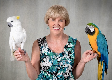
Twenty-four exceptional avian veterinarians from all around the world were nominated for the 2022 T.J. Lafeber Avian Practitioner of the Year. The independent Selection Committee narrowed this list to three finalists and the 2022 T.J. Lafeber Avian Practitioner of the Year, Dr. Sharman Hoppes, was announced during the Plenary Session at the 2022 ExoticsCon.

Sharman Hoppes, DVM, DABVP (Avian Practice) is the owner of Texas Avian & Exotic Hospital in Grapevine, Texas and a Professor Emerita of the zoological medicine service at Texas A&M University.
Share the news
Download and share the press release.
Award recipients
Visit Lafeber.com to learn more about Dr. Hoppes and to see a list of previous Award recipients.
Did you know…?
The T.J. Lafeber Avian Practitioner of the Year is nominated by their peers: YOU. The Awardee is NOT, and has never been, selected by Lafeber Company.
The autonomous Award Selection Committee, consisting of Association of Avian Veterinarians members, is led by Dr. Kenneth Welle.
Bird Basics Teaching Module
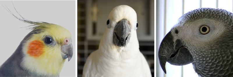
Objectives
What are the basics of avian medicine that a novice veterinary health professional should know before laying hands on the companion parrot patient? Upon completion of this learning aid, the participant will have a basic clinical understanding of avian anatomy, psittacine handling and restraint, history and physical examination, zoonotic concerns, nutrition, basic diagnostic and therapeutic techniques, as well as a few select presenting problems.
The content listed below barely scratches the surface of avian medicine. Use LafeberVet’s list of Avian Quick Links to assist your navigation of additional avian medicine resources on LafeberVet.
Syllabus
Anatomy & Physiology | Handling & Restraint |
|---|---|
|
|
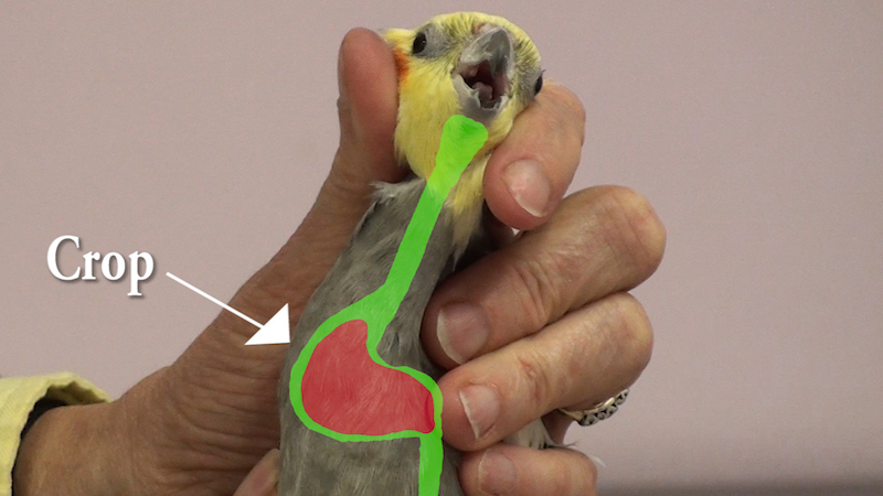 |  |
History & Exam | Zoonotic Concerns |
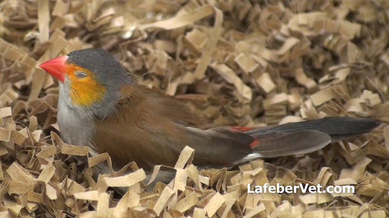 | 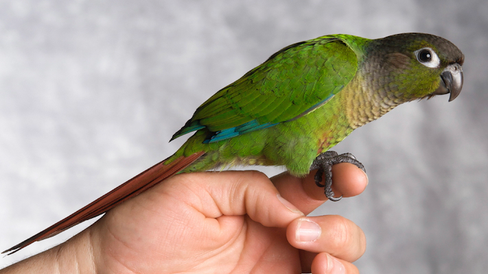 |
Housing & Nutrition | Supportive Care Techniques |
|
|
 | 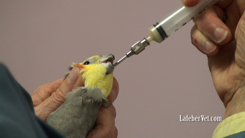 |
Minimum Database | Basic Techniques |
|
|
 Photo credit: Dr. Minh Huynh Photo credit: Dr. Minh Huynh | 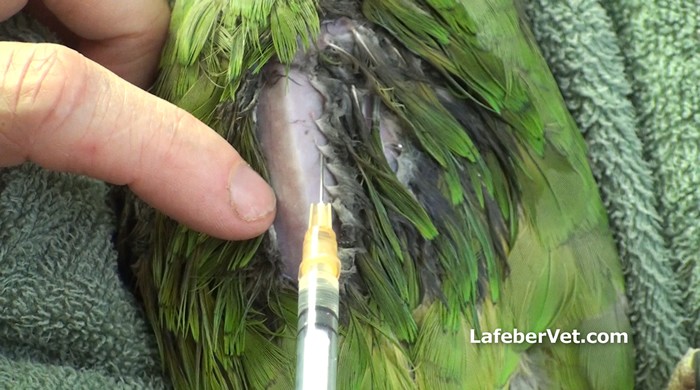 |
Select Clinical Problems |
|
Approval pending
Stay Tuned: This teaching module will be submitted for approval by the American Association of Veterinary State Boards for continuing education credit.
Avian Neonatology and Pediatrics
RACE approval has expired for this webinar recording by Lorenzo Crosta, med Vet, PhD, DECZM, EBVS European Veterinary Specialist in Zoo Health Management, however, renewal of this course is pending. The objective of this presentation was to assist the exotic animal practitioner, with little or no experience in avian neonatology and pediatrics. This presentation reviews the logical diagnostic plan in the young bird. The approach to common pediatric conditions, ranging from developmental and orthopedic problems to common traumatic injuries and infectious diseases, is also explored. Practical clinical examples are presented . . .
Pour les vétérinaires. Par les vétérinaires.
Le site Lafervet.com est conçu pour une utilisation par les vétérinaires. Il est ouvert aux vétérinaires diplômés, aux techniciens vétérinaires diplômés, aux animaliers et aux étudiants dans ces domaines.
Créer un compte pour accéder à des articles et des ressources du site.
L'inscription est gratuite.
Para Profesionales Veterinarios. Por Profesionales Veterinarios.
El sitio Lafervet.com es para uso de los profesionales veterinarios. Está abierto a los veterinarios licenciados, técnicos veterinarios licenciados, rehabilitadores licenciados y estudiantes en estos campos.
Cree una cuenta para acceder a los artículos y recursos del sitio.
La registro es gratis.
Already a LafeberVet Member?
Please Login
Avian Quick Links
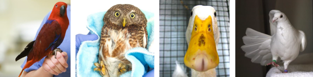
Photo credit: Jill Murray (left) Bothered by Bees (second from right), Ingrid Taylor (right)
Objectives
LafeberVet’s list of avian medicine links will assist your navigation of some of the avian medicine resources on LafeberVet. First, educate yourself before laying hands on the avian patient by reviewing the fundamentals of avian anatomy and physiology as well as important principles of handling and restraint. Then explore content on the examination and history, behavior, housing and nutrition, as well as supportive care techniques, diagnostics, basic therapeutics, and common presenting problems.
Start with content listed in the rows titled, “Begin with…“, then expand your knowledge with other sections, such as “Learn more...”.
Depending on the problem encountered, referral may be in your avian patient’s best interest. Visit LafeberVet’s Referring Non-Traditional Species for additional information.
Syllabus
Common Clinical Problems
| Important Avian Diseases | Taxonomic Groups |
|||
| ||||
| Non-Specific Signs of Illness | Traumatic injury |
|||
| Gastrointestinal Signs | Respiratory Signs |
|||
| Reproductive Tract Disease | Skin and/or Feather Related | |||
| ||||
Basic Information Sheet: Pionus
Pionus spp.
Natural history
Pionus spp. parrots are native to regions of Mexico, Central America, and/or South America. The specific range varies with the species (Table 1).4-7,13,15,18
| Table 1. Geographic range of select Pionus parrots 11,13,15,18 | |||
|---|---|---|---|
| Species | South America | Central America | Mexico |
| Scaly-headed parrot (Pionus maximiliani) | Northern Argentina, through Bolivia and Paraguay to eastern Brazil | ||
| Bronze-winged parrot (P. chalcopterus) | NW Venezuela, western Columbia, Ecuador, NW Peru | ||
| White-headed parrot (P. seniloides) | Mountains of western Venezuela through western Columbia into Andes in Ecuador to NW Peru | ||
| Dusky parrot (P. fuscus) | Extreme northern South America, including SE Venezuela, Guyana and NE Brazil | ||
| Blue-headed parrot (P. menstruus) | Northern South America (Columbia, Ecuador, Peru, SE Brazil, N Bolivia) except the Andes | Costa Rica, Panama | |
| White-crowned parrot (P. senilis) | East coast of central America to Costa Rica and western Panama | Eastern Mexico (mainly Caribbean slope) | |
Taxonomy
Class: Aves
Order: Psittaciformes
Tribe: Arini = New World parrots
Family: Psittacidae
Blue-breasted parrot (Pionus reichenowi)
Blue-headed, blue-hooded, or red-vented parrot (Pionus menstruus)
P. m. menstruus
P. m. rubrigularis (smaller, duller blue, some have red on throat)
P. m. reichenowi (deeper, more extensive blue coloration; horn-colored beak)
Bronze-winged parrot (P. chalcopterus)
Dusky or violet parrot (P. fuscus)
Red or coral-billed parrot (P. sordidus)
Scaly-headed (Maximilian’s) parrot (P. maximiliani)*
Subspecies vary mainly in the color of their upper breast
P. m. melanoblepharus (upper breast mainly darker blue)
P. m. siy (reddish purple)
P. m. lacerus (bluer reddish-purple)
Speckle-faced or plum-crowned parrot (P. tumultuosus)
White-headed, white-capped, or Massena’s parrot (P. seniloides)**
White-crowned parrot (P. senilis)
*Maximilian’s Pionus is a reference to Prince Maximilian of Wied-Neuwied, a nobleman and naturalist who explored southeastern Brazil in the early 19th century.21
** The white-headed or Massena’s parrot has sometimes been described as a subspecies of the plum-crowned parrot: Pionus tumultuosus seniloides (Warden). Some, more recent classification schemes list the white-headed parrot separately as P. seniloides.2
Conservation status
The International Union for Conservation of Nature (IUCN) Red List describes the blue-headed parrot (P. menstruus) population to be stable. The blue-headed parrot is common in a wide range of habitats over an immense area.15,18 Unfortunately, the populations of many other Pionus spp. are decreasing (Table 2). Although many species have a Red List assessment of “least concern”, the blue-breasted parrot (P. reichenowi) is listed as vulnerable by the IUCN.14
When compared to other South American parrots, Pionus spp. are not as common in the pet trade 3, however the blue-headed parrot (Pionus menstruus), white-crowned parrot (P. senilis), dusky parrot (P. fuscus), and bronze-winged parrot (P. chalcopterus) are relatively popular companion birds in the United States.3,4,6,16,22,24 The scaly-headed parrot (P. maximiliani) is also fairly common.7,16,18,22,24 Other Pionus spp., such as the red-billed parrot (P. sordidus), speckle-faced parrot (P. tumultuosus), and white-headed parrot (P. seniloides), are relatively uncommon to rare in aviculture.16,18
Pionus parrots are often described as “small Amazon parrots” because of their stocky builds and often short, square tails.4-7, These medium-sized parrots have a prominent, featherless cere, and a relatively large bill.3, Compared to some psittacine species, their coloration is often quite subdued or subtle, however, under bright lighting, their feathers shimmer with iridescence. Many Pionus also have large, bare eye rings and all species possess bright red undertail coverts.4-7,
| Table 2. Physical characteristics of select Pionus parrots 1,3-7,11,15,18,20,24 | |||||
|---|---|---|---|---|---|
| Pionus species | Blue-headed | Bronze-winged | Dusky | Scaly-headed | White-crowned |
| Click thumbnail image to enlarge |  |  |  |  |  |
| Body weight (average) grams | 230-260 4,18,24 234-295 3 | 180-210 5,18 265-275 24 | 180-230 3,6,18,24 | 180-210 7 230-293 3,18,24 | 180 18 220-230 3,24 |
| Length cm (in) | 23-28 (9-11) | 25-29 (10-11.5) | 24-28 (9-11) | 27-29 (10.5-11) | 24-26 (9-10) |
| Overall | Bright green body, deep blue head | Mostly dark blue-green or blueish-purple; green with bronze back | Mainly dark, brownish-gray | Varying shades of green, feathers are edged with bluish-gray | Mostly dark green and dark blue |
| Head | Bright sapphire blue | Green head; deep blue crown, back of head, upper head; pale chin patch | Dull blue-gray with red patch at base of the bill and purplish blue chin | Primarily brownish-green head feathers edged in gray to blue creating a subtle “scaly” appearance | Dark blue with white forehead and crown |
| Bill | Dark colored with red-pink patches at the base on either side of the upper bill | Pale yellow | Dark gray with a yellow patch at the base of the upper bill | Most birds having a two-tone or mottled beak: yellowish horn turning to dark gray at the base, pale tip | Yellow or pale-colored |
| Eye ring | Blue-tinged | Bare pinkish eye ring becomes dark peach to pink-orange (coral) in adults | Pale | Light colored | White to pale pink becoming pink-orange (coral) in adults |
| Ear coverts | Black | Dark blue to black | Black with streaky pale patch | Green | Dark blue |
| Throat | Bright sapphire blue with a pink-red base | White throat patch above variable pinkish-red patches; upper neck is deep blue | Indistinct or streaked dusky white to pinkish collar | Bluish-purple band running across ventral throat | White patch |
| Breast | Upper breast is green, tinged with olive brown and blue edging | Upper parts of breast dark blue; pale pink to dark peach and orange on upper breast | Brown barred with wine red or reddish-blue | Becoming lighter and more bronze-brown along ventrum | Dark green (upper breast) to bluish green (lower breast) |
| Undertail coverts | Bright red tipped with green | Bright red | Purplish red (tail feathers are also red below with a blue band) | Bright red | Pinkish-red |
| Tail | Cobalt blue | Bright violet | Green | ||
| Wings | Green | Dark bronze-green with iridescent aqua blue underwing coverts | Blue flight feathers with iridescent silver blue underwing coverts | Green with bronze shoulder patches in some individuals | Olive shoulder patch |
| Juvenile | Similar coloring with much less blue, some red on forehead | Less colorful than adults, dark blue-green with paler brown upper wing coverts, may have some red on the forehead and under the chin | Less obvious patches on the cheeks, may have some red on forehead; little or no streaky patch on neck; green on secondaries as well as flight feathers coverts | Less extensive blue on throat, less distinct margins on head | Narrow white band on forehead, green head faintly tinged blue; yellow-green undertail coverts chin, throat, breast/ yellowish-green undertail coverts |
Normal physiologic values
| Lifespan | 25-40 years, maximum 13,16,23 | |
| Puberty | 1.5-4 years 16 | |
| Body weight (average), grams 18 | Coral-billed or sordid parrot | 255 |
| White-headed or Massena’s parrot | 250 | |
| Plum-crowned parrot | 250 | |
| See Physical Description above |
||
Anatomy/physiology
| Musculoskeletal | As seen in other members of Order Psittaciformes, the Pionus parrot possesses a zygodactyl foot with two toes pointed back and two toes pointed forward. | ||
| Gastrointestinal | The normal Pionus spp. bill is longer than most other parrot bills, extending almost to the base of the lower bill or gnathotheca.3,13 The upper bill or rhinotheca also has a distinct notch.3 As seen in other members Order Psittaciformes:
|
||
| Respiratory | An interesting characteristic of Pionus spp. is the Pionus “snarfle, snuffle”, or wheeze, which is exhibited when individuals become upset or frightened.3,16 This display can be misinterpreted as a clinical sign of respiratory disease, but these clinical signs disappear when the bird once again feels safe and secure (see Behavior below for additional information).16,22,23 Pionus possess a prominent naked cere.3 As seen in other members of Order Psittaciformes, the right and left nasal sinuses communicate in Pionus spp. |
||
| Integumentary | Pionus parrots have a distinctive musky (or sweet) odor, that often becomes more prominent during the breeding season.3,16 It has been theorized that this odor serves to attract insects to the nest.16 The uropygial or preen gland is absent in the Pionus parrot.19 See Preventive Medicine (below) for grooming tips. |
||
| Reproductive | To the human eye, Pionus spp. are sexually monomorphic. The age of sexual maturity ranges between 1.5-4 years.16 The breeding age is approximately 3-5 years for blue-headed, scaly-headed, and dusky parrots.4,6,7 Breeding occurs one to two times per year.16 Both male and female birds may cannibalize young in the nest when the parents are visually threatened by larger neighboring species or if the parent birds are stressed by too-frequent nest-box inspection by human care takers or by the presence of predators or vermin.22 |
||
| Pionus parrot neonatology vital statistics 4-7,16,18 | ||||
| Species | Clutch size (ave) | Incubation (days) | Weight of newly hatched chick (g) | Fledging (weeks) |
| Scaly-headed | 3-4 7 4-5 18 | 26 | 9 | 9-10 |
| Bronze-winged | 3-4 | 24-27 | 9 | |
| Blue-headed | 3-4 | 26 | 8-10 | |
| White-crowned | 4-6 | 24-26 | n/a | 8 |
| Dusky | 4 | 24-26 | 9 | 9-10 |
Diet
A healthy diet for a companion Pionus parrot should consist of a formulated diet, supplemented with fresh, vitamin A-rich vegetables and some fruit.22 If table food is offered, the owner should be particularly cautious of dietary fat intake.22 Clinically, these birds are similar to Amazon parrots as they may be prone to obesity, hypercholesterolemia, and other adverse effects associated with high dietary fat.22
A study of free-ranging scaly-headed parrots in Brazil found their diet consisted mostly of native seeds (70.4%), followed by flowers (20.3%), corn from farms near their forest habitat, and fruit pulp.12
Husbandry
Although the enclosure should be as large as possible, at minimum, a Pionus parrot should be provided with a medium-sized parrot cage measuring at least 50 x 60 x 100 cm (20 x 24 x 40 inches).16 Bar spacing should be 2.2 cm (7/8-in) for most Pionus species.16 Smaller birds, such as dusky parrots and white-crowned parrots, should have no more than 1.8 cm (3/4-in) space between bars.16
Bathing is a favorite activity and daily misting, showers, or baths is recommended.
Personality
Pionus parrots tend to have a gentle disposition.3,13,16,23 Pionus have a reputation for being relatively quiet, or least less noisy than larger parrots, but of course Pionus spp. are capable of making loud vocalizations, particularly during the early morning and late afternoon hours or during the breeding season.4-7,16,22,23 Males are believed to be more vocal than females.16 The dusky parrot is also thought to be the noisiest of Pionus spp.3
Pionus also tend to be shy birds.4-7,13 They are often nervous in a hospital setting but are generally not as aggressive or territorial as mature Amazon parrots.17,22
Pionus parrots are relatively independent companion birds.16 They usually do not seek or accept a large amount of handling or cuddling.16 The scaly-headed parrot is believed to be the most sedate or easy going Pionus species.16 The white-crowned parrot (Pionus senilis) may be the most aggressive Pionus spp., requiring a more experienced owner.3,16
As with most psittacine birds, aggression may be more likely to occur during the breeding season.16 An aggressive Pionus may ‘strut’, fanning its tail feathers and raising its head feathers as it slowly stalks back and forth.16 A bird exhibiting this display may bite if approached.16
Response to stress
Pionus parrots are unique among parrots in their response to external stressors.16 When frightened or upset, Pionus produce respiratory noises that can be misinterpreted as snorting and snuffling, wheezing, rattling, hyperventilation, or dyspnea but these signs resolve within minutes when the stressful stimulus is removed.3,16,22 These sounds can be disconcerting for even the experienced avian veterinarian 3, but there should be no nasal discharge or other physical signs of disease.22 This respiratory noise is also produced with the mouth closed.17,23
Mimicry
Pionus parrots are considered intelligent birds but they are not considered the “best talkers”.13,16,23 These birds tend to mimic household sounds or repeat words in a somewhat garbled fashion.13,16 Pionus spp. may also speak in “an exceptionally soft voice”.16 Among Pionus spp., scaly-headed, dusky, and white-crowned parrots are often considered the best talkers.3
Restraint
Pionus may be restrained by holding the head between index and middle fingers. Support the body with palm of the hand as well as the thumb and little finger. Beware of the Pionus “snuffle” (See Behavior above).
Venipuncture
Use a 25-gauge needle and 1 to 3-ml syringe to draw blood from the right jugular vein. Collection of up to 1% of body weight is acceptable in healthy patients.
Important medical conditions
Non-infectious disease
As in Amazon parrots, obesity or malnutrition is a common disorder in Pionus spp.4-7,16,22 Hepatic lipidosis may develop, which can progress to cirrhosis and fibrosis of the liver.22
Like any neotropical parrot, Pionus parrots are also susceptible to respiratory aspergillosis.4-7,16,22
Infectious diseases
Depending on environmental conditions, sinus infections and chronic upper respiratory disease can be common, particularly when the diet is deficient in vitamin A.4-7,22 Bacterial, fungal, and mycoplasma infections are all common causes of chronic upper respiratory diseases in Pionus parrots.22
Like other New World psittacine birds, Pionus spp. are susceptible to Chlamydophila.4-7,16,22
Reported conditions
- A non-Candida albicans species, C. krusei, was identified in six birds exhibiting clinical signs associated with diarrhea, regurgitation, and melena, including one white-crowned parrot.9
- Mycobacteriosis involving the gastrointestinal tract is reportedly seen with greatest frequency in Pionus and Amazona species.10
- Fatal proventricular dilatation disease was described in one captive scaly-headed parrot in Brazil.8
- Prior to 1992 when psittacine birds were commonly imported (or smuggled) into the United States, avian pox virus was a very common finding in Pionus parrots.4-7,22
- Avian polyomavirus infection is often fatal in pre-weaning aged chicks.22
- Feather damaging behavior 4-7
- Toe necrosis 4-7
Preventive medicine
- Obtain a complete history and perform a thorough annual physical examination.
- Establish baseline data with regular clinical testing (complete blood count, protein electrophoresis, and plasma biochemistries.
- Ensure proper husbandry.
- Pionus spp. often fall and injure themselves with severe wing trims. Clip only the primary flight feathers and only enough so the bird can glide to the floor.4-7
For additional information, visit the review article Grooming Companion Birds and the RACE-approved webinar recording Flight Mechanics and Ethical Concerns.
- Recommend quarantine of newly acquired birds.
- Perform additional testing for select diseases based on history and physical exam findings: avian polyomavirus and chlamydiosis.
- Determine the origin and history of newly acquired sick birds to contain and prevent further spread of disease.
- Birds housed in large groups or aviaries are at higher risk of Pacheco’s disease virus and use of the vaccine may be indicated.
- The avian polyomavirus vaccine is recommended for breeding populations.
References
Natasha Omar
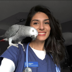
Natasha Omar was born in Nairobi, Kenya, where she lived for 10 years, before moving to Canada in 2012. Since childhood, Natasha has been exposed to all kinds of creatures, from elephants to our beloved domestic pets. Natasha has worked as a veterinary medical assistant in general practice for 4 years and she is now in the process of becoming a registered veterinary technician at Northern Alberta Institute of Technology in Edmonton, Alberta, Canada. Her interests include emergency and exotic animal medicine.
Marianne Wikdahl, RVT
 Marianne Wikdahl graduated with a Bachelor of Science with honors in Veterinary Nursing and Practice Administration from the University of Bristol (UK) in 2011. During her time at university she had the privilege to work as a student veterinary nurse in one of South West England’s largest referral hospitals. After graduation, Marianne moved to Canada where she embarked on her career as an ER/Exotics registered veterinary technician in a busy emergency hospital in Calgary, Alberta. In 2017, Marianne returned to her home country Norway, and is currently working at Sveberg Dyrehospital, a small animal clinic outside of Trondheim. Marianne has a special interest in exotic mammal medicine and has worked with a wide range of exotic species throughout her career so far. She is currently studying the Advanced Programme in Veterinary Nursing of Small Mammals (Girling & Fraser), which she aims to complete in 2022.
Dental Health in Rabbits
This guide to keeping your rabbit’s teeth healthy was awarded first place in the 2022 second annual Association of Exotic Mammal Veterinarians (AEMV) Veterinary Technicians Client Education Materials Contest, sponsored by Lafeber Company.
This client education handout explores the basics of rabbit dental anatomy, before focusing on what owners can do to look after their rabbit’s teeth. Some common dental conditions are also discussed as well as the signs of dental disease in the rabbit.
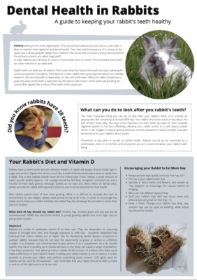
Download the PDF version of this client education handout, or modify the DOCX version for your veterinary hospital.
Chinchilla Nutrition
What to Feed Your Chinchilla:
This client education handout explores the recommendations for a healthy chinchilla diet, including hay, hay-based pellets, produce and greens, and water. The type and amount of treats to offer is also discussed.
This client educational material was awarded first place in the 2022 Association of Exotic Mammal Veterinarians (AEMV) Veterinary Technology Student Client Education Materials Contest, sponsored by Lafeber Company.
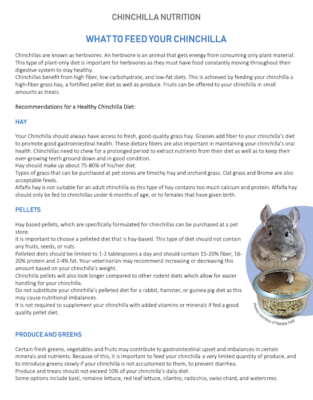
Download the PDF version of this client education handout, or modify the DOCX version for your veterinary hospital.
Marco Di Giuseppe, DVM, PhD, GPCert (ExAP)
 Marco Di Giuseppe currently works as freelance veterinarian in many clinics in Palermo (Italy) offering referral services in rabbit medicine. In the meantime, he is completing a program at the Italian Post-graduate Veterinary School of the Universityof Naples (Italy) to receive the title of Italian Specialist in Laboratory Animals. In 2014, Dr. Di Giuseppe obtained the title of General Practitioner Certificate in Exotic Pet Practice from the European School of Advanced Veterinary Studies. In the same year, he earned a Doctorate in Veterinary Medicine at the University of Padua (Italy) with a thesis on Encephalitozoonosis cuniculi in rabbits. Dr. Di Giuseppe has been an accredited veterinarian in Exotic Mammals, Reptiles and Amphibians Medicine from the Italian Board of Veterinarians since 2014 . In 2013 he founded the first exotic animal clinic in Palermo. Marco is also coauthor of an Italian textbook on rabbit medicine Medicina e Chirurgia del Coniglio da Compagnia
and he is the author of many scientific works on exotic animals with particularly emphasis on rabbit and reptile medicine.
Anneliese Strunk, DVM, DABVP (Avian Practice)
 Anneliese Strunk is the Residency Director and a senior associate at The Center for Bird & Exotic Animal Medicine in Bothell, Washington. Dr. Strunk obtained her undergraduate degree in Biomedical Science from Texas A&M University (TAMU) in 1995, graduating Magna Cum Laude from TAMU College of Veterinary Medicine in 2000. Anneliese spent her first year out of school as an associate at the Research Boulevard Pet and Bird Hospital in Austin, Texas, seeing small animal and exotic cases. Dr. Strunk then completed an internship in exotic animal, wildlife and zoo medicine at the. University of Georgia in 2002, followed by a 3-year residency in companion avian and exotic pet medicine at the University of California at Davis. After completing her residency, Dr. Strunk moved across the country to help start the Avian and Exotics Department at Red Bank Veterinary Hospital in Tinton Falls, New Jersey, where she saw primary care and referral avian and exotics cases. In 2009, she returned to Texas to re-join the team at Research Boulevard Pet and Bird Hospital as the head of Avian and Exotic medicine. She achieved ABVP board certification in Avian Practice in 2010 and has has been enjoying the Pacific Northwest since 2014.
Dr. Strunk is the author and co-author of a number of articles in both professional journals and national conference proceedings. Anneliese is active in both the Association of Avian Veterinarians and the Association of Reptile and Amphibian Veterinarians, and has presented lectures at and been an instructor of hands-on workshops at national and international meetings and veterinary schools.
Alyssa Scagnelli, DVM
 Alyssa Scagnelli is an associate veterinarian at the Avian and Exotic Animal Clinic (AEAC) in Mesa, Arizona. Dr. Scagnelli earned her Doctorate of Veterinary Medicine from the University of Missouri College of Veterinary Medicine in 2015. After graduation, she completed two academic internships at Louisiana State University (LSU) School of Veterinary Medicine. Her last year at LSU was entirely devoted to companion, wildlife, zoo, and aquarium medicine and conservation. In addition to working at AEAC, Dr. Scagnelli is also adjunct faculty at Midwestern University School of Veterinary Medicine in Glendale, Arizona. As adjunct, her predominant focus is on clinical research as it pertains to exotic animal medicine. Alyssa is an active member of the International Iguana Foundation and also volunteers for Arizona’s largest wildlife rehabilitation center. She is most passionate about reptile and aquatic animal medicine. Dr. Scagnelli has published several peer-reviewed journal articles and book chapters on various exotic animal medicine topics.
Byron de la Navarre, DVM
 Byron de la Navarre is chief of staff at Animal House of Chicago, Complete Veterinary Care, which he opened in 2000. Byron attended Cornell University as an undergraduate, where he completed Honors Research in the captive husbandry and breeding of peregrine falcons at the Cornell Lab of Ornithology. Byron then attended the veterinary college at Cornell, earning his DVM degree in 1990. While in veterinary school, he externed at over 12 zoos in North & Central America, Europe, and Israel. After completing an internship, Dr. de la Navarre worked in private practice, including Niles Animal Hospital & Bird Medical Center in the Chicago area for over 7 years.
Dr. de la Navarre has lectured on exotic pet topics at the local, state, national & International levels. Locally, he has served as the President of the Chicago Veterinary Medical Association (CVMA) & on the AVMA’s Membership & Services Committee. Byron is also on the Executive Board of the Association of Reptile and Amphibian Veterinarians (ARAV) and served as the ARAV President in 2005. He has also been active in and served as President of the (AAV) as well as the Association of Exotic Mammals (AEMV).
Reptile Venipuncture Teaching Module
Hematology and biochemistry results serve as an important part of the minimum database for all veterinary patients. Use three videos on blood collection in lizards, snakes, and chelonians (or text with still images) plus one slideshow on general principles to better understand reptile venipuncture and to improve your ability to obtain a clinically useful sample. RACE approval has expired for this teaching module, however, renewal of this course is pending . . .
Pour les vétérinaires. Par les vétérinaires.
Le site Lafervet.com est conçu pour une utilisation par les vétérinaires. Il est ouvert aux vétérinaires diplômés, aux techniciens vétérinaires diplômés, aux animaliers et aux étudiants dans ces domaines.
Créer un compte pour accéder à des articles et des ressources du site.
L'inscription est gratuite.
Para Profesionales Veterinarios. Por Profesionales Veterinarios.
El sitio Lafervet.com es para uso de los profesionales veterinarios. Está abierto a los veterinarios licenciados, técnicos veterinarios licenciados, rehabilitadores licenciados y estudiantes en estos campos.
Cree una cuenta para acceder a los artículos y recursos del sitio.
La registro es gratis.
Already a LafeberVet Member?
Please Login
Charles Cummings, DVM, CertAqV
 Charles Cummings is a Postdoctoral Research Fellow at the Tufts University Clinical and Translational Science Institute, where he focuses on the clinical and epidemiological aspects of zoonotic disease, particularly at the interface of humans and wildlife. From July 2019 to June 2020, Dr. Cummings served as a zoological medicine intern at Louisiana State University School of Veterinary Medicine. Charles earned his Doctorate from Tufts University Cummings School of Veterinary Medicine in 2019. He is also a Certified Aquatic Veterinarian through the World Aquatic Veterinary Medicine Association. He earned a Bachelor of Science in Fisheries and Wildlife from the University of Minnesota in 2015.
Sue Chen, DVM, DABVP (Avian Practice)
Dr. Patricia (Sue) Chen is board certified in Avian Practice by the American Board of Veterinary Practitioners and is the department lead for the avian and exotics department at Gulf Coast Veterinary Specialists. Dr. Chen earned her DVM from Texas A&M College of Veterinary Medicine, where she created a specialized veterinary curriculum track focused on exotic animals. Sue spent time training at zoos and wildlife sanctuaries in Dallas, Seattle, and Australia, as well as a pathology rotation at the Smithsonian’s National Zoo in Washington, D.C. After veterinary school, Dr. Chen completed a specialty internship at Gulf Coast Veterinary Specialists, followed by a residency in avian and exotic pet medicine and surgery at the Animal Medical Center in New York. Dr. Chen has authored publications on avian and exotic medicine and she has a special interest in the use of hyperbaric oxygen therapy in exotic species. In her free time, she enjoys gardening, cooking, and hiking with her family.
2022 AEMV Veterinary Technology Contest
Introduction
Lafeber Company was proud to sponsor the Association of Exotic Mammal Veterinarians (AEMV) Veterinary Technology Client Education Materials Contest. Credentialed veterinary technicians, veterinary technologists, veterinary nurses, as well as students in this field were encouraged to submit a two-page, English-language educational handout (1500 words or less) about a companion exotic mammal health and wellness topic.
Submissions closed April 1. The AEMV Technician Committee evaluated this educational material and they were blinded to the identify of each veterinary technologist or student.
Winners
| 1st Place Technologist
Marianne Wikdahl |
|
| 1st Place Technology Student
Natasha Omar Northern Alberta Institute of Technology |
Chinchilla Nutrition: What to Feed Your Chinchilla
|
Prizes
| 1st Place Technician/Technologist (estimated value $130 USD)  |
3M™ Littmann® Classic II Pediatric Stethoscope Nursing kit: pouch with bandage scissors, hemostat, suture scissors 1 year of FREE Association of Exotic Mammal Veterinarians membership |
| 1st Place Technician/Technology Student (estimated value $130 USD)
|
3M™ Littmann® Classic II Pediatric Stethoscope Nursing kit: pouch with bandage scissors, hemostat, suture scissors 1 year of FREE Association of Exotic Mammal Veterinarians membership |
Learn more
Interested in learning more about exotic companion mammal medicine? Visit AEMV.org or email [email protected] for more information about AEMV Technician Membership, as well as Student Membership and the Student Chapter Program.
Blood Collection in Turtles and Tortoises
Successful venipuncture can be a challenge in turtles and tortoises, however hematology and biochemistry results serve as an important part of the minimum database in chelonians just as they do for all veterinary patients. Use this video, or text with still images, to review the equipment needed and sample handling recommendations as well as the potential complications and proper approach to the jugular vein, brachial vein, subcarapacial vessel, and dorsal coccygeal sinus in the chelonian . . .
Pour les vétérinaires. Par les vétérinaires.
Le site Lafervet.com est conçu pour une utilisation par les vétérinaires. Il est ouvert aux vétérinaires diplômés, aux techniciens vétérinaires diplômés, aux animaliers et aux étudiants dans ces domaines.
Créer un compte pour accéder à des articles et des ressources du site.
L'inscription est gratuite.
Para Profesionales Veterinarios. Por Profesionales Veterinarios.
El sitio Lafervet.com es para uso de los profesionales veterinarios. Está abierto a los veterinarios licenciados, técnicos veterinarios licenciados, rehabilitadores licenciados y estudiantes en estos campos.
Cree una cuenta para acceder a los artículos y recursos del sitio.
La registro es gratis.
Already a LafeberVet Member?
Please Login
2022 AEMV Veterinary Medical Student Case Report Contest

Introduction
Lafeber Company was proud to sponsor the Association of Exotic Mammal Veterinarians (AEMV) Veterinary Medical Student Case Report Contest. Veterinary medical students from all over the world were encouraged to write a 2-page case report (1500 words or less) about an exotic companion mammal seen at their college of veterinary medicine or during a clinical experience.
Submissions closed April 1, 2022. Judges from the AEMV Student Committee evaluating the case reports were blinded to the students, mentors, co-authors, and institutions at which the cases were seen.
Posted below are brief summaries of each winning case report. Each student has been encouraged to submit their paper for peer-reviewed publication.
First place
Typhanie Tan (Student – Université de Montréal, Quebec):
Rabbit gastrointestinal stasis isn’t as straightforward as it seems: unilateral urinoma with ipsilateral hydroureteronephrosis in a domestic rabbit (Oryctolagus cuniculus)
A two-year-old neutered female rabbit (Oryctolagus cuniculus) presented for gastrointestinal stasis of one-day duration. Upon physical examination, a 5-6 cm, mobile, firm abdominal mass was noted cranially to the urinary bladder. Bloodwork was within normal limits. On abdominal tomodensitometry with contrast, a severe hydronephrosis with ipsilateral hydroureter were noted. A voluminous peritoneal cavitary mass attached to the right ureter was non-contrast enhanced. Gastrointestinal stasis was treated medically and an exploratory laparotomy was scheduled. The right kidney and ureter, along with the ureteral mass, were successfully excised and sent for histopathological and bacteriological analysis. A urinoma associated with a severe unilateral hydronephrosis and ipsilateral moderate hydroureter was diagnosed. No associated infection was confirmed. The rabbit recovered uneventfully and recheck bloodwork was unremarkable two-months post-surgery. To the authors’ knowledge, this is the first report of urinoma in a rabbit.
Second place
Ashleigh Pienaar (Student – University of Pretoria, South Africa):
Successful treatment of intestinal obstruction due to lymphoma in a rabbit (Oryctolagus cuniculus)
A 2-year-old female spayed rabbit (Oryctolagus cuniculus) was presented with sudden-onset anorexia and lethargy. Physical examination findings included hypothermia, a distended stomach, and a firm mass in the abdomen caudal to the stomach. Radiographs showed severe gastric dilation and distension of the intestines with gas, which was consistent with the presence of an obstruction. An exploratory laparotomy revealed a large, firm mass in the mid-duodenum. The intestines were resected to remove the mass, and an end-to-end anastomosis was performed. Histopathological examination identified the mass as a lymphoma. The patient was treated with intravenous crystalloid fluids, lignocaine, buprenorphine and meloxicam during the surgery. Post-operatively the patient was treated with buprenorphine for 2 days, as well as meloxicam, metoclopramide, cisapride, and enrofloxacin for 7 days. Support feeding with EmerAid Intensive Care Herbivore was done for the first week post-operatively. Six weeks after the surgery, the patient had fully recovered and showed no signs of metastasis or impaired gastrointestinal function. Based on a literature search, the case presented in this report is one of the first described cases of intestinal obstruction due to lymphoma that was successfully treated surgically.
Third place
Jared Zion (Student – Cornell University, USA):
Insulinoma in a domestic rabbit (Oryctolagus cuniculus domesticus)
A 5-year-old male intact rabbit (Oryctolagus cuniculus domesticus) in a multi-rabbit household presented on an emergent basis for clinical signs consistent with gastrointestinal (GI) stasis. A conservative treatment protocol was implemented but due to the lack of improvement of clinical signs, further diagnostics were employed. Serially low blood glucose values led to the prioritization of synthetic liver failure, sepsis, and insulinoma as differential diagnoses. In conjunction with the hypoglycemic readings, an insulin assay was performed. An abdominal ultrasound revealed a hyperechoic pancreatic nodule. Despite aggressive medical management, the patient failed to clinically improve and the prognosis for surgical success was considered poor. Euthanasia was elected. A necropsy was performed, and the approximately 0.5 cm pancreatic mass was submitted for histopathology. The results confirmed an islet cell pancreatic tumor of insulin origin.
Prizes
| 1st Place: | $100 USD cash prize
Carpenter JW (ed). Exotic Animal Formulary, 5th ed. Elsevier, 2017. Quesenberry KE, Orcutt CJ, Mans C, Carpenter JW (eds). Ferrets, Rabbits, and Rodents: Clinical Medicine and Surgery, 4th ed. St. Louis: Elsevier; 2021. |
| 2nd Place: | $100 USD cash prize
Carpenter JW (ed). Exotic Animal Formulary, 5th ed. Elsevier, 2017. |
| 3rd Place: | $100 USD cash prize |
Permission and privacy concerns
Permission was obtained from the supervising clinician (required) and the owner (optional depending on local privacy laws or facility standards).
Learn more
Interested in learning more about exotic companion mammals? Visit AEMV.org or email [email protected] for more information.

Avian Cardiology Essentials
RACE approval has expired for this webinar recording by Dr. Brenna Fitzgerald, however, renewal of this course is pending. Cardiovascular disease commonly occurs in companion birds and poses a serious threat to the quality of life and longevity of many avian species. Topics discussed in the webinar recording include key features of avian cardiovascular anatomy and physiology, risk factors, as well as specific clinical disease states, including atherosclerosis, congestive heart failure, and pericardial disease and effusion. This lecture also reviewed essential elements of a diagnostic workup, and current treatment approaches that can improve longevity and quality of life for these . . .
Pour les vétérinaires. Par les vétérinaires.
Le site Lafervet.com est conçu pour une utilisation par les vétérinaires. Il est ouvert aux vétérinaires diplômés, aux techniciens vétérinaires diplômés, aux animaliers et aux étudiants dans ces domaines.
Créer un compte pour accéder à des articles et des ressources du site.
L'inscription est gratuite.
Para Profesionales Veterinarios. Por Profesionales Veterinarios.
El sitio Lafervet.com es para uso de los profesionales veterinarios. Está abierto a los veterinarios licenciados, técnicos veterinarios licenciados, rehabilitadores licenciados y estudiantes en estos campos.
Cree una cuenta para acceder a los artículos y recursos del sitio.
La registro es gratis.
Already a LafeberVet Member?
Please Login
Blood Collection in Lizards
Hematology and biochemistry results are an important part of the minimum database for all veterinary patients, including lizards. Proper venipuncture technique is critical for accurate interpretation of laboratory results. Blood samples are most frequently collected from the ventral coccygeal vein and jugular vein in lizards; however, the site selected can depend on a variety of factors including the preferences and experience of the phlebotomist, the volume of blood needed, patient size and temperament, and of course the species involved . . .
Pour les vétérinaires. Par les vétérinaires.
Le site Lafervet.com est conçu pour une utilisation par les vétérinaires. Il est ouvert aux vétérinaires diplômés, aux techniciens vétérinaires diplômés, aux animaliers et aux étudiants dans ces domaines.
Créer un compte pour accéder à des articles et des ressources du site.
L'inscription est gratuite.
Para Profesionales Veterinarios. Por Profesionales Veterinarios.
El sitio Lafervet.com es para uso de los profesionales veterinarios. Está abierto a los veterinarios licenciados, técnicos veterinarios licenciados, rehabilitadores licenciados y estudiantes en estos campos.
Cree una cuenta para acceder a los artículos y recursos del sitio.
La registro es gratis.
Already a LafeberVet Member?
Please Login
Blood Collection in Snakes
Proper patient handling, blood collection technique and sample handling are all critical for accurate interpretation of hematology and biochemistry in all patients, including snakes. Use the video or text with still images to review equipment needed as well as the potential complications and proper approach to the ventral coccygeal vein and the heart, the two most common venipuncture sites in the snake . . .
Pour les vétérinaires. Par les vétérinaires.
Le site Lafervet.com est conçu pour une utilisation par les vétérinaires. Il est ouvert aux vétérinaires diplômés, aux techniciens vétérinaires diplômés, aux animaliers et aux étudiants dans ces domaines.
Créer un compte pour accéder à des articles et des ressources du site.
L'inscription est gratuite.
Para Profesionales Veterinarios. Por Profesionales Veterinarios.
El sitio Lafervet.com es para uso de los profesionales veterinarios. Está abierto a los veterinarios licenciados, técnicos veterinarios licenciados, rehabilitadores licenciados y estudiantes en estos campos.
Cree una cuenta para acceder a los artículos y recursos del sitio.
La registro es gratis.
Already a LafeberVet Member?
Please Login
Brenna Fitzgerald, DVM, DABVP (Avian Practice)
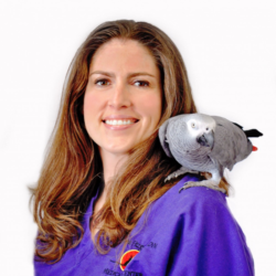
Dr. Brenna Fitzgerald is currently an associate veterinarian at Colorado Exotic Animal Hospital in Denver, Colorado as well as the owner of Avian Exclusive Veterinary Consultation. Dr. Fitzgerald is a 2007 graduate of Texas A&M University College of Veterinary Medicine. She then worked as an associate veterinarian in a private, multi-doctor practice in Dallas, Texas, where she practiced small animal, avian, and exotic animal medicine. Dr. Fitzgerald then completed both a clinical internship and residency at Dr. Brian Speer’s Medical Center for Birds in Oakley, California. She earned Diplomate status through the American Board of Veterinary Practitioners in Avian Practice in 2012. Dr. Fitzgerald stayed on as an associate at the Medical Center for Birds through 2021. She relocated to the Denver area and spent the first half of 2022 providing specialty care to avian patients at Homestead Animal Hospital in Centennial, Colorado. She has since joined the team at Colorado Exotic Animal Hospital where she continues to practice specialty avian medicine. Dr. Fitzgerald is strongly committed to furthering avian medicine, not only through serving her patients and clients in private practice, but also through education and support of the avian veterinary community. She has worked to promote continuing education by speaking at veterinary conferences, designing their educational programs, and authoring a number of scholarly articles.
Test Your Knowledge: Mediterranean Tortoises
. . .
Pour les vétérinaires. Par les vétérinaires.
Le site Lafervet.com est conçu pour une utilisation par les vétérinaires. Il est ouvert aux vétérinaires diplômés, aux techniciens vétérinaires diplômés, aux animaliers et aux étudiants dans ces domaines.
Créer un compte pour accéder à des articles et des ressources du site.
L'inscription est gratuite.
Para Profesionales Veterinarios. Por Profesionales Veterinarios.
El sitio Lafervet.com es para uso de los profesionales veterinarios. Está abierto a los veterinarios licenciados, técnicos veterinarios licenciados, rehabilitadores licenciados y estudiantes en estos campos.
Cree una cuenta para acceder a los artículos y recursos del sitio.
La registro es gratis.
Already a LafeberVet Member?
Please Login
Test Your Knowledge: To Cut or Not to Cut?
. . .
Pour les vétérinaires. Par les vétérinaires.
Le site Lafervet.com est conçu pour une utilisation par les vétérinaires. Il est ouvert aux vétérinaires diplômés, aux techniciens vétérinaires diplômés, aux animaliers et aux étudiants dans ces domaines.
Créer un compte pour accéder à des articles et des ressources du site.
L'inscription est gratuite.
Para Profesionales Veterinarios. Por Profesionales Veterinarios.
El sitio Lafervet.com es para uso de los profesionales veterinarios. Está abierto a los veterinarios licenciados, técnicos veterinarios licenciados, rehabilitadores licenciados y estudiantes en estos campos.
Cree una cuenta para acceder a los artículos y recursos del sitio.
La registro es gratis.
Already a LafeberVet Member?
Please Login
Test Your Knowledge: Rabbit Nutrition
Test Your Knowledge: View the RACE-approved webinar recording on "Rabbit Nutrition and Critical Care Feeding" by Kara Burns, MS, MEd, LVT, VTS (Nutrition), then take the brief post-test to earn 1 hour of continuing education credit . . .
Pour les vétérinaires. Par les vétérinaires.
Le site Lafervet.com est conçu pour une utilisation par les vétérinaires. Il est ouvert aux vétérinaires diplômés, aux techniciens vétérinaires diplômés, aux animaliers et aux étudiants dans ces domaines.
Créer un compte pour accéder à des articles et des ressources du site.
L'inscription est gratuite.
Para Profesionales Veterinarios. Por Profesionales Veterinarios.
El sitio Lafervet.com es para uso de los profesionales veterinarios. Está abierto a los veterinarios licenciados, técnicos veterinarios licenciados, rehabilitadores licenciados y estudiantes en estos campos.
Cree una cuenta para acceder a los artículos y recursos del sitio.
La registro es gratis.
Already a LafeberVet Member?
Please Login
John Chitty BVetMed CertZooMed MRCVS
 John Chitty is a Royal College of Veterinary Surgeons (RCVS) Advanced Practitioner in Zoological Medicine. He qualified from the Royal Veterinary College in 1990 and gained a RCVS Certificate in Zoological Medicine in 2000. Dr. Chitty is co-director of a small animal/exotics practice in Andover, Hampshire (United Kingdom) with 100% avian/exotics/small mammal caseload, both referral and first opinion: Anton Vets. He is consultant to seven zoological collections, a commercial laboratory, and the Great Bustard Reintroduction project. Dr. Chitty has co-edited three texts on avian medicine, one on rabbit surgery, and he is co-author of a textbook on tortoise medicine, Essentials of Tortoise Medicine and Surgery. Dr. Chitty is also the author of various book chapters and papers on a range of species.
John Chitty served as the 2015-2017 President of the European Association of Avian Veterinarians and he was also President of the British Small Animal Veterinary Association from 2017-18. He serves on the editorial board of the Journal of Exotic Pet Medicine and is a Trustee of Vetlife.
Keeping and Treating Mediterranean Tortoises
View this RACE-approved webinar recording by John Chitty BVetMed CertZooMed MRCVS. This seminar explores unique features of this group of terrestrial Chelonia and how these adaptations to a unique biome affect husbandry and disease investigation. This presentation provides an overview on identification and sexing, captive husbandry, hibernation needs and management, reproduction and follicular stasis, clinical investigations, hospitalization needs, and disease prevention . . .
Pour les vétérinaires. Par les vétérinaires.
Le site Lafervet.com est conçu pour une utilisation par les vétérinaires. Il est ouvert aux vétérinaires diplômés, aux techniciens vétérinaires diplômés, aux animaliers et aux étudiants dans ces domaines.
Créer un compte pour accéder à des articles et des ressources du site.
L'inscription est gratuite.
Para Profesionales Veterinarios. Por Profesionales Veterinarios.
El sitio Lafervet.com es para uso de los profesionales veterinarios. Está abierto a los veterinarios licenciados, técnicos veterinarios licenciados, rehabilitadores licenciados y estudiantes en estos campos.
Cree una cuenta para acceder a los artículos y recursos del sitio.
La registro es gratis.
Already a LafeberVet Member?
Please Login
Test Your Knowledge: Avian Radiology
Test Your Knowledge: View the RACE-approved webinar recording on "Avian Radiology: Basic Principles, Reference Values & Clinical Cases" by Minh Huynh, DVM, MRCVS, DECZM (Avian), DACZM, then take the brief post-test to earn 1 hour of continuing education credit . . .
Pour les vétérinaires. Par les vétérinaires.
Le site Lafervet.com est conçu pour une utilisation par les vétérinaires. Il est ouvert aux vétérinaires diplômés, aux techniciens vétérinaires diplômés, aux animaliers et aux étudiants dans ces domaines.
Créer un compte pour accéder à des articles et des ressources du site.
L'inscription est gratuite.
Para Profesionales Veterinarios. Por Profesionales Veterinarios.
El sitio Lafervet.com es para uso de los profesionales veterinarios. Está abierto a los veterinarios licenciados, técnicos veterinarios licenciados, rehabilitadores licenciados y estudiantes en estos campos.
Cree una cuenta para acceder a los artículos y recursos del sitio.
La registro es gratis.
Already a LafeberVet Member?
Please Login
Ultrasound in Birds: From Finch to Goose Quiz
Ultrasound in Birds: From Finch to Goose Quiz . . .
Pour les vétérinaires. Par les vétérinaires.
Le site Lafervet.com est conçu pour une utilisation par les vétérinaires. Il est ouvert aux vétérinaires diplômés, aux techniciens vétérinaires diplômés, aux animaliers et aux étudiants dans ces domaines.
Créer un compte pour accéder à des articles et des ressources du site.
L'inscription est gratuite.
Para Profesionales Veterinarios. Por Profesionales Veterinarios.
El sitio Lafervet.com es para uso de los profesionales veterinarios. Está abierto a los veterinarios licenciados, técnicos veterinarios licenciados, rehabilitadores licenciados y estudiantes en estos campos.
Cree una cuenta para acceder a los artículos y recursos del sitio.
La registro es gratis.
Already a LafeberVet Member?
Please Login
Kevin R. Kazacos, DVM, PhD, DACVM
 Kevin Kazacos is Professor Emeritus of Veterinary Parasitology and former Director of Clinical Parasitology at the Purdue University College of Veterinary Medicine. Dr. Kazacos earned a BS in biology in 1971 from the State University of New York at Albany, a PhD in parasitology in 1974 from the University of Notre Dame, and a DVM in 1979 from Purdue University. He served on the faculty of Purdue University from 1979 to 2014, where he enjoyed teaching veterinary and graduate students.
Dr. Kazacos is a Charter Diplomate of the American College of Veterinary Microbiologists (Parasitology) and was a founding member and Past President of the Companion Animal Parasite Council. Dr. Kazacos has done extensive research on zoonotic helminths, and is best known for his comprehensive studies on Baylisascaris procyonis as a cause of animal and human disease. Dr. Kazacos has been honored with three Distinguished Teaching Awards, the Purdue Veterinary Medicine Alumni Faculty Award for Excellence, the SmithKline Beecham Award for Research Excellence, and a Distinguished Visiting Professorship. He is the author or coauthor of numerous journal articles, book chapters, monographs, research abstracts and proceedings, and has given over 580 presentations at scientific and professional meetings.
Decision Making in Rabbit Gastrointestinal Syndrome
View this RACE-approved webinar recording presented by Joanne Sheen BVM&S CertZooMed DABVP (Exotic Companion Mammal Practice): "To Cut or Not to Cut... Decision Making in Rabbit Gastrointestinal Syndrome". This seminar reviews rabbit gastrointestinal anatomy and physiology as well as the baseline diagnostic workup in RGIS. Treatment is dependent on the underlying etiology. Fluid therapy and analgesia are considered cornerstones in the management of gastrointestinal disease in rabbits, but other specific treatments such as active warming, intestinal promotility agents, antimicrobials, and nutritional support may also be warranted. Surgery may be indicated for some conditions, such as intestinal obstruction, liver . . .
Pour les vétérinaires. Par les vétérinaires.
Le site Lafervet.com est conçu pour une utilisation par les vétérinaires. Il est ouvert aux vétérinaires diplômés, aux techniciens vétérinaires diplômés, aux animaliers et aux étudiants dans ces domaines.
Créer un compte pour accéder à des articles et des ressources du site.
L'inscription est gratuite.
Para Profesionales Veterinarios. Por Profesionales Veterinarios.
El sitio Lafervet.com es para uso de los profesionales veterinarios. Está abierto a los veterinarios licenciados, técnicos veterinarios licenciados, rehabilitadores licenciados y estudiantes en estos campos.
Cree una cuenta para acceder a los artículos y recursos del sitio.
La registro es gratis.
Already a LafeberVet Member?
Please Login
Rabbit Nutrition and Critical Care Feeding
Kara M. Burns, MS, MEd, LVT,VTS (Nutrition) presented this distance-learning event for the veterinary medical students at the University of Minnesota College of Veterinary Medicine as part of the Lafeber Company Student Program. View the RACE-approved webinar recording, then take the brief post-test to earn 1 hour of continuing education credit . . .
Pour les vétérinaires. Par les vétérinaires.
Le site Lafervet.com est conçu pour une utilisation par les vétérinaires. Il est ouvert aux vétérinaires diplômés, aux techniciens vétérinaires diplômés, aux animaliers et aux étudiants dans ces domaines.
Créer un compte pour accéder à des articles et des ressources du site.
L'inscription est gratuite.
Para Profesionales Veterinarios. Por Profesionales Veterinarios.
El sitio Lafervet.com es para uso de los profesionales veterinarios. Está abierto a los veterinarios licenciados, técnicos veterinarios licenciados, rehabilitadores licenciados y estudiantes en estos campos.
Cree una cuenta para acceder a los artículos y recursos del sitio.
La registro es gratis.
Already a LafeberVet Member?
Please Login
Zoonoses Associated with Exotic Pets Quiz
. . .
Pour les vétérinaires. Par les vétérinaires.
Le site Lafervet.com est conçu pour une utilisation par les vétérinaires. Il est ouvert aux vétérinaires diplômés, aux techniciens vétérinaires diplômés, aux animaliers et aux étudiants dans ces domaines.
Créer un compte pour accéder à des articles et des ressources du site.
L'inscription est gratuite.
Para Profesionales Veterinarios. Por Profesionales Veterinarios.
El sitio Lafervet.com es para uso de los profesionales veterinarios. Está abierto a los veterinarios licenciados, técnicos veterinarios licenciados, rehabilitadores licenciados y estudiantes en estos campos.
Cree una cuenta para acceder a los artículos y recursos del sitio.
La registro es gratis.
Already a LafeberVet Member?
Please Login
The Raccoon Roundworm
The raccoon roundworm, Baylisascaris procyonis is widespread in raccoons in North America, however this parasite has also been introduced to Europe and Asia. Use this client education handout to increase awareness of this zoonotic pathogen in caretakers of young children, home owners living in areas where raccoons might be present, as well as workers exposed to raccoons or their feces during the course of their day. This handout provides simple tips that can be used to curb infection caused by the raccoon roundworm, emphasizing avoidance and prevention of exposure to infective eggs.

Download the PDF version of this public health client education handout, or modify the DOCX version version for your veterinary hospital.
Avian Radiology: Basic Principles, Reference Values & Clinical Cases
View this webinar recording by Minh Huynh, DVM, MRCVS, DECZM (Avian), DAZCM, RACE-approved for 1 credit hour. Avian radiography is an invaluable tool to assess internal disorders and to screen for subclinical disease in birds. Coelomic organs can be examined for cardiorespiratory, digestive, or urogenital disease. Radiographs are also extremely useful to diagnose appendicular skeleton lesions, especially in case of trauma. Proper positioning is crucial for accurate interpretation and general anesthesia or sedation is usually recommended for optimal image acquisition. Standard and non-standard views as well as indications and limitations of radiography will be discussed. This presentation also . . .
Pour les vétérinaires. Par les vétérinaires.
Le site Lafervet.com est conçu pour une utilisation par les vétérinaires. Il est ouvert aux vétérinaires diplômés, aux techniciens vétérinaires diplômés, aux animaliers et aux étudiants dans ces domaines.
Créer un compte pour accéder à des articles et des ressources du site.
L'inscription est gratuite.
Para Profesionales Veterinarios. Por Profesionales Veterinarios.
El sitio Lafervet.com es para uso de los profesionales veterinarios. Está abierto a los veterinarios licenciados, técnicos veterinarios licenciados, rehabilitadores licenciados y estudiantes en estos campos.
Cree una cuenta para acceder a los artículos y recursos del sitio.
La registro es gratis.
Already a LafeberVet Member?
Please Login
Zoonotic concern: Baylisascaris procyonis
This zoonotic concern article reviews Baylisascaris procyonis or the raccoon roundworm. Baylisascaris procyonis exhibits a typical ascarid life cycle with adult female worms in the raccoon intestine depositing eggs that are shed in the raccoon feces. Humans can serve as paratenic or accidental hosts of B. procyonis, however more than 150 species of free-ranging and captive wildlife, zoo animals, and domestic animals have also been afflicted. When infective eggs are ingested by paratenic hosts, Baylisascaris larvae can migrate through the brain, eye, and other organs, causing serious health issues. Who is most at risk? What strategies can be implemented . . .
Pour les vétérinaires. Par les vétérinaires.
Le site Lafervet.com est conçu pour une utilisation par les vétérinaires. Il est ouvert aux vétérinaires diplômés, aux techniciens vétérinaires diplômés, aux animaliers et aux étudiants dans ces domaines.
Créer un compte pour accéder à des articles et des ressources du site.
L'inscription est gratuite.
Para Profesionales Veterinarios. Por Profesionales Veterinarios.
El sitio Lafervet.com es para uso de los profesionales veterinarios. Está abierto a los veterinarios licenciados, técnicos veterinarios licenciados, rehabilitadores licenciados y estudiantes en estos campos.
Cree una cuenta para acceder a los artículos y recursos del sitio.
La registro es gratis.
Already a LafeberVet Member?
Please Login
Carla Christman, DVM

Carla Christman is the owner of Healthy Pet Veterinary Clinic in Madison, Wisconsin. Dr. Christman is a 1996 graduate of the University of Wisconsin School of Veterinary Medicine. While she began her veterinary career at a large animal hospital, Dr. Christman opened her small animal practice in 2001. Dr. Christman is a member of the American Veterinary Medical Association, the Wisconsin Veterinary Medical Association, Dane County Veterinary Medical Association, the American Ferret Association, and the Association of Exotic Mammal Veterinarians (AEMV). Dr. Christman currently serves as the Chairperson of the AEMV Student Committee. Dr. Christman also works closely with several animal rescue groups and she shares her home with several pets, including two dogs, three cats, four ferrets, and two guinea pigs.
AEMV Veterinary Medical Student Case Report Contest
Lafeber Company is proud to sponsor the Fifth Annual
Association of Exotic Mammal Veterinarians
Veterinary Medical Student Case Report Contest
Who is eligible?
ALL veterinary students EVERYWHERE
Case report
Submit a two (2) page, English-language case report (1500 words or less) about an exotic companion mammal case seen at your college of veterinary medicine OR during an experience in a clinical setting.
Case reports should include:
- Abstract, maximum 250 words
- History/physical examination findings
- Diagnostic workup, treatment, and outcome
- Discussion
- References, maximum eight
For blinding purposes, please:
- Avoid including the name of the institution at which the work was performed or initials of the authors within the text.
- Remove institution names from illustrations in order to maintain anonymity.
- Include a title page, separate from the case report and include the title of the paper, names and qualifications of all authors, affiliations and full mailing address including email addresses, and contact telephone number of student and clinician.
Sample [download PDF]
See below for additional details.
Prizes
1st Place: |
The famous “pink book”: Quesenberry KE, Orcutt CJ, Mans C, Carpenter JW (eds). Ferrets, Rabbits, and Rodents: Clinical Medicine and Surgery, 4th ed. St. Louis: Elsevier; 2021.
Carpenter JW, Marion C (eds). Exotic Animal Formulary, 5th ed. St. Louis: Elsevier; 2017. Cash prize of $100 USD |
| 2nd Place: | Carpenter JW (ed). Exotic Animal Formulary, 5th ed. Elsevier, 2017.
Cash prize of $100 USD |
| 3rd Place: | Cash prize of $100 USD |
Permission and privacy concerns
Obtain permission from the supervising clinician (required) and the owner (optional depending on local privacy laws or facility standards). The supervising clinician(s) must be included as a case report co-author; but only identified as such in the separate title page as outlined above.
Winning case report abstracts will be posted on the members-only sections of the Association of Exotic Mammal Veterinarians (AEMV) website, the AEMV Facebook page, and LafeberVet.
Deadline
Submissions to [email protected] must be received by 11:59 pm EDT (UTC-4) on Friday, April 1, 2022.
Entries will be evaluated and graded by members of the AEMV Student Committee and winners will be notified April 29, 2022.
Learn more
See the abstracts of the 2021 winning case reports.
Interested in learning more about exotic companion mammal medicine? Visit AEMV.org or email [email protected] for more information about the Association of Exotic Mammal Veterinarians’ Student Chapter Program and Student Membership.