Crystalloids, also called replacement fluids, are the mainstay of rehydration and maintenance fluid therapy, and they can be used together with colloids during resuscitation. Crystalloids are fluids containing sodium chloride and other solutes that are capable of distributing to all body fluid compartments. Replacement fluids have electrolyte concentrations that resemble extracellular fluid, whereas maintenance fluids contain less sodium (40-60 mEq/L) and more potassium (15-30 mEq/L). The most commonly used replacement fluids are 0.9% saline, lactated Ringer’s solution, Normosol-R, or . . .
Pour les vétérinaires. Par les vétérinaires.
Le site Lafervet.com est conçu pour une utilisation par les vétérinaires. Il est ouvert aux vétérinaires diplômés, aux techniciens vétérinaires diplômés, aux animaliers et aux étudiants dans ces domaines.
Créer un compte pour accéder à des articles et des ressources du site.
L'inscription est gratuite.
Para Profesionales Veterinarios. Por Profesionales Veterinarios.
El sitio Lafervet.com es para uso de los profesionales veterinarios. Está abierto a los veterinarios licenciados, técnicos veterinarios licenciados, rehabilitadores licenciados y estudiantes en estos campos.
Cree una cuenta para acceder a los artículos y recursos del sitio.
La registro es gratis.
Already a LafeberVet Member?
Please Login
Erica Mede, CVT

Erica “Air” Mede is the head veterinary technician at Chicago Exotics Animal Hospital and an avid herptoculturist. She is Founder and President of Friends of Scales Reptile Rescue, the only public, not-for-profit 501c3 reptile and amphibian rescue in Illinois. Friends of Scales services the Midwest assisting reptiles, amphibians, and invertebrates in need of medical care and those that need to be rehomed. The rescue works closely with the Illinois Department of Natural Resources and Illinois Conservation Police as well as the local community. Erica serves as a guest lecturer and speaker at higher education institutions, veterinary hospitals, veterinary conferences, and reptile hobbyist events on reptile medicine and husbandry. Erica also served as a past technician liaison for the Association of Reptile and Amphibian Veterinarians.
Erica has shared a variety of photos and brief clips for LafeberVet, primarily featured in Lizard Handling and Restraint, Snake Handling and Restraint, and Chelonian Handling and Restraint.
Emergency and Critical Care Teaching Module
This learning aid is designed to assist the participant in meeting the needs of VECCS-certified facility. The basics of emergency medicine and critical care universal, however veterinarians face a unique set of challenges when caring for birds, exotic companion mammals, and reptiles. Level 1 of this teaching module reviews the basics of exotic animal critical care. To learn more in Level 2, review the key points on critical care or supportive care for each taxonomic group: birds, exotic companion mammals, and reptiles. Each summary page includes a brief quiz that tests your knowledge and reinforces fundamental principles. Delve deeper . . .
Pour les vétérinaires. Par les vétérinaires.
Le site Lafervet.com est conçu pour une utilisation par les vétérinaires. Il est ouvert aux vétérinaires diplômés, aux techniciens vétérinaires diplômés, aux animaliers et aux étudiants dans ces domaines.
Créer un compte pour accéder à des articles et des ressources du site.
L'inscription est gratuite.
Para Profesionales Veterinarios. Por Profesionales Veterinarios.
El sitio Lafervet.com es para uso de los profesionales veterinarios. Está abierto a los veterinarios licenciados, técnicos veterinarios licenciados, rehabilitadores licenciados y estudiantes en estos campos.
Cree una cuenta para acceder a los artículos y recursos del sitio.
La registro es gratis.
Already a LafeberVet Member?
Please Login
Minimum Retail Pricing Program
You won’t be undersold
You’ve recommended Lafeber Company products for years. Now you can sell them without fear of being undersold. Clinical veterinarians have seen their profit margins erode years after year. Now the low prices Lafeber Company’s offers to veterinarians and our new Lafeber minimum retail pricing program gives clinicians the profit on our foods they deserve.
Minimum retail price
As of May 1, 2017, Lafeber Company products have a minimum retail price whether our foods are sold in your hospital, our stores, or online (Table 1). This means you cannot be undersold by online retailers, pet chains, or any other retailer.
Veterinarians can make an 80% to 130% markup on Lafeber’s best-selling products, including Nutri-Berries, Avi-Cakes, Premium Daily Diet Pellets, Hey!Berries and more.
| Table 1. An example of what your practice can earn using the minimum retail price (MRP) | |||||
|---|---|---|---|---|---|
| Product | Size | Your Price | MRP | Your Profit | % Markup |
| Classic Nutri-Berries Cockatiel | 12.5 oz | 4.14 | 8.39 | 4.25 | 103 |
| Classic Nutri-Berries Parrot | 3.25 lb | 15.44 | 27.99 | 12.55 | 81 |
| Tropical Fruit Nutri-Berries Parrot | 10 oz | 3.47 | 7.99 | 4.52 | 130 |
| Avi-Cakes Cockatiel-Parakeet | 8 oz | 2.61 | 5.49 | 2.88 | 110 |
| Hey!Berries Small Mammals | 4 oz | 2.74 | 4.95 | 2.21 | 81 |
| Lafeber Pellets Cockatiel | 5 lb | 10.88 | 22.99 | 12.11 | 111 |
Order
Veterinary hospitals in the United States: To order Lafeber Company products, visit the LafeberVet Online Store or contact the office at (800) 842-6445.
Free shipping
And now there’s more good news: Free Shipping. All orders of $100 of more ship for free within the continental United States beginning July 30, 2017.
Equipment List for Small Mammal Practice
Basic equipment for high-quality exotic companion mammal practice focuses on supportive care, such as fluid therapy, appropriate housing and nutritional support, as well as equipment needed for physical examination and basic techniques. This detailed equipment list also includes supplies recommended for surgical and anesthetic procedures as well as advanced diagnostic techniques . . .
Pour les vétérinaires. Par les vétérinaires.
Le site Lafervet.com est conçu pour une utilisation par les vétérinaires. Il est ouvert aux vétérinaires diplômés, aux techniciens vétérinaires diplômés, aux animaliers et aux étudiants dans ces domaines.
Créer un compte pour accéder à des articles et des ressources du site.
L'inscription est gratuite.
Para Profesionales Veterinarios. Por Profesionales Veterinarios.
El sitio Lafervet.com es para uso de los profesionales veterinarios. Está abierto a los veterinarios licenciados, técnicos veterinarios licenciados, rehabilitadores licenciados y estudiantes en estos campos.
Cree una cuenta para acceder a los artículos y recursos del sitio.
La registro es gratis.
Already a LafeberVet Member?
Please Login
Poisonings in the Avian Patient
Poisonings are relatively uncommon in companion bird emergency medicine, but these conditions do occur and can involve a wide assortment of toxins. In principal, treatment in birds is the same as for other animals. First, stabilize the patient presented with abnormal clinical signs. Establish an airway, initiate respiration, and address cardiovascular needs . . .
Pour les vétérinaires. Par les vétérinaires.
Le site Lafervet.com est conçu pour une utilisation par les vétérinaires. Il est ouvert aux vétérinaires diplômés, aux techniciens vétérinaires diplômés, aux animaliers et aux étudiants dans ces domaines.
Créer un compte pour accéder à des articles et des ressources du site.
L'inscription est gratuite.
Para Profesionales Veterinarios. Por Profesionales Veterinarios.
El sitio Lafervet.com es para uso de los profesionales veterinarios. Está abierto a los veterinarios licenciados, técnicos veterinarios licenciados, rehabilitadores licenciados y estudiantes en estos campos.
Cree una cuenta para acceder a los artículos y recursos del sitio.
La registro es gratis.
Already a LafeberVet Member?
Please Login
Cardiopulmonary Resuscitation in Exotic Animals
There is little empirical information available on cardiopulmonary resuscitation in most exotic animals. Fortunately, the basic principles of CPR are the same for all species, however there are important species-specific considerations. This review article explores techniques for establishing airway control, ventilation and cardiac compression recommendations as well as considerations for emergency drug selection . . .
Pour les vétérinaires. Par les vétérinaires.
Le site Lafervet.com est conçu pour une utilisation par les vétérinaires. Il est ouvert aux vétérinaires diplômés, aux techniciens vétérinaires diplômés, aux animaliers et aux étudiants dans ces domaines.
Créer un compte pour accéder à des articles et des ressources du site.
L'inscription est gratuite.
Para Profesionales Veterinarios. Por Profesionales Veterinarios.
El sitio Lafervet.com es para uso de los profesionales veterinarios. Está abierto a los veterinarios licenciados, técnicos veterinarios licenciados, rehabilitadores licenciados y estudiantes en estos campos.
Cree una cuenta para acceder a los artículos y recursos del sitio.
La registro es gratis.
Already a LafeberVet Member?
Please Login
Reptile Emergency & Critical Care Summary Page
Introduction
The unique challenges of reptile medicine must be balanced against the basic principles of critical care that are universal in all species. Use this emergency and critical care summary page to review the basic approach to the reptile patient and select additional links to supplement your knowledge base, as needed. This page includes two supplemental videos, which are not covered in the brief quiz.

This summary page is a part of the Emergency and Critical Care Teaching Module.
Airway/resuscitation
Reptiles lack an epiglottis and the glottis is ready visualized, making intubation readily accomplished in many species. The tracheal rings are complete in chelonians. Use of an inflated, cuffed endotracheal tube can lead to pressure necrosis because there is no elastic ligament to accommodate tracheal expansion. Although tracheal rings are incomplete in lizards and snakes, an uncuffed endotracheal tube is routinely used in most reptiles, particularly small patients (Video 1). Chelonians possess a cranial tracheal bifurcation, therefore the tube passed should also be relatively short. Secure the endotracheal tube with umbilical tape or a small strip of porous tape
For information on reptile intubation, view timestamp 58:06-59:38 of the R.A.C.E.-approved LafeberVet webinar Spotlight on Anesthesia and Analgesia in Reptiles by Dr. Javier Nevarez.
Reptiles can survive long periods using anaerobic metabolism, therefore it is possible to revive patients with cardiopulmonary arrest. Rising oxygen levels suppress respiration in the reptile, therefore administer low oxygen levels or ventilate with room air using a bag valve mask (i.e. Ambu® bag). The recommended rate for intermittent positive pressure ventilation ranges between 1-6 breaths per minute. Avoid overventilating the reptile patient because this can raise the partial pressure of oxygen (PO2) and further depress respiration. Reptiles should also be maintained at their preferred optimum temperature zone to trigger spontaneous respiration.
Visit Cardiopulmonary Resuscitation in Exotic Animals for additional information.
Signs of illness
Non-specific signs of illness in the reptile can include listlessness, inactivity, weight loss, and anorexia although the significance of a poor appetite can vary with age, season, and reproductive status in the reptile (Fig 1). Sometimes there is a physiologic cause for anorexia in the reptile. For instance, snakes normally go off feed before shedding. If the reptile is emaciated or dehydrated, wrinkled, inelastic skin and sunken eyes can be observed. Debilitated chelonians and lizards may also lack carpal or truncal lift, lying flat rather than lifting up on all four feet. Open-mouth breathing or increased respiratory effort can be observed with advanced lower respiratory tract disease, and an erythematous blush to the ventral belly scutes in squamates or the lower shell (plastron) in chelonians is often associated with septicemia.
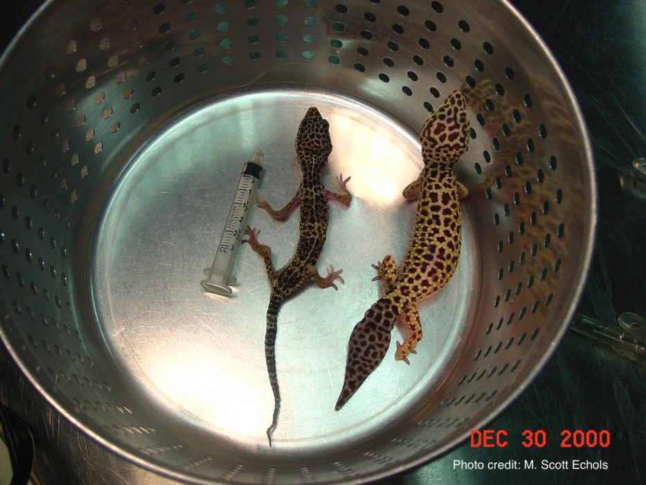
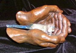
Figure 1. The tail base is a site of fat deposition in the normal leopard gecko (Eublepharis macularius). The gecko tail base can lose its fat depot in the emaciated patient (left). Source: Dr. M. Scott Echols. Click to enlarge
CHELONIANS
The debilitated turtle or tortoise may be unable to retract its head into the shell with the normal degree of strength. Respiratory disease is also an important problem in turtles and tortoises. The normal chelonian subtly moves its head and limbs with each breath, but this pumping movement is more pronounced when there is underlying respiratory disease. Breath sounds can also become audible and the chelonian may stretch the neck and gape its mouth while laboring to breathe. Aquatic turtles with pneumonia can exhibit uneven floating.
LIZARDS
Lizards with systemic disease, malnutrition, or exposure to cold temperatures can exhibit color change, appearing paler, darker, or even duller. Lizards can also appear dull when they are about to shed. Rapid color change is most highly developed in anoles and chameleons. The weak chameleon may be unable to climb or grasp. Musculoskeletal or ophthalmic disease can also prevent a chameleon from perching.
SNAKES
The weak, lethargic, or painful snake can lie listlessly in an uncoiled or “stretched out” position. There may be loss of tongue flicking or lack of interest in the environment. Severe weakness or neurologic deficits may manifest as a loss of the righting reflex. Evidence of retained shed or dysecdysis, including retained spectacles can be seen with ill thrift or deficient husbandry. Snakes can also appear dull when they are about to shed.
History
Improper diet and inadequate housing are often major contributors to illness in reptiles. For this reason, a thorough history is a crucial part of clinical evaluation of the reptile patient.
Listen to The Exotic Animal History podcast (or read the transcript) for additional information.
Obtaining a detailed history is only helpful, when the “correct” answers to your questions are known. The wide variety of reptiles seen in clinical practice can be daunting. Develop a collection of references, both online resources as well as textbooks and journals.
View LafeberVet’s collection of reptile Basic Information Sheets for additional information.
Restraint and handling
CHELONIANS
Most chelonians are easily held by the shell however some aquatic turtles will try to bite and their claws can inflict painful scratches.
LIZARDS
Never grasp any lizard patient by the tail. Some species, like iguanas and geckos, possess tail autotomy, a survival mechanism that allows the animal to escape from predators by dropping the distal tail.
SNAKES
For safety’s sake, there should be one handler for every 3 to 4 feet (0.9-1.2 m) of snake (minimum 5 feet or 1.5 m). Never allow a boa or python to form a complete loop around your neck. Large constrictors are incredibly strong. Simply by tightening muscles to maintain balance, a giant snake can cause injury by halting blood flow to the brain or cutting off air flow, resulting in loss of consciousness. Avoid coiling giant snakes around the torso as well.
Visit LafeberVet’s reptile handling and restraint series for additional information on chelonians, lizards, and snakes.
Housing
Many ill or injured reptiles that present on an emergency basis are hypothermic. Reptiles are ectotherms, which means they derive body heat from their environment. Each reptile species has its own unique preferred optimal temperature zone (POTZ) at which physiology functions optimally and the reptile’s ability to heal or convalesce is most effective. The POTZ is provided as a temperature gradient for the mobile patient, however heat must be provided more cautiously for the collapsed or debilitated reptile through an incubator or brooder. A temperature setting of 82-85°F (28-29.4°C) is suitable for many species.
Visit Exotic ICU: Nursing Care for Reptiles and Basic Husbandry: Hospitalizing Non-Traditional Species for additional information on caging the reptile patient.
Only reptiles of adequate and normal bone quality should be offered vertical space for climbing.
Vascular access
Debilitated reptiles are often dehydrated. Judiciously provide fluid therapy, replacing deficits over 48-96 hours. Maintenance fluid requirements are estimated at 1-3 % of body weight or 10-30 ml/kg/day. Always administer warmed fluids.
Depending on the size of the patient and the species of interest, intravenous and intraosseous catheters are viable options. Intraosseous catheters can be placed into the tibia or femur of the lizard, while the jugular vein is easily accessed in chelonians. Catheterization is challenging in snakes because there are no vessels that are easily accessible.
Visit Catheters in Reptiles for illustrative video and text.
Soaking is an effective way to supplement fluids as reptiles absorb fluids through the cloaca. Never soak a reptile that is so debilitated it is unable to lift its head above water. Subcutaneous fluids can be used to provide maintenance fluids to stable patients and to correct mild dehydration, however the subcutaneous space is relatively limited in reptiles. Only small volumes of subcutaneous fluids can be administered to reptile patients.
Go to the review article Fluid Administration in Reptiles for additional information.
Medical therapy
Most reptile pathogens are opportunistic, Gram-negative bacteria. Pending culture results, select antimicrobials that primarily have a Gram-negative spectrum. The parenteral route is typically used in reptile patients, particularly intramuscular injections. Injections are administered into the cranial half of the body to avoid the renal portal system. The reptile gastrointestinal tract is normally quiescent between meals. Oral medications should only be administered to reptile patients maintained at an appropriate temperature that eat frequently and demonstrate evidence of a functional gastrointestinal tract.
View the LafeberVet video Administration of Medication in Reptiles for additional information.
Analgesia
The painful reptile may exhibit the previously described signs of illness, as well as lameness, a hunched posture, aerophagia, and behavioral changes, such as aggression in a passive animal or apathy in a normally aggressive individual (Video 2).
For information on reptile analgesia, view timestamp 10:02-22:02 of the R.A.C.E.-approved LafeberVet webinar Spotlight on Anesthesia and Analgesia in Reptiles by Dr. Javier Nevarez.
Pain receptors in reptiles are poorly understood, however non-steroidal anti-inflammatory agents like meloxicam (0.5 mg/kg q24h) and opioids are commonly used (Antinoff 2016, Nevarez 2016). Research suggests pure mu opioid receptor agonists, like morphine (1.5-6.5 mg/kg SC), and hydromorphone (0.5 mg/kg SC), are the best choice in reptiles evaluated, although prominent respiratory depression can be observed. Species evaluated include the red-eared slider (Trachemys scripta elegans), green iguana (Iguana iguana), and bearded dragon (Pogona vitticeps) (Mans et al 2012, Kinney et al 2011, Sladky et al 2008). Prominent respiratory depression can be observed.
Nutritional support
Reptile metabolism is approximately one-tenth to one-third lower than similar-sized mammals. This significant lower metabolic rate means that reptiles do not eat as frequently as mammals. Therefore, never rush to feed a reptile patient. As in all species, ensure the patient is warm and hydrated before feeding begins. Nutritional support can be delivered via gavage tube short-term and many lizards can be syringe fed. Esophagostomy tube placement is a popular choice for long-term feeding of anorectic chelonians.
Visit LafeberVet’s Nutritional Support in Reptiles for additional information on feeding the reptile patient.
Test your knowledge
Take the brief quiz Test Your Knowledge: Reptile Critical Care
Common presenting problems
- Bite wounds
- Dystocia
View the R.A.C.E.-approved webinar Five Common Reptile Emergencies for a discussion of five common clinical presentations: trauma, reproductive disease, gastrointestinal foreign body, neurological deficits, and respiratory difficulty.
- Fractures
Fractures are often associated with nutritional secondary hyperparathyroidism and may often require temporary external coaptation.
- Hyperthermia
Hyperthermia can occur if a reptile is left in an aquarium in the sun without shade for as little as 5 minutes. Manage the patient with subcutaneous or intracoelomic fluids and cool, but not cold, water. If signs of coma or cerebral edema are observed, then steroids are indicated (Antinoff 2016).
- Hypothermia
Hypothermia can develop with power failures or when a reptile escapes. The hypothermic reptile is extremely lethargic and evidence of gastrointestinal stasis or bloating may be observed. Slowly warm the patient with warmed fluids and supplemental heat over 4-6 hours. Secondary infections or pneumonia can develop within days or weeks following the hypothermic episode (Antinoff 2016).
- Pneumonia
One of the driving parameters for reptile respiration is the partial pressure of oxygen; significantly elevated oxygen levels can inhibit breathing in the reptile. Therefore supplemental oxygen can actually compromise the breathing of an already ill reptile. If supplemental oxygen is provided, administer no more than 30-40% humidified oxygen.
- Prolapse
- Shell trauma
- Tail trauma or amputation
If a captive animal loses its distal tail and amputation is clean, all that is required is wound management. If bone is exposed, remove this bone segment manually with sedation.
- Thermal burns
Conclusion
The basic principles of emergency medicine and critical care are also the same for all species, however critically ill reptiles pose special challenges due to their unique anatomy, physiology, and behavior. Many ill or injured reptiles are hypothermic, and supplemental heat is mandatory. Provide a quiet, calm environment and an appropriate humidity level. House the collapsed or debilitated reptile in an incubator or brooder. A temperature setting of 28-29.4°C (82-85°F) is suitable for many reptiles.
To learn more…
The medical care of reptile patients is a complex and fascinating topic. To learn more, browse the content featured in LafeberVet’s emergency and critical care teaching module landing page as well as the reptile medicine page on LafeberVet,and continue your education with the Association of Reptilian and Amphibian Veterinarians.
References and further reading
Exotic Companion Mammal Emergency & Critical Care Summary Page
Although the principles of emergency medicine critical care are universal for all species, this approach must be balanced with an understanding of the unique aspects of small mammal medicine. Use this summary page to review the basic approach to the exotic companion mammal patient and select additional links to supplement your knowledge base . . .
Pour les vétérinaires. Par les vétérinaires.
Le site Lafervet.com est conçu pour une utilisation par les vétérinaires. Il est ouvert aux vétérinaires diplômés, aux techniciens vétérinaires diplômés, aux animaliers et aux étudiants dans ces domaines.
Créer un compte pour accéder à des articles et des ressources du site.
L'inscription est gratuite.
Para Profesionales Veterinarios. Por Profesionales Veterinarios.
El sitio Lafervet.com es para uso de los profesionales veterinarios. Está abierto a los veterinarios licenciados, técnicos veterinarios licenciados, rehabilitadores licenciados y estudiantes en estos campos.
Cree una cuenta para acceder a los artículos y recursos del sitio.
La registro es gratis.
Already a LafeberVet Member?
Please Login
Avian Emergency & Critical Care Summary Page
Introduction
Although the principles of emergency medicine and critical care are universal for all species, this approach must be balanced with an understanding of the unique aspects of avian medicine. Use this summary page to review the basic approach to the avian patient and select additional links to supplement your knowledge base.

This summary page is a part of the Emergency and Critical Care Teaching Module.
Airway/resuscitation
Most birds possess complete tracheal rings. Use of a cuffed endotracheal tube can lead to pressure necrosis if the cuff is inflated because there is no elastic ligament to accommodate tracheal expansion. Always select an uncuffed endotracheal tube in small birds. Traditional cuffed tubes can be used in larger birds, but never inflate the cuff. Intubation is a relatively simple process because there is no epiglottis and the tracheal opening is generally very accessible. Once the bird is intubated, secure the tube to the upper or lower beak with tape.
Careful and vigilant patient monitoring is essential for avian patients as cardiac arrest in birds carries a poor prognosis. Presence of the sternal plate or keel means that direct compression of the heart is impossible in birds, however sternal compressions can push air through the bellows-like air sac system.
Delve deeper into this topic in Cardiopulmonary Resuscitation in Exotic Animals.
Signs of illness
Many companion birds are prey species, which tend to hide signs of illness until disease is advanced. Many conditions can produce a very similar clinical picture. A lethargic, “fluffed and ruffled” appearance is a very common non-specific sign of illness in the avian patient. To increase the likelihood that subtle signs of illness will be recognized during the visual examination, first provide prey species with a 5-10-minute acclimation period. Signs of respiratory difficulty in the bird can include open-mouth breathing, increased sternal motion, and tail bobbing.
Review the slideshow Recognizing Signs of Illness in Birds for additional information.
Careful observation is essential. Is the patient strong enough to handle manual restraint and a complete physical examination? Or is the animal so ill that only a cursory examination can be performed? In the debilitated or dyspneic patient, it may be prudent to first place the patient in an incubator or oxygen cage in a dark, quiet room before evaluation. Even after the bird has had time to gather its strength and calm down, it may only be strong enough to handle diagnostics and treatment in stages.
History
The avian history must be detailed and includes not only signalment and recent medical history, but also source of the pet, complete dietary history, caging history–including whether or not the pet is always supervised outside of the cage–exposure to other pets, as well as recent illnesses or deaths of other birds in the household.
Refer to the podcast The Exotic Animal History for additional information.
Restraint & handling
Proper restraint of birds, that does not lead to patient or veterinary staff injury, requires training and practice. Prey species or wild birds will undergo a stress response that can cause catecholamine release and even death due to handling and treatment alone. Never restrain the avian patient for a prolonged period. Always plan a procedure that requires restraint and gather all equipment that may possibly be needed beforehand.
Visit LafeberVet’s Passerine Handling and Restraint and Parrot Handling and Restraint for illustrative videos and text.
Housing
House avian patients in a quiet area away from the sight and sound of predator species like cats, dogs, and ferrets. The cage setup should also physically block the view of one animal from another. Many birds will also benefit from some form of visual security. Drape a towel over part of the incubator or tape newspaper or some other opaque material over part of a treatment cage door. Dim light levels as needed to calm the nervous patient.
A rapid metabolic rate means that small birds have a greater susceptibility to hypothermia. Debilitated birds should be kept warm. Target incubator temperatures range between 80-90°F (26-32°C) for most avian patients. Carefully observe the patient for signs of overheating, such as flat, sleek feathers, outstretched wings, and open-mouth breathing. Use particular caution in overweight birds.
All but the weakest perching birds will be much more comfortable if provided with perch material. Place perches on the cage floor or elevate perches only slightly to minimize the risk of falls.
Visit LafeberVet’s Exotic ICU: Nursing Care for the Avian Patient for additional information.
Vascular access
Even loss of small volumes of blood can leave a tiny animal critically hypovolemic. For these small patients, use small-volume fluid resuscitation with frequent reassessment rather than large fluid boluses. Vascular access sites are limited in the bird. Peripheral veins can be difficult to access, especially during shock, and the vessels are also prone to hematoma formation. Intraosseous catheter placement is generally faster and easier in birds and should be used as a first choice in an emergency situation.
Visit LafeberVet’s Intraosseous Catheter Placement and Intravenous Catheter Placement in the Bird for illustrative videos and text.
Subcutaneous fluids are an excellent way to provide maintenance fluids to stable avian patients and to correct mild dehydration. Subcutaneous fluids may also be the safest route initially for extremely debilitated patients as well as those with respiratory distress or coelomic distension.
Fluids administered should always be warmed. An isotonic crystalloid, like lactated Ringer’s solution, is a good choice for many critical patients.
Medical therapy
Antimicrobial choice is more limited when treating birds. Empirical antibiotics selected in the critically ill patient are generally bactericidal and broad-spectrum, such as cefotaxime 75-100 mg/kg IM, IV q8h (Claforan, Sanofi-Aventis) or piperacillin-taxobactam 100 mg/kg IM q6-12 (Zosyn, Wyeth) (Jenkins 2016, Stout 2016, Hawkins et al 2013).
When oral medications are indicated, commercially available or compounded suspensions are preferable. In-house compounding is not recommended, but can be used for individual dosing until a compounded formulation can be obtained. Drugs can also be administered by the intramuscular or subcutaneous routes. Insulin or tuberculin syringes provide more accurate dosing than larger syringes.
Corticosteroid use is controversial in birds, and there are many contraindications for its use. Aspergillosis can develop in avian patients during times of stress or immunosuppression, and corticosteroid use is an important predisposing condition. Therefore, corticosteroid use is not recommended as a standard treatment for most clinical conditions. Select another drug with the potential for fewer adverse effects whenever possible.
Analgesia
Clinical signs of pain are often more subtle in birds when compared to those seen in mammals. Behavioral signs of pain in birds can include reduced vocalization, decreased activity, anorexia, isolation from the group, and increased aggression. Physical signs of pain can include tachycardia, hypertension, arrhythmias, tachypnea, hypoxemia, hypercapnia, acidosis, abnormal posture and/or lameness, and weight loss.
As in all veterinary patients, provide pre-emptive analgesia and multimodal analgesic agents whenever possible. Non-steroidal anti-inflammatory drugs (NSAIDS), such as meloxicam, and opioids are frequently used. Kappa-agonists opioids, like butorphanol, are commonly used in birds however growing evidence suggest mu-agonists, such as hydromorphone and fentanyl, are more effective in some avian species.
Nutritional support
The rapid metabolic rate of small avian patients leads to rapid depletion of glucose reserves. Fasting should be avoided and when required, generally should be less than 6 hours.
Nutritional support is essential in these patients. Provide familiar food items ad libitum. After the patient has been warmed and hydrated, tube or gavage feeding is often an essential part of avian supportive care. Tube feeding is a relatively straightforward technique in the bird, however there are serious potential complications, including aspiration, laceration of the oropharynx, cellulitis, and even death. Therefore, this technique should be practiced beforehand so tube feeding can be performed efficiently, safely, and gently in the clinical patient. Closely monitor patient body weight and droppings.
Visit Tube Feeding Birds for additional information.
Test your knowledge
Take the brief quiz Test Your Knowledge: Avian Critical Care
Common emergencies or presenting problems
- Anorexia
- Broken blood feather
- Cloacal prolapse
- Diarrhea
- Dyspnea
- Dystocia, egg binding
- Fluffed and ruffled, lethargic appearance
- Regurgitation
- Seizure activity
- Toxic exposure
- Trauma
Conclusion
Although the principles of critical care are universal in all species, their application can be quite challenging in birds. Many companion birds are prey species. Provide these patients with a 5-10-minute acclimation period whenever possible, and minimize the time spent handling the bird. Supportive care relies upon supplemental heat provided in calm, quiet environment. Fluid therapy can be provided by an intraosseous catheter in select avian patients. Once the patient is warm and hydrated, begin nutritional support.
To learn more…
Like all segments of veterinary medicine, the medical care of birds is a complex and fascinating topic. This brief summary merely scratches the surface of what every clinician should know when they touch a bird. To learn more, browse the content featured in LafeberVet’s emergency and critical care teaching module landing page as well as the avian medicine page on LafeberVet, then continue your education with the Association of Avian Veterinarians.
References and further reading
The Exotic Animal History
Although patient history is important in all species, improper diet and substandard housing are often major contributors to illness in non-traditional pets. This means that a detailed and accurate history is often one of the most critical diagnostic tools for the exotic animal patient. This review focuses on birds, reptiles, and small exotic companion mammals, beginning with the signalment and presenting complaint, before moving onto the environmental history, dietary history, and of course the medical history . . .
Pour les vétérinaires. Par les vétérinaires.
Le site Lafervet.com est conçu pour une utilisation par les vétérinaires. Il est ouvert aux vétérinaires diplômés, aux techniciens vétérinaires diplômés, aux animaliers et aux étudiants dans ces domaines.
Créer un compte pour accéder à des articles et des ressources du site.
L'inscription est gratuite.
Para Profesionales Veterinarios. Por Profesionales Veterinarios.
El sitio Lafervet.com es para uso de los profesionales veterinarios. Está abierto a los veterinarios licenciados, técnicos veterinarios licenciados, rehabilitadores licenciados y estudiantes en estos campos.
Cree una cuenta para acceder a los artículos y recursos del sitio.
La registro es gratis.
Already a LafeberVet Member?
Please Login
Analgesia and Sedation in Exotic Companion Mammals
The approach to analgesia and sedation in exotic companion mammals faces special challenges, including small patient size and unique features of the prey species mentality. Recognition of pain is more difficult in rabbits and rodents because many small mammals are very good at hiding the signs of pain commonly observed in predator species. Instead pain in a rabbit or rodent is often inferred from the patient’s clinical condition as well as the absence of normal behaviors. The diagnostic and therapeutic plan frequently requires some form of chemical restraint in exotic mammal medicine. When compared to general anesthesia, sedation is . . .
Pour les vétérinaires. Par les vétérinaires.
Le site Lafervet.com est conçu pour une utilisation par les vétérinaires. Il est ouvert aux vétérinaires diplômés, aux techniciens vétérinaires diplômés, aux animaliers et aux étudiants dans ces domaines.
Créer un compte pour accéder à des articles et des ressources du site.
L'inscription est gratuite.
Para Profesionales Veterinarios. Por Profesionales Veterinarios.
El sitio Lafervet.com es para uso de los profesionales veterinarios. Está abierto a los veterinarios licenciados, técnicos veterinarios licenciados, rehabilitadores licenciados y estudiantes en estos campos.
Cree una cuenta para acceder a los artículos y recursos del sitio.
La registro es gratis.
Already a LafeberVet Member?
Please Login
Melinda Cowan, BVSc (hons) FANZCVS (Avian Medicine)
 Dr. Melinda Cowan graduated from the University of Sydney in 2007 with first class honors. After initially working in a busy small animal clinic, she took a position at a specialized bird and exotic pet practice in Brisbane, Australia and completed a residency in avian medicine. Melinda completed final examinations in 2016 to become a bird specialist and Fellow of the Australian and New Zealand College of Veterinary Scientists. Dr. Cowan currently practices at the Small Animal Specialist Hospital in Sydney, where she cares for a wide range of exotic animals. She has also worked for the Royal Society for the Prevention of Cruelty to Animals (RSPCA) Queensland wildlife hospital, treating a variety of native animals in addition to the domestic birds admitted to the shelter.
Test Your Knowledge: Reptile Critical Care
Test your knowledge after completing the reptile portion of the LafeberVet Emergency and Critical Care teaching module . . .
Pour les vétérinaires. Par les vétérinaires.
Le site Lafervet.com est conçu pour une utilisation par les vétérinaires. Il est ouvert aux vétérinaires diplômés, aux techniciens vétérinaires diplômés, aux animaliers et aux étudiants dans ces domaines.
Créer un compte pour accéder à des articles et des ressources du site.
L'inscription est gratuite.
Para Profesionales Veterinarios. Por Profesionales Veterinarios.
El sitio Lafervet.com es para uso de los profesionales veterinarios. Está abierto a los veterinarios licenciados, técnicos veterinarios licenciados, rehabilitadores licenciados y estudiantes en estos campos.
Cree una cuenta para acceder a los artículos y recursos del sitio.
La registro es gratis.
Already a LafeberVet Member?
Please Login
Test Your Knowledge: Exotic Companion Mammal Critical Care
Test your knowledge after completing the exotic companion mammal portion of the LafeberVet Emergency and Critical Care teaching module . . .
Pour les vétérinaires. Par les vétérinaires.
Le site Lafervet.com est conçu pour une utilisation par les vétérinaires. Il est ouvert aux vétérinaires diplômés, aux techniciens vétérinaires diplômés, aux animaliers et aux étudiants dans ces domaines.
Créer un compte pour accéder à des articles et des ressources du site.
L'inscription est gratuite.
Para Profesionales Veterinarios. Por Profesionales Veterinarios.
El sitio Lafervet.com es para uso de los profesionales veterinarios. Está abierto a los veterinarios licenciados, técnicos veterinarios licenciados, rehabilitadores licenciados y estudiantes en estos campos.
Cree una cuenta para acceder a los artículos y recursos del sitio.
La registro es gratis.
Already a LafeberVet Member?
Please Login
Andrea Hubbard, DVM, DACLAM
 Dr. Andrea Hubbard is Assistant Director of Quality Assurance and Training for the Institute of Comparative Medicine at Columbia University in the City of New York. Dr. Hubbard served as a research associate at the University of Massachusetts Medical School, where she was responsible for maintaining a transgenic mouse colony, before earning her Doctorate of Veterinary Medicine at Ross University School of Veterinary Medicine. She completed a residency in laboratory animal medicine at Columbia University in 2013 and continued her career as a clinical veterinarian at Columbia University before becoming an Assistant Director in 2016. She is a Diplomate of the American College of Laboratory Animal Medicine.
Nichole Parand née Arbona DVM
 Nichole Parand née Arbona graduated from The University of Arizona with a Bachelor of Science in Ecology and Evolutionary Biology before earning her Doctor of Veterinary Medicine from Kansas State University College of Veterinary Medicine. While in school, Dr. Parand was a Lafeber Company student ambassador and assisted with various LafeberVet articles. In school, she was highly involved in research and primary authored a paper in the Journal of Feline Medicine and Surgery on a common southern fungal disease (valley fever) in cats. She also performed research in Madagascar on critically endangered lemurs. Dr. Parand joined Irvington Pet Hospital and the VetnCare team in February 2021 and soon after became clinical director and partner. Nichole has a special interest in surgery and exotic animals, particularly pocket pets.
Test Your Knowledge: Avian Critical Care
Test your knowledge after completing the avian portion of the LafeberVet Emergency and Critical Care teaching module . . .
Pour les vétérinaires. Par les vétérinaires.
Le site Lafervet.com est conçu pour une utilisation par les vétérinaires. Il est ouvert aux vétérinaires diplômés, aux techniciens vétérinaires diplômés, aux animaliers et aux étudiants dans ces domaines.
Créer un compte pour accéder à des articles et des ressources du site.
L'inscription est gratuite.
Para Profesionales Veterinarios. Por Profesionales Veterinarios.
El sitio Lafervet.com es para uso de los profesionales veterinarios. Está abierto a los veterinarios licenciados, técnicos veterinarios licenciados, rehabilitadores licenciados y estudiantes en estos campos.
Cree una cuenta para acceder a los artículos y recursos del sitio.
La registro es gratis.
Already a LafeberVet Member?
Please Login
Recognizing Signs of Illness in Birds
Signs of illness in birds are often quite subtle until disease is advanced. Fortunately, quite a bit of information can be gleaned from a detailed history and careful observation. View this brief slideshow for tips on the visual examination . . .
Pour les vétérinaires. Par les vétérinaires.
Le site Lafervet.com est conçu pour une utilisation par les vétérinaires. Il est ouvert aux vétérinaires diplômés, aux techniciens vétérinaires diplômés, aux animaliers et aux étudiants dans ces domaines.
Créer un compte pour accéder à des articles et des ressources du site.
L'inscription est gratuite.
Para Profesionales Veterinarios. Por Profesionales Veterinarios.
El sitio Lafervet.com es para uso de los profesionales veterinarios. Está abierto a los veterinarios licenciados, técnicos veterinarios licenciados, rehabilitadores licenciados y estudiantes en estos campos.
Cree una cuenta para acceder a los artículos y recursos del sitio.
La registro es gratis.
Already a LafeberVet Member?
Please Login
2017 Avian Practitioner of the Year
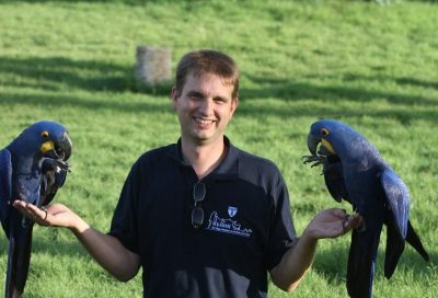
Dr. Michael Lierz named
2017 T.J. Lafeber Avian Practitioner of the Year

When nominations closed for the 2017 T.J. Lafeber Avian Practitioner of the Year, a list of 26 outstanding avian veterinarians were submitted for consideration. The independent Selection Committee, hosted through Louisiana State University, narrowed the list to five finalists and the Award recipient, Dr. Michael Lierz, was announced during the Opening Session at the 2017 Annual Conference of the Association of Avian Veterinarians .
Michael Lierz, DZooMed, DECZM (WPH), DECPVS is a Full Professor and Director of the Klinik für Vögel, Reptilien, Amphibien und Fische (Clinic for Birds, Reptiles, Amphibians and Fish) at the Justus-Liebig University of Giessen [MORE].
Award recipients
Visit Lafeber.com for a list of previous Award recipients.
Did You Know…?
 The T.J. Lafeber Avian Practitioner of the Year is nominated by their peers: YOU.
- The Awardee is NOT, and has never been, selected by Lafeber Company.
This autonomous committee, consisting of Association of Avian Veterinarians members, is led by Dr. Tom Tully, Professor and Chief of the Zoological Medicine Service at Louisiana State University School of Veterinary Medicine.
A Lafeber Company Veterinary Consultant, who does NOT vote on the Award recipient, manages committee paperwork and scheduling only.
Emergency Preparedness Plan for Exotic Pets
Get ready now to care for exotic pets during an accident or natural catastrophe that causes great damage or even loss of life, such as blizzard, earthquake, fire, flood, hurricane, mud slide, or tornado. This disaster relief client education handout was revised and posted with permission from “Ready-Pets-Go!” by the Humane Society of Greater Rochester . . .
Pour les vétérinaires. Par les vétérinaires.
Le site Lafervet.com est conçu pour une utilisation par les vétérinaires. Il est ouvert aux vétérinaires diplômés, aux techniciens vétérinaires diplômés, aux animaliers et aux étudiants dans ces domaines.
Créer un compte pour accéder à des articles et des ressources du site.
L'inscription est gratuite.
Para Profesionales Veterinarios. Por Profesionales Veterinarios.
El sitio Lafervet.com es para uso de los profesionales veterinarios. Está abierto a los veterinarios licenciados, técnicos veterinarios licenciados, rehabilitadores licenciados y estudiantes en estos campos.
Cree una cuenta para acceder a los artículos y recursos del sitio.
La registro es gratis.
Already a LafeberVet Member?
Please Login
Brendan Carmel, BVSc, MVS MANZCVS (Unusual Pets) GDipComp
 Dr. Brendan Carmel is the owner and Senior Veterinarian at Warranwood Veterinary Centre, which provides care for unusual and exotic pets in Melbourne, Australia. A graduate of the University of Melbourne, Brendan served as zoo veterinarian for 3 years at Healesville Sanctuary, Victoria where he completed a Masters of Veterinary Studies in Australian Native Wildlife. He is the 2017-2018 President of the Unusual Pet and Avian Veterinarians, special interest group of the Australian Veterinary Association. Dr. Carmel is also a founding member of both the Association of Exotic Mammal Veterinarians and the Association of Reptilian and Amphibian Veterinarians. He is a member of the Australian and New Zealand College of Veterinary Scientists in Unusual Pet Medicine and Surgery, an Associate Editor of the Australian Veterinary Journal, and a frequent speaker at national and international veterinary conferences. Dr. Carmel is also the recipient of the 2015 Oxbow Exotic Mammal Health Award for excellence in exotic mammal health and care. Dr. Carmel is also the co-author of A Guide to Health & Disease in Reptiles & Amphibians.
What Parrots Want: The Importance and Use of Foraging and Environmental Enrichment for Birds Post Test
Post test for What Parrots Want: The Importance and Use of Foraging and Environmental Enrichment for Birds webinar . . .
Pour les vétérinaires. Par les vétérinaires.
Le site Lafervet.com est conçu pour une utilisation par les vétérinaires. Il est ouvert aux vétérinaires diplômés, aux techniciens vétérinaires diplômés, aux animaliers et aux étudiants dans ces domaines.
Créer un compte pour accéder à des articles et des ressources du site.
L'inscription est gratuite.
Para Profesionales Veterinarios. Por Profesionales Veterinarios.
El sitio Lafervet.com es para uso de los profesionales veterinarios. Está abierto a los veterinarios licenciados, técnicos veterinarios licenciados, rehabilitadores licenciados y estudiantes en estos campos.
Cree una cuenta para acceder a los artículos y recursos del sitio.
La registro es gratis.
Already a LafeberVet Member?
Please Login
What Parrots Want: The Importance and Use of Foraging and Environmental Enrichment for Birds
This webinar recording is RACE-approved for 1 hour of continuing education. Despite parrots being popular pets, much of the information regarding their nutritional and behavioral needs is still unknown. Unlike dogs and cats, most psittacine species are not domesticated and have therefore likely retained most, if not all, of their wild instincts and behavioral needs. In captivity, however, most parrots have little to no opportunity to perform these species-typical behaviors. This will not only reduce their welfare, but can also result in the onset of abnormal repetitive behaviors, including feather damaging behavior, and oral or locomotor stereotypies . . .
Pour les vétérinaires. Par les vétérinaires.
Le site Lafervet.com est conçu pour une utilisation par les vétérinaires. Il est ouvert aux vétérinaires diplômés, aux techniciens vétérinaires diplômés, aux animaliers et aux étudiants dans ces domaines.
Créer un compte pour accéder à des articles et des ressources du site.
L'inscription est gratuite.
Para Profesionales Veterinarios. Por Profesionales Veterinarios.
El sitio Lafervet.com es para uso de los profesionales veterinarios. Está abierto a los veterinarios licenciados, técnicos veterinarios licenciados, rehabilitadores licenciados y estudiantes en estos campos.
Cree una cuenta para acceder a los artículos y recursos del sitio.
La registro es gratis.
Already a LafeberVet Member?
Please Login
Bearded Dragon Infectious Disease Slideshow
Inland bearded dragons are native to Australia and are a popular companion animal. Private breeders often select for desirable temperaments and various color morphologies in an effort to provide an ever-increasing variety to the pet trade. They are also bred commercially and sold by large pet retailers making them a widely available pet reptile. As general husbandry for exotic companion animals continues to improve and as owners develop strong bonds with their reptile companions, presentation for infectious disease is expected to become more commonplace. This infectious disease slideshow is intended to serve as a quick reference guide for the . . .
Pour les vétérinaires. Par les vétérinaires.
Le site Lafervet.com est conçu pour une utilisation par les vétérinaires. Il est ouvert aux vétérinaires diplômés, aux techniciens vétérinaires diplômés, aux animaliers et aux étudiants dans ces domaines.
Créer un compte pour accéder à des articles et des ressources du site.
L'inscription est gratuite.
Para Profesionales Veterinarios. Por Profesionales Veterinarios.
El sitio Lafervet.com es para uso de los profesionales veterinarios. Está abierto a los veterinarios licenciados, técnicos veterinarios licenciados, rehabilitadores licenciados y estudiantes en estos campos.
Cree una cuenta para acceder a los artículos y recursos del sitio.
La registro es gratis.
Already a LafeberVet Member?
Please Login
Avian Respiratory Anatomy, Physiology & Diseases: An Overview Post Test
The Avian Respiratory Tract Overview webinar was reviewed and approved by the American Association of Veterinary State Boards (AAVSB) Registry of Approved Continuing Education (R.A.C.E.) program for 1 hour of continuing education, in jurisdictions which recognize AAVSB R.A.C.E. approval . . .
Pour les vétérinaires. Par les vétérinaires.
Le site Lafervet.com est conçu pour une utilisation par les vétérinaires. Il est ouvert aux vétérinaires diplômés, aux techniciens vétérinaires diplômés, aux animaliers et aux étudiants dans ces domaines.
Créer un compte pour accéder à des articles et des ressources du site.
L'inscription est gratuite.
Para Profesionales Veterinarios. Por Profesionales Veterinarios.
El sitio Lafervet.com es para uso de los profesionales veterinarios. Está abierto a los veterinarios licenciados, técnicos veterinarios licenciados, rehabilitadores licenciados y estudiantes en estos campos.
Cree una cuenta para acceder a los artículos y recursos del sitio.
La registro es gratis.
Already a LafeberVet Member?
Please Login
The Avian Neurological Exam
Introduction
As a part of the Lafeber Company Student Program, Dr. Susan Orosz presented an exclusive presentation to the University of Illinois College of Veterinary Medicine Non-Traditional Species Club as a distance learning event.
Photo credit: Dr. Susan Orosz
Lecture objectives included:
|
|
Webinar recording
A recording of Dr. Orosz’s presentation will be posted as soon as possible. The videos featured during Dr. Orosz’s presentation are also posted below.
Videos
Video credit: Dr. Natalie Antinoff
Video credit: Dr. Natalie Antinoff
Video credit: The Raptor Center – University of Minnesota
Video credit: Dr. Susan Orosz
Video credit: Dr. Natalie Antinoff
Video credit: Dr. Susan Orosz
Video credit: Dr. Susan Orosz
Video credit: Dr. Susan Orosz
Video credit: Dr. Natalie Antinoff
Video credit: Dr. Natalie Antinoff
Video credit: Dr. Natalie Antinoff
Video credit: Dr. Susan Orosz
Video credit: Dr. Susan Orosz
Nicholas Crossland, DVM, DACVP

Dr. Nicholas Crossland is a Diplomate of the American College of Veterinary Pathologists. He currently serves as a T32 Post-Doctoral fellow and PhD student at Tulane National Primate Research Center in Covington, Louisiana. His research focuses on Borrelia burgdoferi and mechanisms of persistence in host tissues utilizing the non-human primate, rat, and mouse models. Nick earned a Bachelor of Science degree in Animal Sciences at Kansas State University and subsequently a Doctorate of Veterinary Medicine graduating Cum Laude. Dr. Crossland also successfully completed an anatomical pathology residency at Louisiana State University School of Veterinary Medicine in 2016. His residency exposed him to a vast and diverse case load of exotic animal species that influenced his passion for this field . He has co-authored a variety of veterinary manuscripts including the Veterinary Clinical Pathology article “Nannizziopsis guarroi infection in 2 Inland Bearded Dragons (Pogona vitticeps): clinical, cytologic, histologic, and ultrastructural aspects”.
Avian Respiratory Anatomy, Physiology & Diseases: An Overview
This live webinar event was presented by James Morrisey, DVM, DABVP (AvianPractice). View a recording of this AAVSB R.A.C.E.-approved web-based seminar, then take the brief post-test to earn 1 hour of continuing education credit. The avian respiratory system has several unique and fascinating adaptations for flight that are important to clinicians. This webinar overviews the anatomy and physiology of the avian respiratory tract. Clinical correlates are pointed out as the presenter goes through anatomy and physiology. Clinical signs of respiratory disease in birds are then discussed along with how the clinician can use these . . .
Pour les vétérinaires. Par les vétérinaires.
Le site Lafervet.com est conçu pour une utilisation par les vétérinaires. Il est ouvert aux vétérinaires diplômés, aux techniciens vétérinaires diplômés, aux animaliers et aux étudiants dans ces domaines.
Créer un compte pour accéder à des articles et des ressources du site.
L'inscription est gratuite.
Para Profesionales Veterinarios. Por Profesionales Veterinarios.
El sitio Lafervet.com es para uso de los profesionales veterinarios. Está abierto a los veterinarios licenciados, técnicos veterinarios licenciados, rehabilitadores licenciados y estudiantes en estos campos.
Cree una cuenta para acceder a los artículos y recursos del sitio.
La registro es gratis.
Already a LafeberVet Member?
Please Login
A Guide to Nasotracheal Intubation in Rabbits
Rabbit intubation can be accomplished using either an orotracheal or nasotracheal technique. Both intubation methods can be challenging in rabbit patients and require patience and practice. Nasotracheal intubation may be the preferred approach in situations where maximum access and maneuverability is required in the oral cavity. Nasotracheal intubation is also preferred where an extended recovery is expected . . .
Pour les vétérinaires. Par les vétérinaires.
Le site Lafervet.com est conçu pour une utilisation par les vétérinaires. Il est ouvert aux vétérinaires diplômés, aux techniciens vétérinaires diplômés, aux animaliers et aux étudiants dans ces domaines.
Créer un compte pour accéder à des articles et des ressources du site.
L'inscription est gratuite.
Para Profesionales Veterinarios. Por Profesionales Veterinarios.
El sitio Lafervet.com es para uso de los profesionales veterinarios. Está abierto a los veterinarios licenciados, técnicos veterinarios licenciados, rehabilitadores licenciados y estudiantes en estos campos.
Cree una cuenta para acceder a los artículos y recursos del sitio.
La registro es gratis.
Already a LafeberVet Member?
Please Login
Presenting Problem: Dyspnea in Ferrets
This presenting problem article reviews the basic approach to the dyspneic ferret beginning with clinical signs of the dyspneic ferret, key points of urgent care, as well as case management. This latter section reviews tips on taking the history, performing the physical exam, important differential diagnoses, as well as the diagnostic/therapeutic approach . . .
Pour les vétérinaires. Par les vétérinaires.
Le site Lafervet.com est conçu pour une utilisation par les vétérinaires. Il est ouvert aux vétérinaires diplômés, aux techniciens vétérinaires diplômés, aux animaliers et aux étudiants dans ces domaines.
Créer un compte pour accéder à des articles et des ressources du site.
L'inscription est gratuite.
Para Profesionales Veterinarios. Por Profesionales Veterinarios.
El sitio Lafervet.com es para uso de los profesionales veterinarios. Está abierto a los veterinarios licenciados, técnicos veterinarios licenciados, rehabilitadores licenciados y estudiantes en estos campos.
Cree una cuenta para acceder a los artículos y recursos del sitio.
La registro es gratis.
Already a LafeberVet Member?
Please Login
Five Common Reptile Emergencies Post Test
The Five Common Reptile Emergencies webinar was reviewed and approved by the American Association of Veterinary State Boards (AAVSB) Registry of Approved Continuing Education (R.A.C.E.) program for 1 hour of continuing education, in jurisdictions which recognize AAVSB R.A.C.E. approval . . .
Pour les vétérinaires. Par les vétérinaires.
Le site Lafervet.com est conçu pour une utilisation par les vétérinaires. Il est ouvert aux vétérinaires diplômés, aux techniciens vétérinaires diplômés, aux animaliers et aux étudiants dans ces domaines.
Créer un compte pour accéder à des articles et des ressources du site.
L'inscription est gratuite.
Para Profesionales Veterinarios. Por Profesionales Veterinarios.
El sitio Lafervet.com es para uso de los profesionales veterinarios. Está abierto a los veterinarios licenciados, técnicos veterinarios licenciados, rehabilitadores licenciados y estudiantes en estos campos.
Cree una cuenta para acceder a los artículos y recursos del sitio.
La registro es gratis.
Already a LafeberVet Member?
Please Login
Feather Destructive Behavior in Psittacine Birds Post Test
Categories: Avian, Parrot,
The Feather Destructive Behavior in Psittacine Birds webinar was reviewed and approved by the American Association of Veterinary State Boards (AAVSB) Registry of Approved Continuing Education (R.A.C.E.) program for 1 hour of continuing education, in jurisdictions which recognize AAVSB R.A.C.E. approval . . .
Pour les vétérinaires. Par les vétérinaires.
Le site Lafervet.com est conçu pour une utilisation par les vétérinaires. Il est ouvert aux vétérinaires diplômés, aux techniciens vétérinaires diplômés, aux animaliers et aux étudiants dans ces domaines.
Créer un compte pour accéder à des articles et des ressources du site.
L'inscription est gratuite.
Para Profesionales Veterinarios. Por Profesionales Veterinarios.
El sitio Lafervet.com es para uso de los profesionales veterinarios. Está abierto a los veterinarios licenciados, técnicos veterinarios licenciados, rehabilitadores licenciados y estudiantes en estos campos.
Cree una cuenta para acceder a los artículos y recursos del sitio.
La registro es gratis.
Already a LafeberVet Member?
Please Login
Best Practices: Cytodiagnosis in Exotic Pet Practice Post Test
Take the test for R.A.C.E. approved credit . . .
Pour les vétérinaires. Par les vétérinaires.
Le site Lafervet.com est conçu pour une utilisation par les vétérinaires. Il est ouvert aux vétérinaires diplômés, aux techniciens vétérinaires diplômés, aux animaliers et aux étudiants dans ces domaines.
Créer un compte pour accéder à des articles et des ressources du site.
L'inscription est gratuite.
Para Profesionales Veterinarios. Por Profesionales Veterinarios.
El sitio Lafervet.com es para uso de los profesionales veterinarios. Está abierto a los veterinarios licenciados, técnicos veterinarios licenciados, rehabilitadores licenciados y estudiantes en estos campos.
Cree una cuenta para acceder a los artículos y recursos del sitio.
La registro es gratis.
Already a LafeberVet Member?
Please Login
Anatomy & Physiology of the Avian Gastrointestinal Tract: Clinical Applications Webinar Post Test
Anatomy and Physiology of the Avian Gastrointestinal Tract: Clinical Applications Webinar was reviewed and approved by the American Association of Veterinary State Boards (AAVSB) Registry of Approved Continuing Education (R.A.C.E.) program for 1 hour of continuing education, in jurisdictions which recognize AAVSB R.A.C.E. approval . . .
Pour les vétérinaires. Par les vétérinaires.
Le site Lafervet.com est conçu pour une utilisation par les vétérinaires. Il est ouvert aux vétérinaires diplômés, aux techniciens vétérinaires diplômés, aux animaliers et aux étudiants dans ces domaines.
Créer un compte pour accéder à des articles et des ressources du site.
L'inscription est gratuite.
Para Profesionales Veterinarios. Por Profesionales Veterinarios.
El sitio Lafervet.com es para uso de los profesionales veterinarios. Está abierto a los veterinarios licenciados, técnicos veterinarios licenciados, rehabilitadores licenciados y estudiantes en estos campos.
Cree una cuenta para acceder a los artículos y recursos del sitio.
La registro es gratis.
Already a LafeberVet Member?
Please Login
Quality Exotic Small Mammal Anesthesia Post Test
Quality Exotic Small Mammal Anesthesia was reviewed and approved by the American Association of Veterinary State Boards (AAVSB) Registry of Approved Continuing Education (R.A.C.E.) program for 1 hour of continuing education, in jurisdictions which recognize AAVSB R.A.C.E. approval . . .
Pour les vétérinaires. Par les vétérinaires.
Le site Lafervet.com est conçu pour une utilisation par les vétérinaires. Il est ouvert aux vétérinaires diplômés, aux techniciens vétérinaires diplômés, aux animaliers et aux étudiants dans ces domaines.
Créer un compte pour accéder à des articles et des ressources du site.
L'inscription est gratuite.
Para Profesionales Veterinarios. Por Profesionales Veterinarios.
El sitio Lafervet.com es para uso de los profesionales veterinarios. Está abierto a los veterinarios licenciados, técnicos veterinarios licenciados, rehabilitadores licenciados y estudiantes en estos campos.
Cree una cuenta para acceder a los artículos y recursos del sitio.
La registro es gratis.
Already a LafeberVet Member?
Please Login
Barbara Ambros, DrMedVet, MVetSc, DECVAA
![]()
Dr. Ambros is an Associate Professor of anesthesiology at the Western College of Veterinary Medicine (WCVM). A native of Austria, Dr. Ambros received her Doctor of Veterinary Medicine degree from the University of Vienna. She completed her MVSc degree and residency in anesthesia at the WCVM before attaining Diplomate status in the European College of Veterinary Anaesthesia and Analgesia in 2010. Dr. Ambros’ research interests are focused on pain management in small animals. She also has a strong interested in pain management and anesthesia in exotic pets.
Katrina Lafferty, RLAT, VTS (Anesthesia/Analgesia)
 Katrina Lafferty is a 2001 graduate of DePaul University with a Bachelor of Fine Arts in Theatre. Katrina received her degree in Veterinary Technology in 2005. Katrina is a registered laboratory animal technician (RLAT) and she earned her veterinary technician specialty (VTS) in Anesthesia in 2009. She is one of less than 200 technicians worldwide to have that designation. Katrina spent 2005 through 2016 as a Senior Technician in the Anesthesia and Pain Management Department at the University of Wisconsin-Madison School of Veterinary Medicine. In 2016 she moved to the Wisconsin National Primate Research Center (WNPRC), also part of the University of Wisconsin-Madison, as a lead technician in the Surgery and Anesthesia Department. She spent almost 5 years at the WNPRC, working to improve and innovate the anesthetic and analgesic care of a number of non-human primate species. In 2021, Katrina returned to the School of Veterinary Medicine surgical services department. There she is responsible for pre-and post-operative care in a wide variety of domestic, exotic, and wildlife species. She is also involved in the education of veterinary and veterinary technician students. Katrina is involved in the education of all members of the veterinary community and she has written numerous articles and textbook chapters. She has presented at over 50 continuing education seminars on international level. Her passion is anesthesia and pain management, most particularly in exotic species.
Five Common Reptile Emergencies
View the recording of this webinar presented by Eric Klaphake, DVM, DACZM, DABVP (Avian Practice), DABVP (Reptile & Amphibian). This presentation explores five common reptile clinical presentations in detail: trauma, gastrointestinal foreign body, neurological deficits, respiratory difficulty, and reproductive problems . . .
Pour les vétérinaires. Par les vétérinaires.
Le site Lafervet.com est conçu pour une utilisation par les vétérinaires. Il est ouvert aux vétérinaires diplômés, aux techniciens vétérinaires diplômés, aux animaliers et aux étudiants dans ces domaines.
Créer un compte pour accéder à des articles et des ressources du site.
L'inscription est gratuite.
Para Profesionales Veterinarios. Por Profesionales Veterinarios.
El sitio Lafervet.com es para uso de los profesionales veterinarios. Está abierto a los veterinarios licenciados, técnicos veterinarios licenciados, rehabilitadores licenciados y estudiantes en estos campos.
Cree una cuenta para acceder a los artículos y recursos del sitio.
La registro es gratis.
Already a LafeberVet Member?
Please Login
Esophagostomy Tube Placement in Birds
Placement of an enteral feeding tube is a recognized method of supportive care, and the esophagostomy tube is an accepted route that is generally well tolerated by avian patients and relatively easy to place. In clinical patients, esophagostomy tube placement has been described in psittacine birds, raptors, and ostriches.
Esophagostomy tube placement is indicated in cases of severe beak trauma or disease, as well as diseases of the oral cavity or proximal esophagus, such as abscesses and neoplasia. Esophagostomy tubes may also be used to . . .
Pour les vétérinaires. Par les vétérinaires.
Le site Lafervet.com est conçu pour une utilisation par les vétérinaires. Il est ouvert aux vétérinaires diplômés, aux techniciens vétérinaires diplômés, aux animaliers et aux étudiants dans ces domaines.
Créer un compte pour accéder à des articles et des ressources du site.
L'inscription est gratuite.
Para Profesionales Veterinarios. Por Profesionales Veterinarios.
El sitio Lafervet.com es para uso de los profesionales veterinarios. Está abierto a los veterinarios licenciados, técnicos veterinarios licenciados, rehabilitadores licenciados y estudiantes en estos campos.
Cree una cuenta para acceder a los artículos y recursos del sitio.
La registro es gratis.
Already a LafeberVet Member?
Please Login
The Parrot Brain On Shapes: Similarities with Human Visual Processing
Introduction
Objects are often not fully visible in everyday life. Human beings are capable of processing the complex visual information related to “incompleteness” because our visual environment is primarily composed of opaque objects that can overlap and partially hide each other (e.g., Pepperberg and Nakayama 2016). Scientists believe that many nonhuman species are also able to deal with “incompleteness”. For instance, processing partial clues about a potential predator and reacting is safer than not, even when false alarms arise (Fig 1).

Figure 1. Objects are not always fully visible in everyday life, yet the ability to process partial clues, like a potential predator, can be crucial in everyday life. Photo credit: Tony Alter via Flickr Creative Common
Amodal completion
When a partially covered object is still easily seen and identified, like a cat in grass or a square behind a circle (Fig 2), this process is an example of amodal completion (e.g., Pepperberg and Nakayama 2016). Whereas previous knowledge and memory generally play a role in the recognition of non-occluded objects, additional perceptual processes (e.g., an understanding of depth perception, that the circle is in front of the square) seem to be required for amodal completion (Vallortigara 2006).

Figure 2. When a partially covered object is still easily seen and identified, this is described as amodal completion. Despite the missing lower right corner, the partially occluded object is quickly recognizable as a square.
A number of species have been shown to perceive something about occluded objects, including chicks, mynahs, magpies and monkeys (Vallortigara 2006; reviewed in Pepperberg and Nakayama 2016, Plowright et al 1998, Funk 1996, Pepperberg and Funk 1990). Circumstantial evidence for amodal completion has also been recently evaluated by testing the reaction in wild birds (Paridae spp.) to occluded or amputated models of a predator (Accipiter nisus) attached to a feeder (Tvardíková and Fuchs 2010). However, alternative explanations exist for the behavior of all these species: the design of their tasks were such that subjects might be focusing on simple aspects of the stimuli without understanding what they were really seeing, for example, matching the 90-degree angle in the upper left of Figure 1 to one in a test shape without necessarily recognizing that the figure is actually an occluded square or, in the case of the wild birds, having experience with a partially hidden raptor that flies out for an attack.
Nevertheless, the concept of amodal completion appears to be an ecologically valid and ubiquitous task. For instance, recognition of a partially occluded mother would be useful when you can move by yourself to rejoin her in order to reinstate social contact. This is probably why recognition of partly occluded objects emerges early in precocial chicks but not for highly altricial species like the human newborn (Vallortigara 2006).
Pigeons, and some other animals, may have evolved with a visual system that does not actively complete occluded stimuli, but recognizes them as two separate figures (Fig 3) (Fujita 2006). Pigeons appear to respond to visual stimuli on the basis of local, visible features, and fail to complete—or possibly even perceive—continuation of the figure behind the occluded object (Fujita 2006). Moreover, completion theoretically requires more processing time. It may be adaptive for pigeons not to complete stimuli, at least not in the feeding tasks commonly used in their tests (Fujita 2006).

Figure 3. Pigeons have been shown not to complete partially occluded figures in studies using a variety of stimuli and procedures and view them as two items that, when separated, appear as in this figure.
Plowright et al (1998) found that pigeons lost interest in food when it became invisible behind a screen. Fujita 2001 hypothesized that this was related to this species’ nutritional strategy. Pigeons are grain eaters, and because grain is usually abundant, the animal is not required to search behind obstacles. Amodal completion may be important for other avian species that engage in finding and eating a wider variety of food items, including worms and insects that often hide under leaves or soil and may be only partly visible.
Modal completion
A different type of visual task involves subjective contours, or seeing figures that are actually imaginary. The formal term is “modal completion”. The most well-known example of modal completion is the Kanizsa triangle, in which one can see a triangle bounded by three “pac-men” figures positioned in such a way that the open angles of 60-degrees all point inwards to the same region (Fig 4). In addition to the subjective or illusory contours that create the edges of a triangle, a second illusory component consists of the triangle appearing brighter than the surrounding region even though it has the same physical luminance. Bees and barn owls, among other species, have also been shown to perceive something about subjective contours, but for the same reasons described for amodal completion tasks, the data are ambiguous (reviewed in Pepperberg and Nakayama, 2016).

Figure 4. Subjective or illusory contours are visual illusions that evoke the perception of an edge without a difference in luminance, color, or texture across that edge.
The experiment
We thus decided to see what would happen if we could test a nonhuman exactly the same way that humans are tested: by showing stimuli as in the figures above, and simply asking what is seen. We had the perfect subject: Griffin, a 16-year old, male Grey parrot (Psittacus erithacus), who was previously taught English-language labels for colors and shapes of various solid objects (Fig 5). Unlike many animal subjects, Griffin could vocally describe the items in his environment, offering a unique opportunity to compare human and nonhuman data (Pepperberg and Nakayama 2016). He had, however, never been tested on two-dimensional pictures of objects.

Figure 5. Griffin the Grey parrot (Psittacus erithacus) used his abilities to produce labels for colors and shapes (calling them 1, 2, 3, 4, 6, 8-corner). Eight-cornered shapes were excluded in testing.
The experiment was designed to answer three questions (Pepperberg and Nakayama 2016). Can a Grey parrot…
- Recognize two-dimensional stimuli after having been trained on three-dimensional objects?
- Perform identification tasks using novel images to show the presence of modal and amodal completion?
- Perform recognition tasks on objects for which he has not been trained and under very different circumstances from his training?
Despite the lack of training on two-dimensional items such as those in Figures 4, 6 and 7, as well as having limited exposure to occluded objects and none to subjective contours, Griffin immediately transferred from training on three-dimensional objects to testing on two-dimensional stimuli. Griffin could perceive and appropriately label occluded and Kanizsa figures, thereby demonstrating both amodal and modal completion (Pepperberg and Nakayama 2016).
Each of the 2D stimuli were held vertically, approximately 15-20 cm from one of Griffin’s eyes to attract his attention (Video 1). Monocular vision was used because the extent of binocular overlap in the Grey parrot is unknown. The Senegal parrot, a species that is not very closely related to Greys, has approximately 30% binocular overlap (Demery et al 2011).
Video 1. Two-dimensional stimuli were held vertically towards one of Griffin’s eyes. The experimenter manually tracked the position of the test object with respect to the parrot’s head to maintain presentation in front of one eye.
To test for amodal completion, Pepperberg’s students asked Griffin to label various colored polygons occluded by black circles (Fig 6). For evaluation of modal completion, Griffin was shown Kanizsa figures constructed using black “pac-men” to form regular polygons on colored paper (Fig 7) (Pepperberg and Nakayama 2016).

Figure 6. “What shape blue?” was one question used to test Griffin for amodal completion. Note: No Arabic numbers were on Griffin’s stimuli. We used 38 different stimuli, including five probes (one for each possible shape) of the type shown in Figure 3.
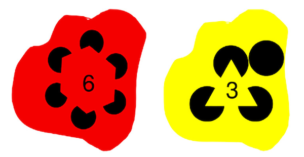
Figure 7. Twenty-eight different Kanizsa figures were used in this study, plus nine probes of the type shown in the right of this figure. Griffin was asked to describe how many corners were present in the object created by “pac-men” figures. Note: Most experiments in the non-human literature use one identical figure for testing. Again, no numbers were present on Griffin’s stimuli.
The colors used were those tested in a previous study to ensure Griffin would interpret the label for these printed colors correctly (Pepperberg et al 2008). Orange and purple were adjusted within appropriate ranges as these colors can be problematic for Greys to identify (Pepperberg 1994; 2006). The ability of psittacine birds to see in the ultraviolet spectrum can cause their color perception to differ somewhat from that of humans (Pepperberg and Nakayama 2016, Carvalho et al 2011, Goldsmith and Butler 2005, Bowmaker et al 1994).
Test results showed high accuracy (Table 1). Griffin was correct on the first trial of each modal and amodal task, which suggested he immediately transferred his responses—without training—from non-occluded three-dimensional items to drawings of occluded or imaginary shapes. Griffin responded to the true shape of figures, despite the fact that all were partially hidden or not physically real. Most errors occurred for one-and three-cornered polygons and triangles, which he seemed to confuse in both modal and amodal tasks (Pepperberg and Nakayama 2016). However, these errors were few overall, and did not suggest that he was focusing solely on the 60-degree angles involved.
| Table 1. Summary of Griffin’s test results | ||
|---|---|---|
| Parameter measured | Percent accuracy * | # of trials correct |
| Modal completion | 76 | 29/38 |
| Amodal completion | 70 | 23/33 |
| *Chance is associated with 20% accuracy | ||
Conclusion
In the human visual experience, when an object is partially concealed by an obstacle, we do not perceive only the pieces or fragments of that object. The parts that are directly visible usually suffice for recognition of the whole object. Humans also tend to see objects that are created by illusions, as in the Kanizsa figures. At least one Grey parrot, Griffin, responds similarly and these results—particularly his transfer from solid three-dimensional figures to occluded and imaginary two-dimensional stimuli, without any training—imply that he has a sophisticated concept of shape. Although this information itself is unlikely to be directly applicable when planning environmental enrichment, it reminds us of the intelligence of these birds and the need for such stimulation in their environment.
References
2016 Avian Practitioner of the Year
Dr. Gregory Rich named 2016 T.J. Lafeber Avian Practitioner of the Year
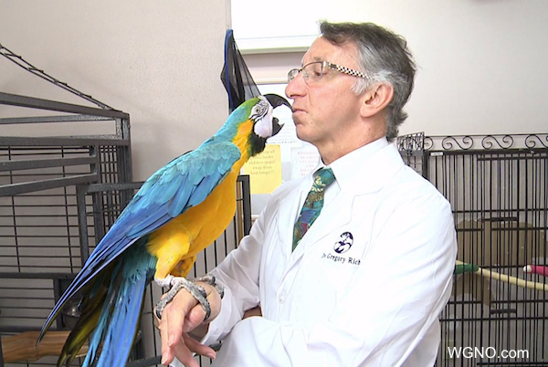
When nominations closed for the 2016 T.J. Lafeber Avian Practitioner of the Year, a list of 19 outstanding avian veterinarians were submitted for consideration. The independent Selection Committee, hosted through Louisiana State University, narrowed the list to five finalists and the Award recipient, Dr. Gregory Rich, was announced during the Opening Session at ExoticsCon 2016 on Monday, August 29.
Dr. Rich truly embodies the spirit of this Award, exhibiting dedication and compassion for his avian patients as well as clinical excellence. [MORE]

Award recipients
Visit Lafeber.com for information on previous Award recipients.
Did You Know…?
The T.J. Lafeber Avian Practitioner of the Year is nominated by their peers: YOU.
- The Awardee is NOT, and has never been, selected by Lafeber Company.
- This autonomous committee, consisting of Association of Avian Veterinarians members, is led by Dr. Tom Tully, Professor and Chief of the Zoological Medicine Service at Louisiana State University School of Veterinary Medicine.
- A Lafeber Company Veterinary Consultant, who does NOT vote on the Award recipient, manages committee paperwork and scheduling only.
Don Harris, DVM
 Don Harris is the owner of Miami’s first exotics-only veterinary hospital, Avian & Exotic Animal Medical Center. Don is a graduate of Louisiana State University School of Veterinary Medicine. Although his Cajun background in the swamps of Louisiana did nothing to prepare him for life in Miami, it did provide for him a wealth of experience in handling wildlife and exotic animals. In 1990, after several years in mixed animal practice, his practice became exclusively avian and exotic. The Avian & Exotic Animal Medical Center was opened in 1996. Don serves as a consulting veterinarian for the Georgia Aquarium Research Center, Miami MetroZoo and Jungle Island in Miami and he is on the Advisory Board of Abaxis Veterinary Diagnostics. Don has also served as Past-President of the South Florida Veterinary Medical Association, the Association of Avian Veterinarians (AAV), and the North American Veterinary Community, organizer of the North American Veterinary Conference, the largest veterinary conference in the world. Dr. Harris has presented at veterinary conferences both nationally and internationally, and he has also received many commendations over the years from various institutions and associations including AAV Speaker of the Year (2014).
Irene Pepperberg, PhD
Irene Pepperberg, PhD is a Research Associate and Lecturer at Harvard University in Cambridge, Massachusetts. Dr. Pepperberg’s work is in the field of animal cognition, specifically African Grey parrots. The main focus of her work is to determine the cognitive and communicative abilities of these birds, and compare their abilities with those of great apes, marine mammals, and young children. She is studying the mechanisms of their learning as well as the outcomes.
In 1977, Dr. epperberg began her work with Alex, her first Grey parrot research subject. For 30 years, Irene and Alex investigated concepts such as shapes, colors, size, and numbers, revolutionizing what the world knows about avian cognition. Following in his footsteps are the Grey parrots, Griffin and Athena. Currently, Dr. Pepperberg’s work is entirely supported by The Alex Foundation. Visit The Alex Foundation website or their Facebook page.
Kirk Klasing, PhD
Dr. Kirk Klasing is a Professor in the Department of Animal Science at the University of California at Davis . . .
Pour les vétérinaires. Par les vétérinaires.
Le site Lafervet.com est conçu pour une utilisation par les vétérinaires. Il est ouvert aux vétérinaires diplômés, aux techniciens vétérinaires diplômés, aux animaliers et aux étudiants dans ces domaines.
Créer un compte pour accéder à des articles et des ressources du site.
L'inscription est gratuite.
Para Profesionales Veterinarios. Por Profesionales Veterinarios.
El sitio Lafervet.com es para uso de los profesionales veterinarios. Está abierto a los veterinarios licenciados, técnicos veterinarios licenciados, rehabilitadores licenciados y estudiantes en estos campos.
Cree una cuenta para acceder a los artículos y recursos del sitio.
La registro es gratis.
Already a LafeberVet Member?
Please Login
Best Practices: Cytodiagnosis in Exotic Pet Practice
View a recording of this web-based seminar presented by Terry Campbell, MS, DVM, PhD. Cytology is a simple, rapid diagnostic procedure requiring little in terms of equipment and cost to the veterinarian. Most clinical veterinarians are familiar with sample collection techniques for domestic mammals; which also apply to the small exotic mammals. Common cytological specimens used in avian and reptilian medicine include: aspirates, imprints of biopsy material, tracheal washes, crop (ingluvies) aspirates or washes in birds, gastric washes in reptiles, sinus aspirates, lung washes in reptiles, aspiration of coelomic fluid, and fecal smears . . .
Pour les vétérinaires. Par les vétérinaires.
Le site Lafervet.com est conçu pour une utilisation par les vétérinaires. Il est ouvert aux vétérinaires diplômés, aux techniciens vétérinaires diplômés, aux animaliers et aux étudiants dans ces domaines.
Créer un compte pour accéder à des articles et des ressources du site.
L'inscription est gratuite.
Para Profesionales Veterinarios. Por Profesionales Veterinarios.
El sitio Lafervet.com es para uso de los profesionales veterinarios. Está abierto a los veterinarios licenciados, técnicos veterinarios licenciados, rehabilitadores licenciados y estudiantes en estos campos.
Cree una cuenta para acceder a los artículos y recursos del sitio.
La registro es gratis.
Already a LafeberVet Member?
Please Login
Rabbit Anatomy Basics Slideshow
Part of LafeberVet’s Rabbit Basics Teaching Module, the Rabbit Anatomy Basics slideshow is a 22-minute recording designed to impart a basic understanding of rabbit anatomy for the veterinary technician and veterinary nurse. This slideshow may also be of use as a basic learning aid for veterinary medical students and as a basic refresher for the clinician . . .
Pour les vétérinaires. Par les vétérinaires.
Le site Lafervet.com est conçu pour une utilisation par les vétérinaires. Il est ouvert aux vétérinaires diplômés, aux techniciens vétérinaires diplômés, aux animaliers et aux étudiants dans ces domaines.
Créer un compte pour accéder à des articles et des ressources du site.
L'inscription est gratuite.
Para Profesionales Veterinarios. Por Profesionales Veterinarios.
El sitio Lafervet.com es para uso de los profesionales veterinarios. Está abierto a los veterinarios licenciados, técnicos veterinarios licenciados, rehabilitadores licenciados y estudiantes en estos campos.
Cree una cuenta para acceder a los artículos y recursos del sitio.
La registro es gratis.
Already a LafeberVet Member?
Please Login
Bret Meckel, DVM

Bret Meckel, DVM is a Professor and serves as Department Chair for the Veterinary Technology Program at State University of New York in Delhi. Dr. Meckel has worked extensively with exotics during 10 years of private practice and he teaches an Exotic Animal Nursing elective for veterinary technicians interested in exotic animal practice. Dr. Meckel has also developed a study abroad program where veterinary technician students can participate in a 3-week wildlife management program in South Africa.
Rabbit Breed Basics Slideshow
The domestic or European rabbit, Oryctolagus cuniculus, is descended from wild rabbits of Europe and northwestern Africa, where free-ranging Oryctolagus are still found. Rabbits come in many different shapes, sizes, and colors. The American Rabbit Breeder Association currently recognizes 49 rabbit breeds, and the number listed by the British Rabbit Council is even higher. Many house rabbits have several different breeds in their background. View this 8-minute slideshow to review common rabbit breeds seen in clinical practice as well as their associated disease predispositions. This slideshow is part of the Rabbit Basics Teaching Module . . .
Pour les vétérinaires. Par les vétérinaires.
Le site Lafervet.com est conçu pour une utilisation par les vétérinaires. Il est ouvert aux vétérinaires diplômés, aux techniciens vétérinaires diplômés, aux animaliers et aux étudiants dans ces domaines.
Créer un compte pour accéder à des articles et des ressources du site.
L'inscription est gratuite.
Para Profesionales Veterinarios. Por Profesionales Veterinarios.
El sitio Lafervet.com es para uso de los profesionales veterinarios. Está abierto a los veterinarios licenciados, técnicos veterinarios licenciados, rehabilitadores licenciados y estudiantes en estos campos.
Cree una cuenta para acceder a los artículos y recursos del sitio.
La registro es gratis.
Already a LafeberVet Member?
Please Login
Basic Rabbit Care Handout and Infographic
A sound rabbit diet consists of fresh hay, good-quality pellets, fresh vegetables, and water. Anything beyond this is considered a treat and should be offered in limited quantities only. This client education handout reviews not only diet, but also housing, including “bunny proofing”, and handling.
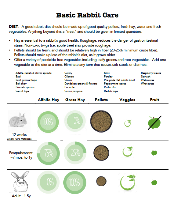
Download the Rabbit Care PDF Handout
Test your knowledge
This Basic Rabbit Care handout and infographic are part of LafeberVet’s Basic Rabbit Care Teaching Module. Please take this brief quiz to test your knowledge.
Green circle leaf image credit: glopphy via 123RF Stock Photo
Pigeon Disease Primer
The “Pigeon Disease Primer” explores important differential diagnoses for common clinical problems observed in pigeons and doves. Although the clinical approach to the columbiform relies on the same concepts of “One Medicine” used in all species, many of the infectious diseases of pigeons are relatively unique to this taxonomic group, or at least much more prevalent when compared to psittacine birds or songbirds . . .
Pour les vétérinaires. Par les vétérinaires.
Le site Lafervet.com est conçu pour une utilisation par les vétérinaires. Il est ouvert aux vétérinaires diplômés, aux techniciens vétérinaires diplômés, aux animaliers et aux étudiants dans ces domaines.
Créer un compte pour accéder à des articles et des ressources du site.
L'inscription est gratuite.
Para Profesionales Veterinarios. Por Profesionales Veterinarios.
El sitio Lafervet.com es para uso de los profesionales veterinarios. Está abierto a los veterinarios licenciados, técnicos veterinarios licenciados, rehabilitadores licenciados y estudiantes en estos campos.
Cree una cuenta para acceder a los artículos y recursos del sitio.
La registro es gratis.
Already a LafeberVet Member?
Please Login
Dave Rupiper, DVM

Dr. Dave Rupiper is the owner of East Petaluma Animal Hospital in Petaluma, California. After graduating from the University of California at Davis and completing four externships in avian medicine at various specialty institutions throughout California, Dr. Rupiper has been practicing veterinary medicine since 1991. Dr. Rupiper was board certified in avian practice from 2000 through 2013, and he maintains a strong interest in avian medicine as well as exotic animal practice. In addition to companion birds, he provides professional services for pigeon racing, performing and showing, poultry flocks, ratites, waterfowl collections, and cage bird breeding. Dr. Rupiper served as a co-authored on two earlier editions of the Exotic Animal Formulary and he is a frequent speaker for local bird clubs, veterinary organizations, and colleges.
Order Columbiformes: Species and Breeds
Introduction
Pigeons and doves belong to order Columbiformes and family Columbidae. Within family Columbidae, there are five subfamilies consisting of 42 genera and 308 species (Box 1) (Fig 1-Fig 5). Pigeons and doves are found on every continent except Antarctica, and they live in virtually all types of terrestrial habitats (Camfield 2004, Hooimeijer and Dorrestein 1997).
| Box 1. Family Columbidae includes... | |
|---|---|
| Subfamily Columbinae | Doves and pigeons |
| Subfamily Didunculinae | Tooth-billed pigeon or Manumea |
| Subfamily Gourinae | Crowned pigeons |
| Subfamily Otidiphabinae | Pheasant pigeons |
| Subfamily Treroninae | Fruit doves, imperial pigeons |

Figure 1. Pigeons and doves, like these racing pigeons shown here, belong to subfamily Columbinae. Image by “kellinahandbasket” via Flickr Creative Commons. Click image to enlarge.

Figure 2. The tooth-billed pigeon (Didunculus strigirostris) of subfamily Didunculinae. Photo by Museum de Genève via Wikimedia Commons. Click image to enlarge.
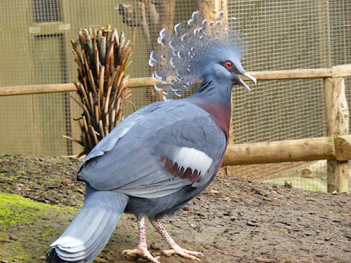
Figure 3. Standing up to 75 cm (30 in) in height, the Victoria crowned pigeon (Goura victoria) of subfamily Gourinae is considered the largest surviving species of pigeon on earth. Photo by Marie Hale via Flickr Creative Commons. Click image to enlarge.

Figure 4. Pheasant pigeons, like the green-naped pheasant pigeon (Otidiphaps nobilis), are large, terrestrial pigeons found primarily in the rainforests of New Guinea and nearby islands. Photo by Drew Avery. Click image to enlarge.
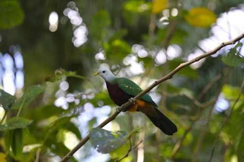
Figure 5. Fruit doves, like the Wompoo fruit dove (Ptilinopus magnificus) shown here, are colorful, fruit-eating doves found in the forests and woodlands of Southeast Asia and Oceania. Photo by cuatrok77 via Flickr Creative Commons. Click image to enlarge.
Columbids tend to be stocky birds with relatively small heads, short beaks, as well as a fleshy cere and a bare ring of skin around the eyes. Columbiforms also tend to have short, squat legs and long keels. Small columbiforms are generally called “doves” while larger birds are often called “pigeons”. Granivorous species tend to be understated beige, brown, or grey colors while frugivores are more brightly colored (Camfield 2004).
TRUE PIGEONS
True pigeons belong to subfamily Columbinae, which consists of 21 genera and 46 species, including the extinct passenger pigeon (Ectopistes migratorius).
Genus Columba: Pigeons
The rock dove (Columba livia) is the common ancestor of all domestic pigeons (Columba livia domestica). Domesticated pigeons that have returned to the wild are also descended from the rock dove and are called city or street pigeons or incorrectly feral pigeons (Hooimeijer and Dorrestein 1997). Street pigeons are found almost exclusively in urban areas near human beings (Pigeon Control Resource Centre 2009).
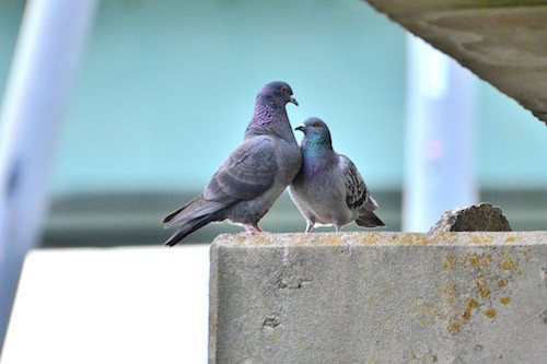
Figure 6. The rock dove (Columba livia) is the common ancestor of all domestic pigeons, including the street pigeons shown here. Photo by Toshihiro Gamo via Flickr Creative Commons. Click image to enlarge.
The common wood pigeon (Columba palumbus) is the most common pigeon found in the United Kingdom (Pigeon Control Resource Centre 2009, BBC News 2005). The common wood pigeon is a large bird. It is primarily grey with white on its neck and wing and pink-tinged feathers on its breast. The common wood pigeon is abundant in southern and western Europe, and it migrates to northern and eastern parts of Europe and western Asia. This species is classified as Least Concern on the International Union for Conservation of Nature (IUCN) Red List.

Figure 7. The common wood pigeon (Columba palumbus) is the most common pigeon in the United Kingdom. Photo by Tim Dutton via Flickr Creative Commons. Click image to enlarge.
Genus Zenaida: Zenaida doves
Mourning doves (Zenaida macroura) are common throughout the United States. Their range extends into southern Canada and down into Panama, and this highly adaptable species is found in a variety of habitats. The mourning dove is the most widespread and abundant game bird in North America, and hunters kill approximately 45 million birds on an annual basis. Fortunately this species is categorized by the IUCN with a Conservation Status of Least Concern.
The mourning dove possesses a long, narrow tail and a small head (Fig 8). This species also exhibits sexual dimorphism. The male tends to be larger and slightly brighter in color than the female. Male mourning doves also possess a bluish crown and a rosy breast.

Figure 8. The mourning dove (Zenaida macroura) is a well-managed game bird in North America. Photo by slodocents via Flickr Creative Commons. Click image to enlarge.
Genus Streptopelia: Turtle doves
The ringed turtle dove (Streptopelia risoria), also known Barbary dove, originates from the savannahs and dry woodlands of Africa, however release of captive bred specimens has also led to feral colonies in California, southern Florida, and parts of Illinois USA (Pappas 2002). The ringed turtle dove is a warm creamy buff color with a black semi-collar on the back of its neck. The chin, belly, and tail coverts are white-tinged.

Figure 9. The ringed turtle dove (Streptopelia risoria) is also known as the Barbary dove. Photo by Derek Keats via Flickr Creative Commons. Click image to enlarge.
The Eurasian collared dove (Streptopelia decado) is native to Asia and Europe. The species was introduced to the Bahamas in the 1970s. From there, feral populations made their way to Florida by the 1980s and then rapidly colonized most of North America. The Eurasian collared dove possesses white tail feathers, dark-tipped wings, as well as a black half-collar on the nape.

Figure 10. Feral colonies of the Eurasian collared dove (Streptopelia decado) are found in North America. Photo by Dawn Beattie via Flickr Creative Commons. Click image to enlarge.
Genus Geopelia: Ground doves
The zebra dove (Geopelia striata) and diamond dove (Geopelia cuneata) are native to the open country and scrubland of southeast Asia and Australia. These small doves, with long narrow tails are commonly kept in captivity.

Figure 11. Diamond doves (Geopelia cuneata) are commonly kept in captivity. Photo by Leonardo DaSilva via Flickr Creative Commons. Click image to enlarge.
Genus Gallicolumba: Island ground doves
Island ground doves include the Indo-Pacific ground doves and the bleeding heart doves. These mid-sized columbids are found in the rainforests of the Philippines, Indonesia, and in the Pacific region.
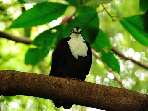
Figure 12. The white-breasted ground dove (Gallicolumba jobiensis) is an island ground dove belonging to genus Gallicolumba. Photo by Kierean Palmer via Flickr Creative Commons. Click image to enlarge.
Genus Caloenas: Nicobar pigeon
The Nicobar pigeon (Caloenas nicobarica) is native to the small islands of Malaysia and Polynesia. This beautiful bird has iridescent green feathers over its dorsum and wing as well as distinctive long feathers that stream down its neck. The Nicobar pigeon is the closest living relative of the dodo (Raphus cucullatus), a large, flightless bird that went extinct in the 1600s (WAZA). The IUCN Red List status of the Nicobar pigeon is Near Threatened.
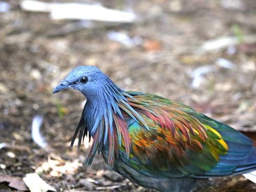
Figure 13. The Nicobar pigeon (Caloenas nicobarica) is a frugivorous species native to the islands of Malaysia and Polynesia. Photo by cuatrok77 via Flickr Creative Commons. Click image to enlarge.
FANCY BREEDS OF PIGEONS
Pigeon fanciers have bred domestic varieties of the rock pigeon for many different colors, patterns, shapes, sizes, and behaviors. There are over 300 known breeds or varieties of domestic pigeon; birds are often exhibited at pigeon shows, fairs, and livestock exhibits. The body weight of mature birds ranges from 150-180 grams in toy breeds like the African owl to greater than 1400 grams in the American giant runt. Depending on their breed and time of birth, pigeons reach maturity between 5 to 12 months of age. Toy breeds mature faster than late-hatch giant breeds (Harlin 2000).
Utility pigeons
Originally bred for meat production, utility pigeon breeds include the French mondain (Fig 14), the king (Fig 15), and the American giant runt (Fig 16). The giant runt is a very large, heavy bird. The average length of the giant runt is approximately 0.5 m (20 in) with a 1 m (40 in) wingspan. The body weight of this breed has been known to exceed 1.4 kg (3 lb) (Whitby 2013).

Figure 14. The French mondain is a breed of utility or meat production pigeon. A “blue grizzle” is shown here. Image by Jim Gifford via Wikimedia Commons. Click image to enlarge.
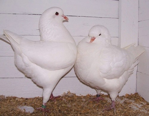
Figure 15. King pigeons are another meat production pigeon. Image by Graham Manning via Wikimedia Commons. Click image to enlarge.

Figure 16. American giant runt. Image by Graham Manning via Wikimedia Commons. Click image to enlarge.
Homing pigeons
Homing pigeons, also known as “homer pigeons” or “messenger or carrier pigeons”, are essentially the marathon athlete of the pigeon world (Rupiper 1998). These birds are used to carry messages and are bred specifically for their ability to find their way home over extremely long distances (Fig 17). Flights as long as 1800 km (1100 miles) have been recorded in competitive races. The average flying speed documented is 80 kilometers per hour (50 mph).

Figure 17. Homing pigeons are bred specifically for their ability to find their way back to their home or loft. Image by Mary Witzig via Wikimedia Commons. Click image to enlarge.
Homing pigeons include breeds designed specifically for show including the English carrier, Dragoon, and German beauty homer (Fig 18). Most racing “homers” weigh between 360 to 480 grams.

Figure 18. Stuffed specimen of a black English carrier pigeon at the Chicago Field Museum. Image by ‘UCFFool’ via Wikimedia Commons. Click image to enlarge.
Tumbler pigeons
Tumbler pigeons, such as high flyers, sustained flyers, flying tipplers, and rollers, are bred for show purposes but can also be used in flying competitions for their acrobatic abilities (Pigeon Control Resource Centre 2009, Hooimeijer and Dorrestein 1997). Some exhibition tumblers, including nuns, English short faced tumblers, and magpie pigeons were originally bred for their acrobatic abilities but crossbreeding has created show birds (Fig 19-Fig 22) (Pigeon Control Resource Centre 2009).

Figure 19. Nun pigeon (black) by Jim Gifford via Wikimedia Commons
Figure 20. Komorner tumbler by Jim Gifford via Wikimedia Commons
Figure 21. Danish tumbler red magpie by Omar Runólfsson
Figure 22. Australian saddleback tumbler by Jim Gifford via Wikimedia Commons
Asian feather and voice pigeons
Specialized breeds, including the fantail, trumpeter and Jacobin, possess extensive feathering as well as a laughing or ‘trumpeting’ voice (Fig 23, Fig 24) (Pigeon Control Resource Centre 2009). Fantail pigeons have rounded bodies and large, flat tails that look like tiny peacocks. These docile birds are a very popular and very old breed.

Figure 23. Fantail fancy pigeons. Photo credit: Todd.vision

Figure 24. Red Jacobin pigeon. Photo credit: UCFFool
Color pigeons
There are many different varieties of pigeons bred for their beauty with specific coloring or markings (Fig 25). These varieties may display pastel colors of soft blues, grays, striking black and white, and many different patterns. The swallow, Medena, Danish Suabian (Fig 26), and archangel are just a few examples of color pigeon breeds. Archangels are black pigeons with a coppery sheen. Medenas are stocky, gaily marked pigeons with short tails (Pigeon Control Resource Centre 2009).

Figure 25. Blue barred spot swallow with feathered toes known as “muffs”. Photo credit: “UCFFool” via Flickr Creative Commons
Figure 26. Danish Subian. Photo credit: Omar Runólfsson
Pouters and croppers
Pouters and croppers, such as the English pouter, Norwich cropper, and pygmy pouter, possess long, pencil-like legs and the ability to inflate the crop with air so that this region appears like a balloon. Some pouters are crested, others have feathered feet and legs (Fig 27) (Pigeon Control Resource Centre 2009).

Figure 27. Shown here, a reversewing pouter by Omar Runólfsson via Flickr Creative Commons. Click image to enlarge.
Frills and owls
Breeds like the old German owl, Oriental frill, and Aachen Lacquer shilled owl possess stunted beaks and extraordinary chest feathers (Fig 28-Fig 30).
Figure 28. Shown here, Oriental frills -satinette by Ómar Runólfsson via Flickr Creative Commons. Click image to enlarge.
Figure 29. African owl pigeon by Ómar Runólfsson via Flickr Creative Commons. Click image to enlarge.
Figure 30. Shown here, African owl pigeon by Ómar Runólfsson via Flickr Creative Commons. Click image to enlarge.
Conclusion
Pigeons and doves belong to order Columbiformes and family Columbidae. There are many possible species, breeds, and varieties that may be seen in clinical practice however some of the most common specimens are members of Genus Columba such as the racing pigeon, fancy pigeon, and street pigeon.
References
Pigeon Fancy: Talking the Talk
This practice of pigeon fancy is generally called “pigeon racing” in North America and “pigeon play” in Europe. The principles of pigeon fancy are intricate and there are a variety of vocabulary terms that seem quite mysterious. There is also a history of resistance to veterinary involvement among many pigeon breeders so it can be intimidating for even experienced avian veterinarians to interact with the pigeon fancier . . .
Pour les vétérinaires. Par les vétérinaires.
Le site Lafervet.com est conçu pour une utilisation par les vétérinaires. Il est ouvert aux vétérinaires diplômés, aux techniciens vétérinaires diplômés, aux animaliers et aux étudiants dans ces domaines.
Créer un compte pour accéder à des articles et des ressources du site.
L'inscription est gratuite.
Para Profesionales Veterinarios. Por Profesionales Veterinarios.
El sitio Lafervet.com es para uso de los profesionales veterinarios. Está abierto a los veterinarios licenciados, técnicos veterinarios licenciados, rehabilitadores licenciados y estudiantes en estos campos.
Cree una cuenta para acceder a los artículos y recursos del sitio.
La registro es gratis.
Already a LafeberVet Member?
Please Login
Madeline Rae, DVM, MS, DABVP (Avian Practice)

Dr. Madeline Rae received her Bachelor’s degree in Microbiology in 1981 from Oregon State University and her DVM in 1985 from Oregon State University. After a few years of clinical practice, she returned to academia for a residency in veterinary pathology and a Master’s of Science degree. In 1993, she became certified in Avian Practice by the American Board of Veterinary Practitioners, re-certifying in 2003 and 2013. For the last 22 years, she has been the owner of a private diagnostic pathology service focusing on birds (exotic pet birds and wildlife), small mammals and reptiles. Madeline has been a reviewer for the Journal of Avian Medicine and Surgery for 20 years and she has presented several talks at the Association of Avian Veterinarians Annual Conference. She has also taught avian medicine to veterinary medical students at Oregon State University since 1991.
How Did We Get Off the Goo?
Introduction
Many people have been curious about the way we at International Bird Rescue were able to clean the birds affected by the San Francisco Bay Mystery Goo Spill in January 2015 (Fig 1).
Figure 1. Surf scoter (Melanitta perspicillata) treated with baking soda paste during removal of the mystery goo. Photo credit: International Bird Rescue.
Before the wash
When faced with an unknown contaminant that needs to be removed from an animal, we first try what is known to work well for other contaminants on a few plucked feathers. A typical and effective wash for petroleum contamination involves Dawn® dish soap (Procter and Gamble) with or without pre-treatment with a non-toxic solvent called methyl soyate, made from soybeans. Pre-treatment agents help break up dry or weathered asphalt-like material. Unfortunately Dawn plus methyl soyate did not do the job in the case of “goo” birds.
When our typical wash procedure does not do the trick, we have to get creative, however a wash procedure must show good odds of success before we will subject a live bird to the stress of washing. We are also limited in what solvents can be used because many agents are too toxic for use on live animals, such as non-polar solvents like hexane. Hence, we generally only use products that are safe for human use.
We initially thought the “mystery goo” might be a silicone-based product, which we know from experience is very hard to remove. Our “bird-washing wizards” (i.e. IBR senior rehabilitation staff) tried a variety of products like baby oil, vinegar, and other options on “gooed” feathers from birds that did not arrive alive. A variety of procedures and application of products in different orders, using different methods for varying lengths of time were tried.
After much experimentation, we decided to try the most effective procedure on a few live birds, and then see if the birds were able to become waterproof afterwards. We wanted to know how the first group did before inflicting the long procedure on more birds. Thankfully, despite taking about twice as long as a wash for an oiled bird, this technique worked acceptably well and we were able to proceed with cleaning all the birds. Below is the procedure we used (Table 1).
CAVEAT: As in all animal care, the condition of the animal is of supreme importance. First and foremost any bird being cleaned must be medically stable before any attempts at cleaning, lest the stress of the washing kill the bird. Extreme care must be taken to prevent the bird from becoming chilled or overheated during washing, rinsing, or drying. All cleaning products must be thoroughly rinsed off at the end of the wash, as these products also contaminate the plumage.
Cleaning “mystery goo” birds
This goo removal technique first involved application of “pre-treatment” agents worked into the feathers, then birds were washed and finally rinsed until water began to bead up and roll off the plumage (Fig 2-Fig 4). While resting in a clean cage, birds are dried, while being carefully monitored for evidence of overheating (Table 1).
|
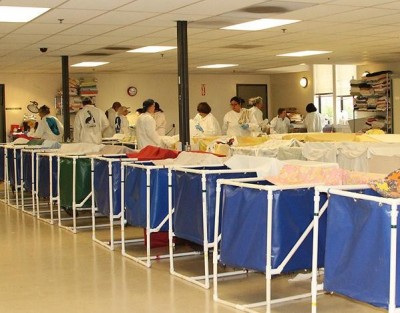
Figure 2. When housed out of water, seabirds are kept in soft sided, net-bottom pens to prevent captivity-related lesion formation. These are standard housing for non-waterproof seabirds during oil spills. Photo credit: International Bird Rescue.

Figure 3. Methyl soyate being applied to a horned grebe (Podiceps auritus), and worked into the feathers with a soft toothbrush. Photo credit: International Bird Rescue.
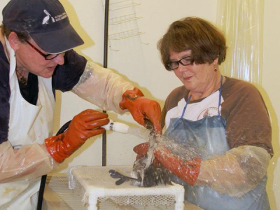
Figure 4. All cleaning products must be thoroughly rinsed off before the bird will be able to become waterproof again. Photo credit: International Bird Rescue.
After the “mystery goo” was removed, the birds entered the waterproofing process, a completely separate and complex process in which birds are managed so they can begin to preen and realign feathers.
Further reading
Massey JG. Summary of an oiled bird response. J Exotic Pet Med 15(1):33-39, 2006.
Miller E, Bryndza H, Milionis C, et al. An evaluation of the efficacy of eighty-six products in the removal of petrochemicals from feathers. Proc Sixth International Effects of Oil on Wildlife Conference 2000: 55-66.
Munaweera K, Ngeh LN, Bigger SW, Orbell JD. Towards a rational choice for pre-treatment agents for the cleansing of oiled wildlife. Proc Annu Conf Australian Wildlife Rehabilitation Conference. 2012. Available at http://www.awrc.org.au/uploads/5/8/6/6/5866843/orbell_kasup_oiled_2012.pdf. Accessed Sep 12, 20015.
Ratliff C, Gentry J, Schmalz S, Heatley JJ. Veterinary response to animal contamination. J Exotic Pet Med. Published electronically Aug 21, 2015.
U.S. Fish and Wildlife Service. Best Practices for Migratory Bird Care During Oil Spill Response. November 2003. Available at http://www.fws.gov/contaminants/Documents/best_practices.pdf. Accessed September 8, 2015.
Ziccardi M. Oil spill: Oil effects on wildlife & general medical concerns. Proc Annu Conf American Board Vet Practitioners 2011. Available at http://www.vin.com/members/cms/project/defaultadv1.aspx?id=4832946&pid=11330&catid=&. Accessed on Sep 7, 2015.
Teresa Bradley Bays, DVM, CVA, DABVP (Exotic Companion Mammal Practice), CVMMP, CTPEP
 Teresa Bradley Bays is an American Board of Veterinary Practitioners Diplomate in Exotic Companion Mammal Practice and a Certified Veterinary Acupuncturist. She is also certified in Veterinary Medical Manipulation and Palliative and End-of-Life Care. Dr. Bradley Bays was the owner of a veterinary practice for almost 30 years. Today, she is semi-retired and currently owns 4 Paws Rejuvenation, which offers alternative veterinary therapies. Dr. Bradley Bays has lectured nationally and internationally and she has authored numerous articles in exotic animal medicine as well as several chapters for Veterinary Clinics of North America: Exotic Animal Practice. Teresa is also the senior editor and coauthor of Exotic Pet Behavior – Birds, Reptiles and Small Mammals, and the author of behavior chapters in both Ferrets, Rabbits, and Rodents and Mader’s Reptile and Amphibian Medicine and Surgery, Third Edition. Teresa is a Past-President of the Association of Reptile and Amphibian Veterinarians and a Past-President of the Missouri Academy of Veterinary Practitioners.
Lauren Thielen, DVM, DABVP (Avian Practice)
 Lauren Thielen is currently a Clinical Assistant Professor in the exotic animal medicine section at Cornell University College of Veterinary Medicine in New York. Dr. Thielen also serves as an adjunct professor for the University of Miami Miller School of Medicine Comparative Pathology Avian & Wildlife Laboratory, providing clinical pathology case consultations. Lauren graduated from Texas A&M College of Veterinary Medicine in 2013. She then completed an internship at Broward Avian and Exotic Animal Hospital in Florida and an avian residency at the Veterinary Center for Birds & Exotics in New York. After completing her training, she was the founding Medical Director of Texas Avian & Exotic Hospital in Grapevine, Texas. Dr. Thielen became a Diplomate of the American Board of Veterinary Practitioners in avian practice in 2020. Dr. Thielen is featured on the television shows “Dr. T, Lone Star Vet” and “Dr. K’s Exotic Animal ER” on NatGeo, her platform for educating people all over the world about exotic animal care.
External Coaptation in Birds: Bandages and Splints
Traumatic orthopedic injuries are relatively common in the avian patient. Although bird bones are strong when intact, they tend to shatter on impact as the cortices are thin and brittle. A lack of abundant soft tissue coverage often leads to open fractures . . .
Pour les vétérinaires. Par les vétérinaires.
Le site Lafervet.com est conçu pour une utilisation par les vétérinaires. Il est ouvert aux vétérinaires diplômés, aux techniciens vétérinaires diplômés, aux animaliers et aux étudiants dans ces domaines.
Créer un compte pour accéder à des articles et des ressources du site.
L'inscription est gratuite.
Para Profesionales Veterinarios. Por Profesionales Veterinarios.
El sitio Lafervet.com es para uso de los profesionales veterinarios. Está abierto a los veterinarios licenciados, técnicos veterinarios licenciados, rehabilitadores licenciados y estudiantes en estos campos.
Cree una cuenta para acceder a los artículos y recursos del sitio.
La registro es gratis.
Already a LafeberVet Member?
Please Login
Feeding the Hospitalized Bird of Prey
Introduction
Raptors or birds of prey are a diverse group that include owls, falcons, eagles, hawks, kites, and vultures (Fig 1).

Figure 1. The group known as raptors or birds or prey are a diverse group. Photo credits: Dr. Elizabeth Daut (left), Beka Weiss via Flickr Creative Commons (FCC) (middle), and John Haslam via FCC (right). Click image to enlarge.
All raptors consume a meat-based diet ranging from the specialist diet of the fish-eating osprey (Pandion haliaetus) to a generalist diet that can include insects, mammals, birds, reptiles, amphibians, and even carrion. Other than poultry, the exact nutritional requirements of birds are unknown, however the natural raptor diet is always relatively high in protein and fat and low in carbohydrates. Whole prey diets have a calcium/phosphorus ratio of 1.5:1 as the bird actually consumes the bones as well as the meat (Kubiak 2012, Bird 1976).
Gastrointestinal specializations of the raptor
The raptor upper gastrointestinal tract differs substantially from that seen in omnivorous and granivorous birds like parrots and chickens.
- Most diurnal raptors possess a spindle-shaped crop that is particularly well developed in vultures. Owls (Order Strigiformes) lack crops, however food can be stored throughout the esophagus (Murray 2014, Klasing 1998, Duke 1997, McLelland 1979).
- The stomach can expand to hold large prey items with the assistance of mucosal ridges on the serosal surface of the proventriculus. The ventriculus is thin-walled and poorly muscled particularly in comparison to granivorous birds that use the ventriculus to grind seeds and grains (Murray 2014, Denbow 2000, Duke 1997, McLelland 1979).
- Indigestible material, like fur, feathers, and bones, are retained within the ventriculus, compacted into a pellet, and regurgitated or egested (Fig 2) (Forbes 2015, Murray 2014, Duke 1997).

Figure 2. Indigestible material is compacted into pellets or casts in the raptor ventriculus and egested. Photo credit: Dr. Elizabeth Daut. Click image to enlarge.
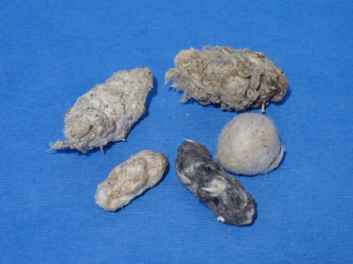
Evaluation of nutritional status
Body condition scoring
Body condition scoring (BCS) reflects changes in body composition and nutritional status much better than body weight alone. Scoring relies upon palpation of pectoral musculature, visualization of subcutaneous fat stores, and palpation of coelomic fat. The BCS scale in birds is sometimes ranked from 1 (emaciation) to 5 (obesity). Energy excess or obesity is most commonly seen in captive birds housed in permanent breeding aviaries or zoological parks (Forbes 2015, Chitty 2008). Although pectoral musculature can be atrophied from lack of use due to young age or injury, the emaciated patient has typically lost at least one-third of normal body weight and appears abnormally thin (Fig 3).
Emaciation is a common presentation in raptors that have not learned to hunt successfully during their first year. Birds can also present in poor condition due to a variety of causes such as inability to hunt or eat food, inclement weather, chronic injury, or when they become trapped in a building or some other man-made structure (Murray 2014, Handrich 1993, Garcia-Rodriguez 1987, Shapiro 1981). Energy deficits can also be observed in falconry birds as a result of inappropriate training practices (Chitty 2008).

Figure 3. Emaciation (BCS 1) in a first-year red-tailed hawk (Buteo jamaicensis). Photo credit: Dr. Elizabeth Daut. Click image to enlarge.
Blood work
Delay comprehensive testing until the patient is stable enough to handle the stress of restraint and venipuncture. Initial laboratory tests often include hematology, examination of a blood smear for parasites, total protein, packed cell volume, blood glucose, and ideally creatine kinase, lactate dehydrogenase, aspartate transaminase, and electrolytes. Anemia and hypoproteinemia are common findings in the chronically malnourished bird of prey (Fig 4) (Murray 2014, Redig 2009).
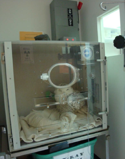
Figure 4. A snowy owl (Bubo scandiacus) rehabilitated at The Wildlife Center of Virginia. Initial testing revealed total protein 0.6 mg/dL, packed cell volume 16% and glucose 101 mg/dL. Photo credit: Dr. Elizabeth Daut. Click image to enlarge.
Normal blood glucose levels can be quite variable in raptors. Levels usually exceed 150 mg/dL (8.33 mmol/L) in healthy birds and can reach as high as 800 mg/dL (44.44 mmol/L) in some smaller species. Seizure activity can be observed if glucose levels fall below 80 mg/dL (4.44 mmol/L). Fortunately blood glucose tends to be maintained within normal limits for a relatively long period in the fasting raptor (O’Donnell 1978).
Ancillary testing
Perform fecal parasite testing (wet mount cytology/flotation), particularly for thin birds. Administer an anthelmintic as needed (Murray 2014). Case history and physical examination findings may also dictate additional diagnostic tests such as radiographs and/or endoscopy.
Metabolic needs during starvation versus stress
Starvation
Starvation is associated with a gradual decrease in the metabolic rate, resulting in hypometabolism. During starvation, the patient attempts to maintain normal blood glucose levels by increasing glycogenolysis and gluconeogenesis, while reducing glycogen stores. When glycogen stores are depleted, muscle and fat are broken down to provide substrate for gluconeogenesis. Starvation eventually affects visceral protein mass and the function of vital organs (Parrish 2005).
Glycogen reserves are usually depleted within 24 hours of fasting in the granivorous bird, however research has shown raptors may be relatively tolerant of acute food deprivation. Despite an average loss of 28% and 26% body mass during 8 and 13 days of fasting respectively, barn owls (Tyto alba) and common buzzards (Buteo buteo) were still able to fly and self-feed when food was reintroduced (Handrich 1993, Garcia-Rodriguez 1987). No significant changes in hematocrit or total protein were seen in the fasting buzzards (Garcia-Rodriguez 1987). In contrast, American kestrels (Falco sparverius) that were deprived of food for 3 days, lost 17 –20% body mass and both metabolism and body temperature decreased. Investigators concluded the kestrels would have died if fasting had lasted 5 days (Shapiro 1981).
Hypermetabolism
During physiologic stress like critical illness, the preferred energy source is lean body mass, resulting in increased body protein catabolism. Energy requirements are increased by 30–50% to sustain tissue repair, inflammatory processes, and immune function. Release of catecholamines, glucagon and glucocorticoids increases the rate of gluconeogenesis and glycogenolysis, further increasing the metabolic rate (Labato 1992). Very little is known about the hypermetabolic state in birds in general or raptors in particular, but these same principles are believed to hold true.
Supportive care
Supportive care for the chronically malnourished or emaciated raptor should include fluid therapy, supplemental heat, and measures to minimize stress. In addition to standard stress reduction measures when hospitalizing special species*, raptors can also be calmed by lightly covering the head with a towel or use of a falconry hood (Fig 5).

Figure 5. Falconry hoods are commonly used to calm birds of prey. Photo credit: Andreas Praefcke via Wikimedia Commons. Click image to enlarge.
*Editor’s note: For additional information visit Basic Husbandry: Hospitalizing Non-Traditional Pets and Exotic ICU: Nursing Care for the Avian Patient.
Fluids
Most patients presented to wildlife centers are at least 5% dehydrated, and many thin or malnourished raptors are up to 12% dehydrated. Normalize hydration status gradually over 24–48 hours, although ongoing losses need to be replaced more rapidly. Visit Fluid Therapy in the Avian Patient for additional information.
Subcutaneous fluids are an effective route of administration for mildly ill patients. Place an intravenous or intraosseous catheter for more debilitated patients, but be mindful of the risk for fluid overload in the hypoproteinemic patient. Supplement patients with thiamine and other B vitamins before and during initial re-feeding (thiamine 1–2 mg/kg SC every 24 hours) (Murray 2014). Maintenance fluid support is often continued for 3–5 days after feeding is initiated. Blood transfusion is also an option for the select critical patient with severe anemia and hypoproteinemia.
Supplemental heat
Hypothermia is very common in debilitated, emaciated raptors. Provide supplemental heat to prevent the bird from expending energy on thermoregulation. Warmed intraosseous or intravenous fluids and a heated intensive care unit can elevate both core and peripheral body temperature.
Indications for nutritional support
Indications for nutritional support range from profound weight loss and anorexia to severe illness or injury (Table 1).
| Table 2. Indications for nutritional support |
|---|
|
Calculating energy requirements
The basal metabolic rate (BMR) represents the minimum amount of energy or kilocalories (kcal) necessary to maintain the body at rest or the energy to stay alive. An estimate of BMR is based on metabolic scaling where BMR = K (Weight in kg0.75). The K factor is a theoretical constant for kilocalories used over 24 hours. The K value for non-passerine birds is 78 (Murray 2014).
Maintenance energy requirements (MER) equal BMR plus the energy needed for normal physical activity, digestion, and thermoregulation. MER is estimated as 1.5 x BMR (Forbes 2015, Murray 2014, Kirkwood 1981). Larger birds eat more food but require a significantly smaller percentage of their body mass as daily food intake. (Forbes 2015).
Tube feeding
As long as the patient possesses a functional gastrointestinal tract, enteral nutrition can be started once the patient is warm and adequately hydrated (Murray 2014). Oral electrolyte fluids like Pedialyte (Abbott) are sometimes offered before tube feedings. Avoid solutions rich in glucose as raptors have little glucose in their normal diets, and research indicates glucose solutions can hasten death in debilitated birds of prey (Dobbs 1983). View the algorithm shared by Dr. Scott Ford for a helpful summary of the approach to nutritional support for the bird of prey (Fig 6).

Figure 6. Raptor fluid resuscitation and refeeding algorithm. Click image to enlarge. Source: Dr. Scott Ford. Click here to download a PDF version (112 KB)
* Editor’s note: The European Union has recommended a ban on hetastarch (hydroxyethyl starch or HES) use in human patients and the US Food and Drugs Administration has instituted a ‘black box warning’. These colloids have been associated with acute renal injury and coagulopathy in some septic patients. A recent veterinary study has also found an increased risk of an adverse outcome including death or acute renal injury in dogs with HES therapy (Hayes 2016). Some veterinarians have completely eliminated colloid use in their practices while others select lower molecular weight HES, such as Voluven™ (Hospira) or Vetstarch™ (Abbott Labs).
Tube feeding formulas commonly fed to birds of prey include the intensive care diet Emeraid Carnivore (Emeraid LLC), as well as recovery diets like Carnivore Care (Oxbow), a/d Canine/Feline Critical Care (Hill’s), and Maximum-Calorie (Iams). Divide the patient’s caloric needs by the energy content of the tube feeding formula (calories per ml) to determine the total volume of formula needed daily. Gradually increase the total amount fed daily over at least 2–3 days until the patient is fed MER (Box 1). Debilitated patients are typically fed small volumes of formula by three or four times daily (Murray 2014). In well-trained falconry birds, where restraint is not required, this can be achieved with the use of an esophagostomy tube (Huynh et al 2014).
| Box 1. Tube feeding formula calculations |
|---|
| A 900-gram red-tailed hawk (Buteo jamaicensis) presents with a fractured right humerus. BCS 2/5. BMR = 78 (0.9)0.75 = 72 kcal/day Estimate MER as 1.5 x BMR = 108 kcal/day Emeraid Carnivore = 1.67 kcal/ml 108 kcal/day ÷ 1.67 kcal/ml = 65 ml daily |
Although calculation of caloric needs is crucial, feedings must also take estimated crop or stomach volume into account. The initial volume fed should be no more than approximately 1–1.5% of body weight, increasing to 2–3% at the next feeding, and the volume fed should not exceed 5% of body weight (Huynh 2014). When using an esophagostomy tube, a smaller volume not exceeding 1.5% is fed with an increased frequency of administration (N. Forbes, written communication, Sep 2015). Deliver formula via rubber tubing or a metal gavage tube or feeding needle (Fig 7) (Box 2).
| Box 2. Crop volume |
|---|
| Full crop volume in a 900-gram hawk is estimated as 5% of body weight in grams or 50 ml/kg = 45 ml Initial tube feedings are often started at 2.5% to 3% of body weight. 25 ml/kg = 22.5 ml 30 ml/kg = 27 ml Therefore the volume of the first tube feeding should range between 23-27 ml. |
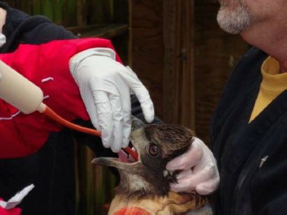
Figure 7. Tube feeding a red-tailed hawk (Buteo jamaicensis) with a red rubber catheter. Photo credit: Dr. Elizabeth Daut. Click image to enlarge.
Feeding prey foods
Some ill or injured birds of prey can self-feed upon presentation although meals should not begin until the patient is warm and rehydrated. Adult raptors are normally fed once daily and birds fed whole prey should not be re-fed until they have cast (Forbes 2015). Remove casting material, like fur and feathers, if the bird is in poor condition or if medication is given multiple times daily (Forbes 2015).
Once hydration and electrolyte abnormalities have been resolved and the patient maintains body weight well and appears enthusiastic to eat, gradually make the transition from tube feedings to solid foods. The initial meal for such patients often consists of three to four bite-sized pieces of mice or other whole prey, devoid of fur or feathers and soaked in warm water. If the patient passes stool normally (see monitoring below) and no complications are observed, then the amount of food offered can be increased gradually. Work up to several small meals daily, allowing the crop to empty between feedings. Provided the bird has regained normal body condition, continues to demonstrate a good appetite, and is passing normal feces, gradually introduce indigestible material (Murray 2014). Once the patient is stable, offer the entire prey item, including internal organs, to ensure the diet is nutritionally balanced (Fig 8) (Murray 2014, Klasing 1998). Prey should be of appropriate size so the bird can consume the entire item.

Figure 8. Whole prey items are often first offered as a “chopped plate”. Photo credit: Dr. Elizabeth Daut. Click image to enlarge.
If the meal is not eaten immediately, do not allow the food item to sit out for a long period of time as bacterial proliferation and contamination can occur (Chitty 2008). Ideally the food item should be left out no longer than 30–60 minutes (Chitty 2008), particularly when placed outdoors during warm weather months. If the bird does not begin to self-feed, continue tube feedings, offer solid food later in the day or the next day, or assist feed the patient, as clinical status dictates (Fig 9).

Figure 9. Assist feeding a hawk chopped, moistened pieces of whole prey. Photo credit: Dr. Elizabeth Daut. Click image to enlarge.
Feeding growing birds
When hand feeding juvenile birds, offer small pieces of food at a time using tongs or hemostats. Slightly moisten food items with water and carefully monitor crop emptying. Young vultures can be particularly challenging to hand raise because of their tendency to regurgitate with stress. Chitty (2008) recommends partially pre-digesting food items by first soaking food in a commercial pancreatic supplement for 30 minutes.
Young chicks are unable to egest indigestible material or ‘casting’ and are at risk for proventricular impaction and death (Forbes 2015). Do not feed indigestible material to any chick less than approximately 12 days old. Some species like the merlin (Falco columbarius) should not be fed casting until approximately 20 days of age (Forbes 2015). While whole prey diets have a calcium/phosphorus ratio (Ca:P) equivalent to 1.5:1; meat without bone has a Ca:P ratio of 1:20 (Kubiak 2012, Bird 1976). Like all growing animals, raptor chicks have increased calcium and vitamin D3 requirements and if fed a diet without adequate bone or alternative vitamin/mineral supplementation the chick is at risk for nutritional secondary hyperparathyroidism (Fig 10, Fig 11) (Chitty 2008). Deviation of long bones and multiple folding fractures are common findings in raptors exclusively fed meat-only diets (Kubiak 2012).
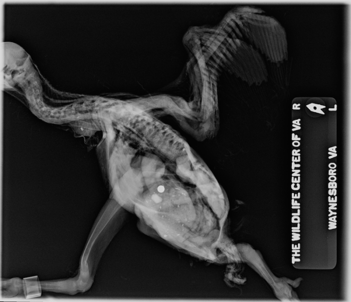
Figure 10. Lateral radiographic view of a nestling black vulture (Coragyps atratus) with nutritional secondary hyperparathyroidism or metabolic bone disease. Photo credit: Dr. Dave McRuer, The Wildlfe Center of Virginia. Click image to enlarge.
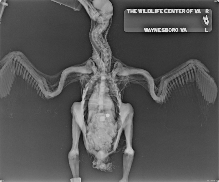
Figure 11. Ventrodorsal view of a nestling black vulture (Coragyps atratus) with metabolic bone disease. Photo credit: Dr. Dave McRuer, The Wildlfe Center of Virginia. Click image to enlarge.
Prey food options
The best diet for captive birds of prey is one that resembles the natural diet as closely as possible (Murray 2014, Chitty 2008, Cooper 2002). Fortunately most raptors in a rehabilitation setting will readily take any appropriate meat source when their preferred food is unavailable (Murray 2014, Chitty 2008), particularly when the food item is sliced open or skinned. Ospreys are a notable exception and hand feeding is often required in captivity (Murray 2014).
The nutritional composition of whole vertebrate prey is generally complete and fairly constant across different types of prey items although the fat content can vary (Murray 2014, Klasing 1998).
Mammals
Rabbits and rodents are commonly fed in captivity to raptors that feed on mammals. Although relatively expensive, laboratory mice and rats are popular food items. Captive-raised rats and mice tend to be higher in fat and lower in protein than their free-living counterparts (Fig 12) (Murray 2014, Chitty 2008, Crissey 1999, Douglas 1994). To minimize the risk of excessive dietary fat, offer sub-adult rats and mice that weigh approximately 75% of adult weight (N. Forbes, written communication, Sep 2015).
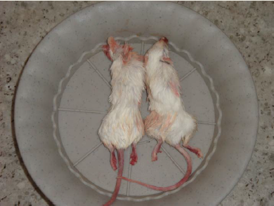
Figure 12. An adequate source of nutrition, mice are commonly fed to captive birds of prey, however laboratory-raised mice tend to be relatively high in fat. Photo credit: Dr. Elizabeth Daut. Click image to enlarge.
Birds
Birds commonly fed in a rehabilitation setting include day-old-chicks (DOC), Coturnix quail, chicken, and turkey (Forbes 2015, Murray 2014, Chitty 2008). Chicks are readily available and relatively inexpensive. Good-quality DOC contain adequate amounts of protein as well as good levels of calcium and fat-soluble vitamins. Unfortunately DOC and other avian-derived diets carry an increased risk of transmitting Salmonella spp. and other infectious agents when compared to mammalian diets. The yolk sac is the primary source of calcium and fat-soluble vitamins, therefore DOC should generally be fed with the yolk sac intact (Chitty 2008, Bird 1976) Unfortunately yolk is also a rich source of cholesterol and chicks with intact yolk sacs should not be fed long term more than twice weekly (N. Forbes, written communication, Sep 2015).
Pigeons and doves should not be fed to raptors, as several infectious agents, including Trichomonas* spp., falcon herpes virus, and tuberculosis can potentially be transmitted (Forbes 2015, Murray 2014, Redig 2009, Cooper 2002).
*Editor’s note: With regards to trichomoniasis, some clinicians report successful reduction or prevention of risk by feeding pigeons and doves that have been frozen and thawed. At this time of this posting, there are no primary references that have actually shown that protozoa are killed after freezing. Additionally in one study T. gallinae was very resistant to damage by freezing and was the only trichomonad that survived freezing and thawing for 6 months even without the aid of glycerol as a protectant (McEntegart 1954).
There is one study by Bailey et al, in which freezing T. gallinae-infected pigeon carcasses for 24h inactivated trichomonads, which might reduce the risk of disease transmission (Bailey et al 2000). Several sources also recommended that the crop, cervical esophagus, and head be removed from frozen and thawed pigeon/dove carcasses prior to feeding to decreasing the risk of transmission, although this practice cannot completely eliminate risk as trichomonads can be present in other tissues in the disease state (Ford 2010, Ford et al 2009, Forbes 2008).
Fish
Wild-caught, fresh whole trout is ideal for fish-eating birds of prey as farmed fish is generally much higher in fat (Chitty 2008). Supplement thawed frozen fish with vitamin B1, as activation of thiaminase will destroy available vitamin (Chitty 2008). Supplement thiamine at 30 mg/kg fish as fed orally every 24–48 hours (Murray 2014, Joseph 2006, Cooper 2002).
Storage and handling of prey items
Maintain all food storage tools and equipment in a hygienic manner (Chitty 2008). Utensils, cutting boards, food containers, tables, and gloves should be properly cleaned and sanitized daily (Crissey 2001). Operations that regularly feed prey items, should maintain refrigerators and freezers dedicated to meat storage (Fig 13) (Crissey 2001).
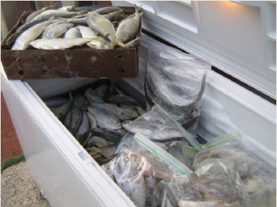
Figure 13. Store prey food in a freezer dedicated to meat storage. Photo credit: Wildlife Center of Virginia. Click image to enlarge.
Freezing food items
Partially eviscerate killed prey immediately prior to freezing, as bacteria within the gastrointestinal tract are most likely to contaminate the meat. Leave other viscera such as liver, kidney, and heart in place (Chitty 2008). Quickly freeze the carcass to prevent overgrowth of bacteria (Forbes 2015), and store carcasses in a freezer kept between –30 to –18°C (–22 to 0°F) (Crissey 2001, AZA NAG). Temperatures above –9°C (16°F) but below the freezing point can adversely affect food appearance and nutrient content (Crissey 2001). Stored meat can be kept for prolonged periods (i.e. up to a year) in a freezer with temperatures maintained at –23°C (–10°F) or lower. There are no studies reporting shelf-life recommendations for a particular species of whole prey (Crissey 2001), however frozen fish should ideally be kept no longer than 4–6 months (Bernard 1997).
Thawing food items
Freezing tends to break down tissues, making prey food more susceptible to bacterial invasion after thawing (Crissey 2001). Incorrect thawing increases the potential for not only bacterial proliferation, but also nutrient loss, loss of palatability, and lipid peroxidation or rancidity (Crissey 2001). NEVER thaw meat at room temperature (Crissey 2001, Bernard 1997). Use running water to thaw meat only in emergency situations as this method increases nutrient loss. The safest and preferred method for thawing prey foods is use of a clean refrigerator at 2°C–3.5°C (36°F–38°F) over 24 –48 hours (Forbes 2015, Chitty 2008, Crissey 2001, AZA NAG). Relative humidity should be maintained at 85-90% in refrigerated spaces (Crissey 2001). Fresh meat or thawed meat should be used within 24 hours (Crissey 2001). Never thaw, eviscerate, and re-freeze food items.
Monitoring the patient
Careful patient monitoring will allow the amount fed to be adjusted as needed (Dierenfeld 2014). Evaluate body weight and body condition score regularly and carefully observe pellets and droppings closely. Remember that a reduction in pectoral muscle mass can be attributed to reduced activity in the face of an appropriate diet.
Raptor droppings or “mutes” generally consist of dark brown to black feces surrounded by white urates and a small amount of clear urine. Feces may be tan colored in birds fed day-old chicks (Fig 14) (Redig 2009, Joseph 2006). Stool may be more watery when casting material is not fed (Cooper 2002).

Figure 14. A relatively large amount of urine and tan feces produced in a bird fed day-old chicks. Photo credit: Dr. Elizabeth Daut. Click image to enlarge.
The volume, appearance and timing of cast or pellet production vary with the species, the diet, and the individual bird (Fig 2)(Chitty 2008). Owls generally egest one pellet per meal. Hawks or falcons often egest one pellet for every two to three meals, depending on the diet fed, less often with chicks, more often with rodents. Multiple pellets can be produced if the meal is very large (Duke 1996, Duke 1976, Balgooyen 1971). Raptors fed white laboratory mice produce tan-colored pellets, while pellets from free-ranging birds tend to be darker. Fresh pellets may be covered with mucus and occasionally some bile staining on the surface. The normal pellet should be odorless.
Failure to produce a pellet can indicate dysfunction of the gastrointestinal tract. Pellet production can occur within 12–16 hours after feeding (Chitty 2008), however the range can vary widely by species, individual health status, and meal. A healthy adult red-tailed hawk may not cast a pellet for 24–48 hours after a large meal (D. McRuer, written communication, Dec 2015).
Water
Free-ranging raptors acquire most of their daily water needs through their diet, however captive raptors should always have access to fresh drinking water (Forbes 2015, Chitty 2008). Consider removing the water dish before flying a bird to avoid waterlogged feathers (Chitty 2008). Ensure water containers can be easily disinfected (Forbes 2015).
Summary
The raptor gut is a fairly simple system with a poorly developed ventriculus and a relatively underdeveloped large bowel or ceca. Nevertheless the functional raptor gastrointestinal tract is very metabolically demanding, requiring high energy and moisture, due to the formation of compacted indigestible material or pellets. Management of weak, emaciated raptors includes minimizing stress, supplemental heat, fluid therapy, as well as supplementation of thiamine and other B vitamins. Nutritional support should not be started until the patient is warm and hydration/electrolyte deficits have been addressed. Initial diagnostic testing may also need to be postponed if the patient is extremely debilitated. Very weak raptors are often tube fed initially. When the patient is carefully transitioned to solid food, rabbits, rodents like mice and/or rats, day-old chicks, and commercially available Coturnix quail are common food items offered.
References
Behavior Essentials: Clinical Approach to the Rabbit
The approach to a prey species like the rabbit often calls for a profound paradigm shift for clinicians used to dealing only with cats and dogs. Rabbits can stress very easily in a clinical setting and the challenge of managing a small mammal like the rabbit increases exponentially when they are presented for illness or injury . . .
Pour les vétérinaires. Par les vétérinaires.
Le site Lafervet.com est conçu pour une utilisation par les vétérinaires. Il est ouvert aux vétérinaires diplômés, aux techniciens vétérinaires diplômés, aux animaliers et aux étudiants dans ces domaines.
Créer un compte pour accéder à des articles et des ressources du site.
L'inscription est gratuite.
Para Profesionales Veterinarios. Por Profesionales Veterinarios.
El sitio Lafervet.com es para uso de los profesionales veterinarios. Está abierto a los veterinarios licenciados, técnicos veterinarios licenciados, rehabilitadores licenciados y estudiantes en estos campos.
Cree una cuenta para acceder a los artículos y recursos del sitio.
La registro es gratis.
Already a LafeberVet Member?
Please Login
Christine Higbie, DVM

Dr. Christine Higbie received her DVM from the University of Florida in 2009. Following graduation, she completed a rotating internship in small animal medicine and surgery at Garden State Veterinary Specialists in New Jersey. She then worked as a general practice and emergency veterinarian at Red Bank Veterinary Hospital until 2013, when she completed an internship in zoo medicine at Kansas State University. Christine is currently a resident in zoological medicine at Louisiana State University School of Veterinary Medicine.
David Brust, DVM
David Brust is a 1984 graduate of Texas A&M College of Veterinarian Medicine and has been practicing exotic animal medicine for the last 31 years. Dr. Brust currently serves as President of the Association of Sugar Glider Veterinarians, which is composed of nearly 2000 veterinarians in 30 countries. He is the Past-President of the West Houston VMA and past host of the radio talk show “Ask the Vet”. Dr. Brust is the author of Sugar Gliders the Complete Veterinary Care Guide, and the Exotic DVM article “What Every Veterinarian Needs to Know about Sugar Gliders“. He has contributed to the Exotic Animal Formulary and has taught several Veterinary Information Network courses on sugar gliders. He has also published several clinical studies on sugar glider diets and husbandry. As an outspoken advocate for sound husbandry practices, Dr. Brust has filmed over 30 educational videos for the public regarding proper sugar glider care and husbandry.
Presenting problem: Head Tilt and Rolling in Rabbits
Head tilt or torticollis, also known as “wry neck” and uncontrolled or episodic rolling are common presentations in the pet rabbit (Oryctolagus cuniculus). There are two common causes of head tilt or torticollis and rolling in the rabbit . . .
Pour les vétérinaires. Par les vétérinaires.
Le site Lafervet.com est conçu pour une utilisation par les vétérinaires. Il est ouvert aux vétérinaires diplômés, aux techniciens vétérinaires diplômés, aux animaliers et aux étudiants dans ces domaines.
Créer un compte pour accéder à des articles et des ressources du site.
L'inscription est gratuite.
Para Profesionales Veterinarios. Por Profesionales Veterinarios.
El sitio Lafervet.com es para uso de los profesionales veterinarios. Está abierto a los veterinarios licenciados, técnicos veterinarios licenciados, rehabilitadores licenciados y estudiantes en estos campos.
Cree una cuenta para acceder a los artículos y recursos del sitio.
La registro es gratis.
Already a LafeberVet Member?
Please Login
Quality Exotic Small Mammal Anesthesia

Updated video recording available
In 2023, Ms. Nugent-Deal shared an updated video recording on this topic: Exotic Small Mammal Anesthesia. This new webinar recording is RACE-approved for 1 hour of continuing education.

Dave McRuer, DVM
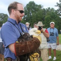
Dr. Dave McRuer obtained a Bachelor of Science degree in biology at Mount Allison University in Sackville, New Brunswick (1999), and then a Master’s degree in ecology and evolutionary biology from Carleton University in Ottawa, Ontario (2001). From 2001–2005, Dave attended the Atlantic Veterinary College and earned his veterinary degree. After graduating, he completed an internship in zoological medicine, exotics, and wildlife from the Western College of Veterinary Medicine in Saskatoon, Saskatchewan (2005–2006). Dave then completed training in preventive medicine at the Wildlife Center of Virginia (WCV) in July 2006, becoming Director of Veterinary Services in 2007. He is responsible for overseeing the WVC teaching hospital, treating and supervising treatment of native wildlife, and he also supervised the creation of an innovative software program designed to monitor trends in wildlife health: WILD-One. Dr. McRuer is a Diplomate of the American College of Veterinary Preventive Medicine and his research interests include zoonotic disease, infectious diseases of wildlife, and avian medicine and surgery.
Elizabeth Daut, DVM, PhD

Dr. Elizabeth Daut is currently a postdoctoral fellow at the National Socio-Environmental Synthesis Center (SESYNC), a National Science Foundation-funded think tank in Annapolis, Maryland. She started working with wildlife during her undergraduate studies at Virginia Tech and continued during veterinary school at Cornell University where she received her DVM degree in 1996. After working at an avian clinic in Oregon, Dr. Daut joined Peace Corps and spent most of the next 10 years in southern Ecuador where she set up an animal welfare organization. It was in Ecuador where she became interested in the consequences of and motivations for illegal wildlife trade and worked with the Environmental Ministry to develop strategies to combat the trade. Upon returning to the United States, Dr. Daut worked at the Wildlife Center of Virginia, but eventually returned for a PhD at Texas A&M University where she investigated various aspects of illegal wildlife trade, particularly domestic trade of native birds, in Peru. Her current research at SESYNC evaluates the disease risk to native wildlife from importation of animals (wild-caught and captive-bred) for the exotic pet industry.
Jeffrey Rhody, DVM, DABVP (Exotic Companion Mammal Practice)
Jeffrey Rhody is the owner and chief veterinarian at Lakeside Veterinary Center, which celebrated its 26th anniversary in June 2015. Jeff graduated from The Ohio State University College of Veterinary Medicine cum laude in 1983. He opened Lakeside Veterinary Center in 1988 and since the mid-1990s, he has seen an increasing number of non-traditional pets. Patients such as rabbits, ferrets, rats, reptiles, and birds make up approximately 40% of the patient base. Dr. Rhody also lectures at regional and national veterinary meetings on the care of exotic companion mammals. He also has served as a consultant on small mammal and exotic animal medicine for the Veterinary Information Network. Jeff became a board-certified specialist in exotic small mammal practice in 2014, making him one of the first 22 such specialists in the world and the first in Maryland.
Joanna Hedley, BVM&S DZooMed (Reptilian) DECZM (Herpetology) MRCVS

Joanna Hedley is a senior lecturer in exotic species and small mammal medicine and surgery at the Royal Veterinary College. Joanna qualified from Royal (Dick) School of Veterinary Studies in 2003 and spent time in mixed, small animal, exotic and wildlife practice before undertaking a residency in exotic animal and wildlife medicine back at R(D)SVS where she obtained her Royal College of Veterinary Surgeons (RCVS) Diploma in Zoological Medicine. Dr. Hedley is a RCVS specialist in zoological and wildlife medicine. She has also had European College of Zoological Medicine specialist status in herpetology since 2014. Joanna joined the Royal Veterinary College in 2014 as Lecturer in Exotic Species and Small Mammal Medicine and Surgery. Dr. Hedley is based at the Royal Veterinary College in Camden, where she leads the exotics clinical service. Her research interests include reptile medicine and exotic pet welfare.
Sugar Glider Disease Update: “Ick”
“Ick”: An emerging disease - A disease syndrome has recently been observed in sugar gliders that includes diarrhea, anorexia, wet, “sticky” joeys, pouch exudate, and joey deaths. The furred, but still largely pouch-dwelling joeys appear poorly groomed or are covered with tacky mucus. Clinically, the joeys are lethargic, dehydrated, with evidence of diarrhea. Some adults appear to be asymptomatic, although mild diarrhea has been noted in many cases . . .
Pour les vétérinaires. Par les vétérinaires.
Le site Lafervet.com est conçu pour une utilisation par les vétérinaires. Il est ouvert aux vétérinaires diplômés, aux techniciens vétérinaires diplômés, aux animaliers et aux étudiants dans ces domaines.
Créer un compte pour accéder à des articles et des ressources du site.
L'inscription est gratuite.
Para Profesionales Veterinarios. Por Profesionales Veterinarios.
El sitio Lafervet.com es para uso de los profesionales veterinarios. Está abierto a los veterinarios licenciados, técnicos veterinarios licenciados, rehabilitadores licenciados y estudiantes en estos campos.
Cree una cuenta para acceder a los artículos y recursos del sitio.
La registro es gratis.
Already a LafeberVet Member?
Please Login
Presenting problem: Pouch Infection and Mastitis in Sugar Gliders
The sugar glider (Petaurus breviceps) is a small marsupial native to Australia and New Guinea. Although sugar gliders lack marsupial bones, also known as epipubic bones or pelvic ribs, female gliders or “dams” possess a pouch or marsupium. Like all marsupials, the glider gives birth to a fetus, which completes development inside the pouch . . .
Pour les vétérinaires. Par les vétérinaires.
Le site Lafervet.com est conçu pour une utilisation par les vétérinaires. Il est ouvert aux vétérinaires diplômés, aux techniciens vétérinaires diplômés, aux animaliers et aux étudiants dans ces domaines.
Créer un compte pour accéder à des articles et des ressources du site.
L'inscription est gratuite.
Para Profesionales Veterinarios. Por Profesionales Veterinarios.
El sitio Lafervet.com es para uso de los profesionales veterinarios. Está abierto a los veterinarios licenciados, técnicos veterinarios licenciados, rehabilitadores licenciados y estudiantes en estos campos.
Cree una cuenta para acceder a los artículos y recursos del sitio.
La registro es gratis.
Already a LafeberVet Member?
Please Login
Presenting problem: Self-Mutilation in Sugar Gliders
Self-mutilation is sometimes observed in isolated sugar gliders or in situations causing social stress. Improper groupings are common in captivity as pet owners often combine a male with one or two females, without realizing that not all individuals get along . . .
Pour les vétérinaires. Par les vétérinaires.
Le site Lafervet.com est conçu pour une utilisation par les vétérinaires. Il est ouvert aux vétérinaires diplômés, aux techniciens vétérinaires diplômés, aux animaliers et aux étudiants dans ces domaines.
Créer un compte pour accéder à des articles et des ressources du site.
L'inscription est gratuite.
Para Profesionales Veterinarios. Por Profesionales Veterinarios.
El sitio Lafervet.com es para uso de los profesionales veterinarios. Está abierto a los veterinarios licenciados, técnicos veterinarios licenciados, rehabilitadores licenciados y estudiantes en estos campos.
Cree una cuenta para acceder a los artículos y recursos del sitio.
La registro es gratis.
Already a LafeberVet Member?
Please Login
What Did You Miss in 2015?
LafeberVet is an ever-growing online library. Although some new content is featured in email campaigns, follow us on Twitter* to keep up on all the latest posts.
Here is a list of new content released in 2015 on LafeberVet.com, EmerAid.com, and our sister sites, Lafeber Pet Birds and Lafeber Conservation.
Avian medicine
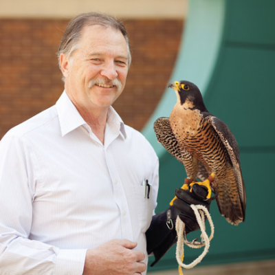
- 2015 T.J. Lafeber Avian Practitioner of the Year: Patrick Redig
- The “Mystery Goo” Bird Event
- Anatomy & Physiology of the Avian Gastrointestinal Tract: Clinical Applications, video
- Medical Management of Psittacines with Bornavirus Ganglioneuritis (PDD), video
- Nutritional Support to the Exotic Animal Patient: Theory & Practical Applications, video
- Feather Destructive Behavior in Psittacine Birds, R.A.C.E.-approved webinar, video
- Wait For It…A Grey Parrot Demonstrates Self Control
- Are You Listed? Find An Avian Veterinarian
Mammals
- Nutritional Support to the Exotic Animal Patient: Theory & Practical Applications, video
- Case Challenge: 5-Year-Old Rabbit with Anorexia & Lethargy
- Bloat and Gastrointestinal Blockage in Rabbits Client Handout
- Information Sheet: European Hedgehog
- Information Sheet: Fennec Fox
Reptiles and amphibians
- Five Common Reptile Emergencies, R.A.C.E.-approved webinar
- Nutritional Support to the Exotic Animal Patient: Theory & Practical Applications, video
- Presenting problem: “Red Leg” in Frogs
- Fast Facts on Chytrid Fungus
- Amphibian Physical Examination Form
- Amphibian History Form
- Palawan Turtle Crisis Slideshow
- Sea Turtle Physical Examination Part 1: Eyes-Ears-Nose-Throat
- Sea Turtle Physical Examination: Part 2
- Body Condition Scoring the Sea Turtle
- Basic Husbandry: Hospitalizing the Sea Turtle
- Sea Turtle Restraint
- Information sheet: Mediterranean Tortoises
- Information sheet: Egyptian Tortoise
- Information sheet: Greek or Spur-Thighed Tortoise
- Information sheet: Hermann’s Tortoise
- Information sheet: Marginated Tortoise
- Information sheet: Russian Tortoise
- Quiz: Can You Keep the Mediterranean Tortoises Straight?
- Mud Turtle Client Handout
- Leopard Tortoise Client Handout
- Chinese Box Turtle Client Handout
- Sulcata or African Spurred Tortoise Client Handout
- A Guide to Esophagostomy Tube Placement in Chelonians
Emergency medicine & critical care
- Five Common Reptile Emergencies, R.A.C.E.-approved webinar
- Animal Bite Checklist
- Surgical Safety Checklist
- Nutritional Support to the Exotic Animal Patient: Theory & Practical Applications, video
- Presenting problem: “Red Leg” in Frogs
- Case Challenge: 5-Year-Old Rabbit with Anorexia & Lethargy
- Bloat and Gastrointestinal Blockage in Rabbits Client Handout
- Critical Care Nutrition, video
- A Guide to Esophagostomy Tube Placement in Chelonians
Contributors in 2015
We are thankful for our contributors, who take time out of incredibly busy schedules to share their time and expertise.
LafeberVet and Emeraid.com contributors featured in 2015
- Kara Burns, MS, MEd, LVT, VTS (Nutrition)
- Bob Dahlhausen, DVM
- Rebecca Duerr, DVM, MPVM
- Neil Forbes, BVetMed DECZM FRCVS
- Ken Harkewicz, VMD
- Marie Kerl, DVM, MPH, DACVIM, DACVECC
- Eric Klaphake, DVM, DACZM, DABVP (Avian Practice), DABVP (Reptile & Amphibian Practice)
- La’Toya Latney, DVM
- Sean McCormack BSc (Hons), MVB, MRCVS
- Anna McRee, DVM
- Terry Norton, DVM, DACZM
- Susan Orosz, PhD, DVM, DABVP (Avian Practice), DECZM
- Aoife O’Sullivan MVB Cert AVP (ECC) MRCVS
- Christal Pollock, DVM, DABVP (Avian Practice)
- Lynne Seibert, DVM, MS, PhD, DACVB
- Jeanette Wyneken, PhD
LafeberVet Reviewers
- Susan Donoghue, MS, VMD
- La’Toya Latney, DVM
- Elisabetta Mancinelli, DVM CertZooMed DECZM (Small Mammals) MRCVS
- Javier Nevarez, DVM, PhD, DACZM, DECZM (Herpetology)
- Timothy Partridge BVSc, MRCVS
- Irene Pepperberg, PhD
Lafeber Pet Birds
Featured content on Lafeber Pet Birds includes contributions by avian veterinarians, Susan Orosz and Gregory Burkett:
- Keep Your Pet Bird Out of the ER This Holiday
- Share These Four Healthy Thanksgiving Foods
- Avian Vets Flock to ExoticsCon
- How Lafeber’s Helping Hands Changed Avian Medicine
- See What Sets Bird Hospitals Apart
- Vaccines & Pet Birds
- Avian Medicine Gets the Spotlight in Paris
- Healthy Gut, Healthier Bird
- Nutri-Berries vs. Standard Avian Diets
- Meet the Princess of Wales Parakeet
- Bird Brains are Not that Simple!
- Parrot Breeding Biology In the Wild & In the Home
- Lineolated Parakeets
- Is Your Pet Bird A Healthy Weight?
Lafeber Conservation

Editor’s note: The Lafeber Conservation website has been discontinued and these links are no longer available. Some conservation medicine content is available on LafeberVet. Also visit Dr. LoraKim Joyner’s Liberating Wings.
- Rearing to be Released: Yellow-headed Parrots in Belize
- Strange, but not Estranged Parrots from Paraguay
- Miskitos Are No Small Thing
- Paraguayan Macaws in Peril
- Team Bat Falcon Wins on a Soccer Field
- Parrot Makes Us Happy (Loros Hacen Feliz)
*For your convenience, there is open access to all LafeberVet content featured on Twitter for a limited time. There will also be open access to all 2015 content for a limited time.
Graham Zoller, DVM, IPSAV (Zoological Medicine)
 Dr. Graham Zoller practices in the Exotic Pet Department at the Fregis Veterinary Hospital Center in Paris, France where he is enrolled as a resident in avian medicine. He is also consultant at the Wildlife rehabilitation center of the National Veterinary School of Alfort. Dr. Zoller graduated from the National Veterinary School of Lyon in 2013 after having successfully defended his thesis on avian cardiovascular disorders. Then he completed an internship in Zoological Medicine at the Faculty of Veterinary Medicine of the Université de Montréal in 2014.
He is a member of the Association of Avian Veterinarians and lectures both nationally and internationally. His main clinical and research interest include avian cardiology, diagnostic imaging and development of prognostic markers in exotic pet.
Dr Zoller has served as a translator for LafeberVet Français.
Minh Huynh, DVM, MRCVS, DECZM (Avian), DACZM
![]() Minh Huynh is Head of the Exotics Department at the Centre Hospitalier Vétérinaire Frégis and Part-time Consultant at Ecole Nationale Vétérinaire d’Alfort. Dr Huynh graduated from Ecole Nationale Vétérinaire d’Alfort in 2006 and completed a rotating internship in small mammal medicine at Centre Hospitalier Vétérinaire Frégis where he stayed for a senior internship in internal medicine. In 2008, he became a resident in Avian and Exotic medicine at Great Western Exotic Referrals in England and passed the European College of Zoological Medicine Board exam in 2014 in the avian subspecialty.
Minh Huynh is Head of the Exotics Department at the Centre Hospitalier Vétérinaire Frégis and Part-time Consultant at Ecole Nationale Vétérinaire d’Alfort. Dr Huynh graduated from Ecole Nationale Vétérinaire d’Alfort in 2006 and completed a rotating internship in small mammal medicine at Centre Hospitalier Vétérinaire Frégis where he stayed for a senior internship in internal medicine. In 2008, he became a resident in Avian and Exotic medicine at Great Western Exotic Referrals in England and passed the European College of Zoological Medicine Board exam in 2014 in the avian subspecialty.
Minh Huynh is the author of numerous international peer reviewed publications in exotic internal medicine. His main areas of research are exploration of gastrointestinal disease and optimization of clinical techniques. He is also focused on determining prognosis and survival factors in common syndromes in exotic pet medicine.
Dr. Huynh has served as a translator for LafeberVet Français.
Laetitia Volait, DVM, Dipl ECZM (Small Mammal)
 Laetitia Volait received her degree in veterinary medicine from the University of Liege, Belgium in 2008. After graduation, she worked in general practice for 3 years in Luxembourg and France. In 2011, she moved in Perth, Australia for 6 months as part of her Masters of Veterinary Studies in Conservation Medicine at Murdoch University. In 2012, she completed an internship in exotic pet medicine at the National Veterinary School of Alfort, France where she is currently pursuing a 3-year residency in small mammals.
Dr. Volait has served as a translator for LafeberVet Français.
Anatomy and Physiology of the Avian Gastrointestinal Tract: Clinical Applications
As a part of the Encore ICARE 2015 Lafeber Symposium Lecture Series, Dr. Susan Orosz provided an introductory presentation on anatomy and physiology of the avian gastrointestinal tract. View a recording of this webinar today . . .
Pour les vétérinaires. Par les vétérinaires.
Le site Lafervet.com est conçu pour une utilisation par les vétérinaires. Il est ouvert aux vétérinaires diplômés, aux techniciens vétérinaires diplômés, aux animaliers et aux étudiants dans ces domaines.
Créer un compte pour accéder à des articles et des ressources du site.
L'inscription est gratuite.
Para Profesionales Veterinarios. Por Profesionales Veterinarios.
El sitio Lafervet.com es para uso de los profesionales veterinarios. Está abierto a los veterinarios licenciados, técnicos veterinarios licenciados, rehabilitadores licenciados y estudiantes en estos campos.
Cree una cuenta para acceder a los artículos y recursos del sitio.
La registro es gratis.
Already a LafeberVet Member?
Please Login
Marie Kerl, DVM, MPH, DACVIM, DACVECC
 Marie Kerl obtained her DVM from Auburn University and Masters of Public Health from the University of Missouri, and completed specialty training at the Animal Medical Center in New York. She is a Diplomate of the American College of Veterinary Internal Medicine and the American College of Veterinary Emergency and Critical Care. Dr. Kerl is a faculty member with the University of Missouri and her duties as Teaching Professor include practicing emergency/ critical care and internal medicine while instructing professional degree students, interns, and residents. Dr. Kerl has received the Norden Distinguished Teaching Award.
Surgical Safety Checklist
Introduction
The Vets Now Surgical Safety Checklist, shared by Head of Edge Programmes, Aoife O’Sullivan, includes a list of safety issues that should be read aloud, with both the veterinarian and veterinary nurse or veterinary technician in attendance. This checklist is divided into three safety categories: induction of anesthesia, before the skin incision, and before the patient leaves the operating room.

Evaluation of the patient before anesthetic induction includes the patient’s physical status classification system, initially created by the American Society of Anesthesiologists (ASA). This grading system is used for record keeping, communications between colleagues, and to create uniformity for statistical analysis. The grading system is not intended for use as a measure to predict operative risk:
- ASA 1. Healthy patient
- ASA 2. Mild systemic disease
- ASA 3. Severe systemic disease
- ASA 4. Severe systemic disease considered a constant threat to life
- ASA 5. A moribund patient not expect to survive without surgical intervention
Surgical safety checklist
Download the Vets Now checklist (223 KB PDF), posted here with permission.
Aoife O’Sullivan MVB Cert AVP(ECC) MRCVS
 Aoife O’Sullivan graduated from University College Dublin in 2001. Initially she spent a few years in equine, mixed practice and then small animal practice. She was introduced to the world of small animal emergency medicine in 2006 and has worked in emergency practice for 8 years. She joined Vets Now in 2009 and became Senior Veterinary Surgeon of the Birmingham clinic in 2010 and progressed to a District Vet role in 2011 until 2013. Aoife currently holds the position of Head of Edge Programmes, with responsibility for Vets Now’s Cutting Edge and Refresh Your Edge courses. Aoife completed her Cert AVP (ECC) with the RVC/RCVS in 2013 and is an RCVS Advanced Veterinary Practitioner in Emergency and Critical Care.
Amphibian Physical Examination Form
The amphibian examination beings with careful visual observation. Use of a small, transparent container can enhance the amphibian visual examination and minimize handling time (Clayton 2009). In an effort to further minimize handling, be prepared to perform both the exam and diagnostics at the same time (Mylniczenko 2009).
During examination of the skin, be sure to include evaluation of the foot pads and rostrum. To assess the patient’s neurologic status, evaluate movement during observed locomotion, the palpebral blink reflex (excluding caecilians), and the righting reflex.
Download the LafeberVet Amphibian Physical Examination Form as a PDF or modify the form to best meet your hospital’s needs: DOCX (90 KB).
References
Amphibian History Form
Introduction
A detailed history is mandatory for the amphibian patient as husbandry needs can have a tremendous impact on amphibian health. During initial contact, encourage the owner to bring the entire enclosure into the clinic for evaluation whenever possible. Alternatively, encourage the client to bring in photographs or a diagram of the habitat as well as any husbandry records. Although husbandry needs can vary considerably among amphibians (Table 1), most species require ambient humidity levels exceeding 70% to 80% (Mylniczenko 2009, Clayton 2007).
Visit LafeberVet’s Clinical Approach to Amphibian Emergencies and Assessing the Sick Frog or Toad for additional information.
| Natural habitat | Recommended range °C (°F) | |
| Tropical lowland, including aquatic species | 24-30 (75-86) | |
| Subtropical, including aquatic species | 21-27 (70-81) | |
| Tropical montane (moist, cool, coniferous), including aquatic species | 18-24 (64-75) | |
| Temperate, summer | 18-24 (64-75) | |
| Aquatic, temperate pond (summer) | 18-24 (64-75) | |
| Aquatic, temperature stream (summer) | 16-21 (61-70) | |
| Hibernation | 10-16 (50-61) |
Download
DOWNLOAD the LafeberVet Amphibian History Form: PDF (58 KB), DOCX (78 KB), DOC (47 KB)
References
Ken Harkewicz VMD

Dr. Ken Harkewicz earned his Veterinary Medicine Doctorate degree from the University of Pennsylvania School of Veterinary Medicine in 1981. After graduation, Dr. Harkewicz completed a 20-month post-doctorate externship in wildlife medicine in Queensland, Australia working with small marsupials, reptiles, and amphibians. Dr. Harkewicz then worked as an exotic animal veterinarian in the Bay Area beginning in 1985 and he was a longtime associate of Berkeley Dog & Cat Hospital (c. 1997). Dr. Harkewicz was a Past-President of the Association of Reptile and Amphibian Veterinarians (2011-2012) and he also served as a consultant for the Veterinary Information Network on reptile and amphibian care. Dr. Harkewicz lectured on reptiles and amphibians for veterinary technology, pre-veterinary, and veterinary medical students at the University of California (UC) Berkeley and UC Davis, as well as at local reptile and amphibian associations. Ken also hosted externs from the Western University College of Veterinary Medicine.
Dr. Harkewicz died suddenly in late August 2021. Please visit the Journal of Herpetological Medicine and Surgery for an In Memoriam by the Association of Reptile and Amphibian Veterinarians.
Medical Management of Psittacines with Bornavirus Ganglioneuritis (PDD)
Did you attend the Lafeber Symposium at the 2015 International Conference on Avian heRpetological and Exotic mammal medicine in Paris? View a recording of this encore, web-based seminar: “Medical Management of Psittacines with Bornavirus Ganglioneuritis (PDD)” by Susan Orosz, DVM, DABVP (Avian Practice), DECZM (Avian). This presentation on avian borna virus contains medium to advanced level content. The novice is encouraged to view the first hour of Dr. Orosz's presentation "Anatomy & Physiology of the Avian Gastrointestinal Tract: Clinical Applications", which includes a helpful review of avian gastrointestinal anatomy and physiology . . .
Pour les vétérinaires. Par les vétérinaires.
Le site Lafervet.com est conçu pour une utilisation par les vétérinaires. Il est ouvert aux vétérinaires diplômés, aux techniciens vétérinaires diplômés, aux animaliers et aux étudiants dans ces domaines.
Créer un compte pour accéder à des articles et des ressources du site.
L'inscription est gratuite.
Para Profesionales Veterinarios. Por Profesionales Veterinarios.
El sitio Lafervet.com es para uso de los profesionales veterinarios. Está abierto a los veterinarios licenciados, técnicos veterinarios licenciados, rehabilitadores licenciados y estudiantes en estos campos.
Cree una cuenta para acceder a los artículos y recursos del sitio.
La registro es gratis.
Already a LafeberVet Member?
Please Login
Nutritional Support to the Critical Exotic Patient
View the recording of this free, interactive webinar, presented by Neil Forbes, BVetMed DECZM (Avian) FRCVS. Many sick or injured exotic animals are presented in critical condition. More of these patients can be saved by appropriate fluids and nutritional support, than by any single medical or surgical procedure. In practical terms, providing this support is often easier said than done. Dr. Forbes’ presentation serves to demystify some of the challenges encountered; practical solutions for all exotic patients are described and discussed . . .
Pour les vétérinaires. Par les vétérinaires.
Le site Lafervet.com est conçu pour une utilisation par les vétérinaires. Il est ouvert aux vétérinaires diplômés, aux techniciens vétérinaires diplômés, aux animaliers et aux étudiants dans ces domaines.
Créer un compte pour accéder à des articles et des ressources du site.
L'inscription est gratuite.
Para Profesionales Veterinarios. Por Profesionales Veterinarios.
El sitio Lafervet.com es para uso de los profesionales veterinarios. Está abierto a los veterinarios licenciados, técnicos veterinarios licenciados, rehabilitadores licenciados y estudiantes en estos campos.
Cree una cuenta para acceder a los artículos y recursos del sitio.
La registro es gratis.
Already a LafeberVet Member?
Please Login
Presenting problem: “Red Leg” in Frogs
Red leg syndrome, also known as “pink belly disease” or bacterial dermatosepticemia, is one of the most common clinical conditions of captive frogs. Associated with peracute to acute bacterial septicemia, red leg is generally a disease of captive animals although the condition has also been implicated in rare mass mortalities of wild amphibians. This presenting problem article reviews clinical findings in red leg syndrome, pathogenesis of disease, as well as key points of urgent care and prognosis. The basics of case management are then reviewed: differential diagnoses, diagnostics, treatment, prevention and control . . .
Pour les vétérinaires. Par les vétérinaires.
Le site Lafervet.com est conçu pour une utilisation par les vétérinaires. Il est ouvert aux vétérinaires diplômés, aux techniciens vétérinaires diplômés, aux animaliers et aux étudiants dans ces domaines.
Créer un compte pour accéder à des articles et des ressources du site.
L'inscription est gratuite.
Para Profesionales Veterinarios. Por Profesionales Veterinarios.
El sitio Lafervet.com es para uso de los profesionales veterinarios. Está abierto a los veterinarios licenciados, técnicos veterinarios licenciados, rehabilitadores licenciados y estudiantes en estos campos.
Cree una cuenta para acceder a los artículos y recursos del sitio.
La registro es gratis.
Already a LafeberVet Member?
Please Login
Animal Bite Checklist
Shared by a Diplomate of the American College of Veterinary Internal Medicine and the American College of Veterinary Emergency and Critical Care, this veterinary hospital checklist provides guidance for the individual as well as the supervisor or clinician in charge. There are also recommendations for monitoring and care of the animal inflicting the bite or scratch.
Download the Animal Bite or Scratch Protocol Checklist PDF (54 KB)
Fast Facts on the Chytrid Fungus
The non-hyphal, zoosporic, chytrid fungus Batrachochytrium dendrobatidis (Bd) has caused widespread and dramatic population declines in both wild and captive amphibians worldwide. Use this table to review the basics of the infectious disease chytridiomycosis . . .
Pour les vétérinaires. Par les vétérinaires.
Le site Lafervet.com est conçu pour une utilisation par les vétérinaires. Il est ouvert aux vétérinaires diplômés, aux techniciens vétérinaires diplômés, aux animaliers et aux étudiants dans ces domaines.
Créer un compte pour accéder à des articles et des ressources du site.
L'inscription est gratuite.
Para Profesionales Veterinarios. Por Profesionales Veterinarios.
El sitio Lafervet.com es para uso de los profesionales veterinarios. Está abierto a los veterinarios licenciados, técnicos veterinarios licenciados, rehabilitadores licenciados y estudiantes en estos campos.
Cree una cuenta para acceder a los artículos y recursos del sitio.
La registro es gratis.
Already a LafeberVet Member?
Please Login
Wait For It…A Grey Parrot Demonstrates Self-Control
Introduction
A series of experiment in the late 1960s and early 1970s led by Stanford University psychology professor, Walter Mischel, were used to measure self-control in 3-5 year old children. Each child was given a treat, like a marshmallow, pretzel, or animal cookie, and promised that if she did not eat the treat right away, she would soon be rewarded with a second delicacy. When similar studies have been tried in animals, not surprisingly most species have not performed very well, often waiting only a few seconds (Hillemann et al 2014). Goffin’s cockatoos (Cacatua goffiniana) evaluated by researchers at the University of Vienna could manage to wait up to 80 seconds at best, although these birds had to hold the reward in their beaks, rather than look at it on the table (Auersperg et al 2013). Dogs, primates, and some birds like crows and ravens have done a bit better (Zielinski 2014). The longest any animal (crows) had managed to wait was 10 minutes until Griffin the grey parrot (see key points above) (Fig 1) (Hilleman et al 2014).
Figure 1. Dr. Irene Pepperberg with Griffin, a 19-year-old grey parrot (Psittacus erithacus)
Why is Griffin’s achievement significant? From a cognitive point of view, self-control is seen as a critical skill for making future-oriented decisions (Hilleman et al 2014). It can be difficult to choose a delayed reward over an immediate one, even when the delay leads to increased pay-offs (Vick et al 2010). The ability to delay gratification has particular advantages for social animals, which may benefit by sharing.
Of course there are trade-offs that exist between self-control and impulsivity with regards to resource exploitation and competition (Stephens 2008). Impulsivity or immediacy can pay off if delaying food intake increases the animal’s risk for survival (Koepke et al 2015). The longer an animal has to wait for a reward in the wild, the higher the risk of losing the desired item. If the bird waits too long, food can walk or fly away and a waiting bird can also be at risk from predators (Madhusoodanan 2014).
The study
A study in Irene Pepperberg’s lab was led by Adrienne E. Koepke of Hunter College and Suzanne L. Gray of Harvard University. The new results of their work with Griffin, a 19-year-old grey parrot (Psittacus erithacus) are now posted online ahead of print by the Journal of Comparative Psychology. To test the grey parrot’s patience, researchers began by learning his favorite foods. Rewards consisted of pairs constructed from seven highly preferred food items: cereal flakes, dried berries, cashews, almonds, crackers, as well as Nestle® Nerds, and Mars® Skittles candy. Researchers also identified three tiers of desirability existed within Griffin’s food preferences with most favored treats being the candies and his least preferred treat (although still preferred to his regular diet) were berries and cereal (Koepke et al 2015).
The test involved two treats placed in cups, one of which Griffin liked better. After placing the cup containing the less preferred food in front of Griffin, the researcher told him to “wait”. The researcher then took the other cup and either stood a few feet away or left the room. The researcher then would returned after a random length of time ranging from 10 seconds to 15 minutes. If the food was still in the cup, Griffin got the treat for which he was waiting. At every time period tested, Griffin successfully waited at least 80% of the time, even at the maximum 15 minutes. This is the best performance ever seen in an animal, comparable to Mischel’s original results with preschoolers (Zielinksi 2014).
In the early Mischel studies, children used a variety of elaborate strategies to distract themselves from the temptation of the first treat. Similarly, Griffin often displayed various coping mechanisms to deal with long delays like preening, sleeping (or at least sitting quietly with eyes closed), talking to himself, and manipulation of the available reward. For instance, sometimes Griffin tossed the cup containing the less favored treat across the room away (Video 1). Also, like children in one study, Griffin occasionally licked the treat, but did not eat it (Koepke et al 2015).
Video
Video 1. Griffin is instructed to wait and then bides his time as he sits before a tempting (but not favorite) treat. Visit Slate to see a second fun, brief video that compares Griffin to children in the Mischel study.
Griffin entered the experiment with some rudimentary knowledge of the vocal label “wait”. The researchers were careful to stress that he did not interpret “wait” as a command, however he did not have to be trained extensively before beginning the experiment (Koepke et al 2015).
I don’t believe Griffin was unique. Members of other bird species would probably be able to accomplish the same thing, given the right circumstances—Suzanne Gray (Zielinksi 2014)
Who is Irene Pepperberg?
Irene Pepperberg, PhD is a cognitive ethologist with an emphasis on animal behavior and comparative psychology. Irene is currently a Research Associate and Lecturer in the Department of Psychology at Harvard University. Her book, The Alex Studies, describes over 20 years of peer-reviewed experiments on grey parrots, received favorable mention from publications in the New York Times and Science. Her memoir, Alex & Me, was a New York Times bestseller and won a Christopher Award. Irene has presented her findings nationally and internationally at universities and scientific congresses, often as a keynote or plenary speaker, and has published numerous journal articles, reviews, and book chapters (Pepperberg 1999).
Dr. Pepperberg works with grey parrots, teaching them to communicate with humans using the sounds of English speech. She then uses this communication code to test the birds in ways similar to those used with young children (Pepperberg 1999).
In June 1977, in a small laboratory at Purdue University, Irene Pepperberg stood with her arm outstretched toward a large bird cage, trying to coax a quivering grey parrot out of the cage and onto her hand. Just one year earlier, Pepperberg had received her doctorate in theoretical chemistry, having devoted years of her life to drawing up mathematical models of complex molecular structures and reactions, first as an undergraduate at MIT and then through her graduate work at Harvard. Yet, here she was, completely spellbound by this trembling, sentient creature, whom she had named “Alex,” an acronym for the “Avian Learning Experiment,” of which he was to be the subject and star. “Here was the bird I hoped—and expected—would come to change the way people think about the minds of creatures other than ourselves…Here was the bird that was going to change my life forever.” –Irene Pepperberg, Alex & Me (2008)
The media eventually picked up Alex’s story and began to follow Pepperberg’s work. Although Irene was not the first to study avian cognition, she and Alex had the widest impact (Fig 2). Studying an animal that can communicate verbally set Pepperberg’s studies apart. Alex had over 100 words in his vocabulary and he set the idea of “bird brain” on its head.
Figure 2. Irene and Alex/Creative Commons
Griffin has been with Irene since he was 7.5 weeks old and he lived with Alex, now departed, most of his life. Athena is Dr. Pepperberg’s first female grey parrot, hatched in 2013 (Andrews 2014, Pepperberg 2008).
How can I support this work?
Support The Alex Foundation (Fig 3). The Alex Foundation works to support research that will expand our understanding of the cognitive and communicative abilities of parrots and through these findings improve the lives of parrots worldwide.
Figure 3. Shown above, a screenshot from The Alex Foundation sponsor page.
References
Feather Destructive Behavior in Psittacine Birds Webinar
This webinar recording was made by Lynne Seibert, DVM, MS, PhD, Dipl. American College of Veterinary Behaviorists. Feather destructive behavior, also called pterotillomania, is one of the most common and frustrating behavior problems seen in captive psittacine birds. The disorder is characterized by feather removal, feather chewing, or self-inflicted trauma to soft tissue, limited to areas that the bird can reach. Behavioral history-taking should include information about the bird’s background, early rearing environment, caregiver expectations, composition of the household, air quality issues, changes in the environment associated with onset of the behavior problem, exposure to chemicals, the . . .
Pour les vétérinaires. Par les vétérinaires.
Le site Lafervet.com est conçu pour une utilisation par les vétérinaires. Il est ouvert aux vétérinaires diplômés, aux techniciens vétérinaires diplômés, aux animaliers et aux étudiants dans ces domaines.
Créer un compte pour accéder à des articles et des ressources du site.
L'inscription est gratuite.
Para Profesionales Veterinarios. Por Profesionales Veterinarios.
El sitio Lafervet.com es para uso de los profesionales veterinarios. Está abierto a los veterinarios licenciados, técnicos veterinarios licenciados, rehabilitadores licenciados y estudiantes en estos campos.
Cree una cuenta para acceder a los artículos y recursos del sitio.
La registro es gratis.
Already a LafeberVet Member?
Please Login